. 1999 Jan 1;19(1):85-95.
doi: 10.1523/JNEUROSCI.19-01-00085.1999.

Opioid receptor subtype expression defines morphologically distinct classes of hippocampal interneurons

Affiliations

Opioid receptor subtype expression defines morphologically distinct classes of hippocampal interneurons

K R Svoboda et al. J Neurosci. .

Abstract

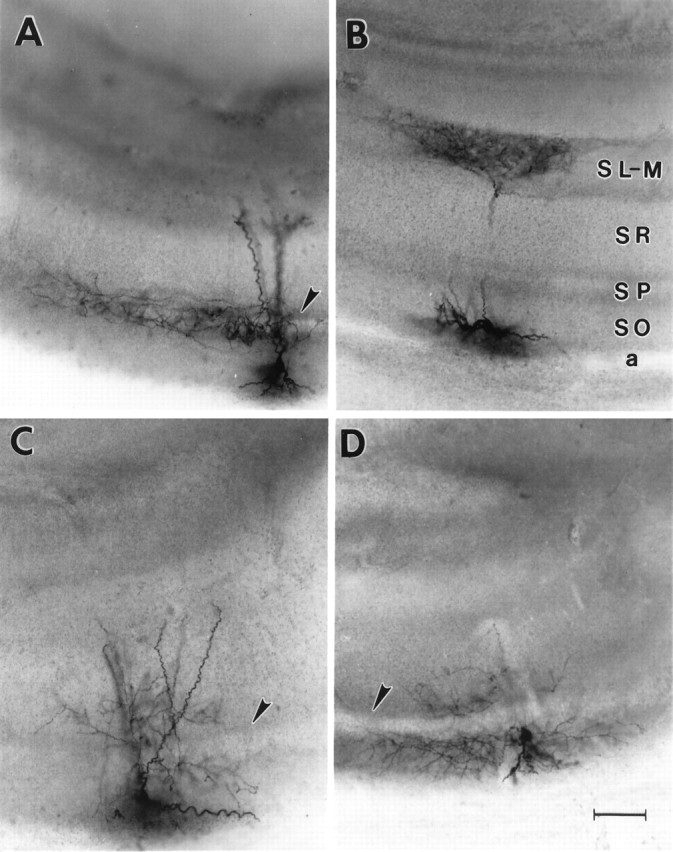
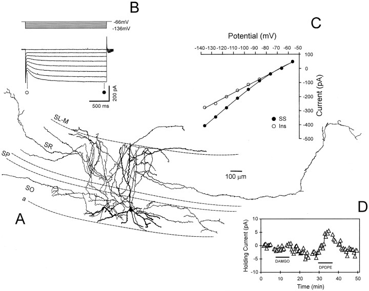
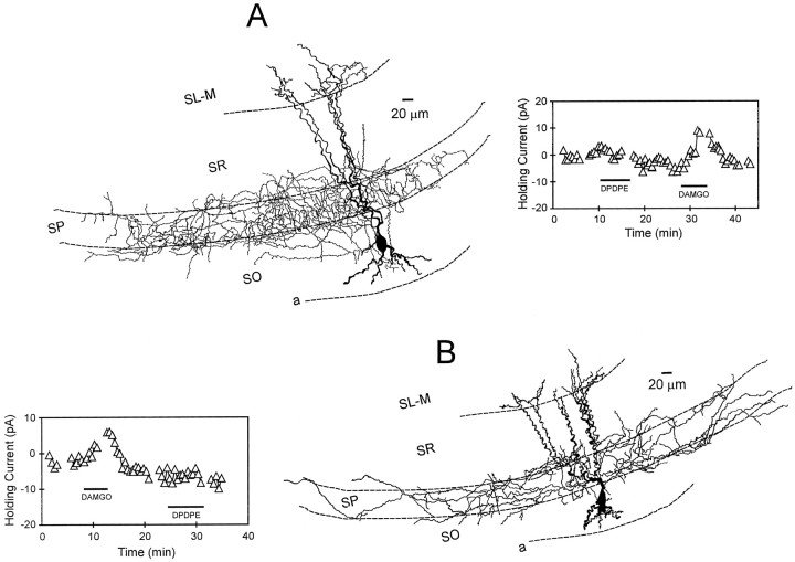
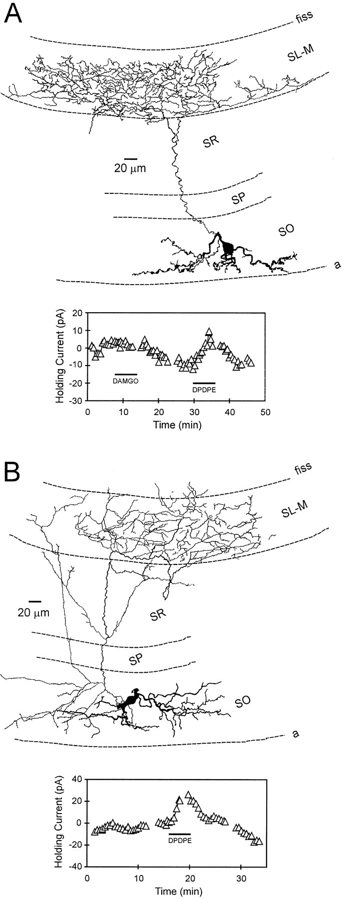
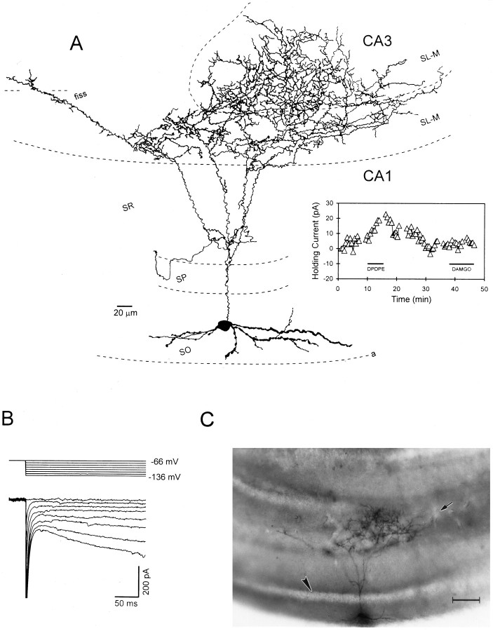
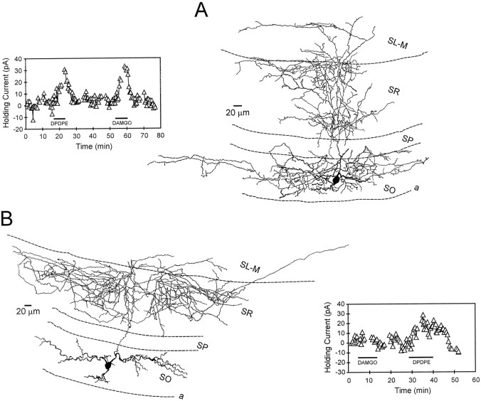
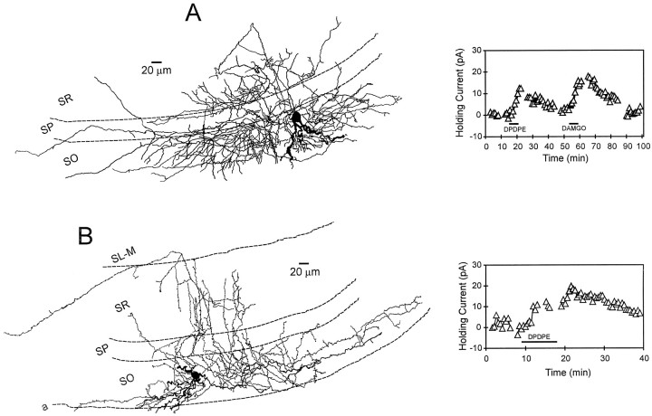
The inhibition of hippocampal pyramidal cells occurs via inhibitory interneurons making GABAergic synapses on distinct segments of the postsynaptic membrane. In area CA1 of the hippocampus, the activation of mu- and delta-opioid receptors inhibits these interneurons, thereby increasing the excitability of the pyramidal cells. Through the use of selective opioid agonists and biocytin-filled whole-cell electrodes, interneurons possessing somata located within stratum oriens of hippocampal slices were classified according to the location of their primary axon termination and the expression of mu- or delta-opioid receptors. Activation of these opioid receptor subtypes resulted in outward currents in the majority of interneurons, which is consistent with their inhibition. Post hoc morphological analysis revealed that those interneurons heavily innervating the pyramidal cell body layer were much more likely to express mu-opioid receptors, whereas cells with axons ramifying in the pyramidal neuron dendritic layers were more likely to express delta-opioid receptors, as defined by the generation of outward currents. This morphological segregation of interneuron projections suggests that mu receptor activation would diminish GABA release onto pyramidal neuron somata, thereby increasing their excitability and output. Conversely, inhibition of interneurons via delta receptor activation would amplify afferent signaling to pyramidal neuron dendrites by reducing GABAergic inhibition of these structures.


Figures

Fig. 1.
Fig. 1.
Morphology of stratum oriens interneurons. Photomicrographs of the four subtypes of biocytin-filled stratum oriens interneurons analyzed in this study. A, An interneuron with its axon almost exclusively innervating stratum pyramidale, in which the CA1 pyramidal cell bodies are located.B, An OLM or horizontal cell. Note the dense axon termination in stratum-lacunosum moleculare and the horizontally oriented dendrites in stratum oriens. These are the defining anatomical characteristics of this cell type. C, An interneuron with its primary axonal ramification in stratum radiatum.D, An interneuron with its primary axonal projection confined to stratum oriens. The arrowheads denote stratum pyramidale in A, C, andD. Scale bar, 100 μm. a, Alveus;SO, stratum oriens; SP, stratum pyramidale; SR, stratum radiatum;SL-M, stratum lacunosum-moleculare.
Fig. 2.
Fig. 2.
Physiological characteristics and opioid sensitivity of a stratum oriens interneuron. A, A camera lucida reconstruction of an interneuron demonstrating extensive axonal ramification in stratum radiatum from which the data shown in panelsBD were obtained. B, Current records obtained during 2 sec hyperpolarizing voltage steps from −56 to −136 mV. The large inward current relaxation near the end of the voltage steps is caused by activation ofIh. C, Current–voltage (IV) relationship demonstrating the instantaneous (○) and steady-state (•) currents obtained at step potentials between −136 and −56 mV. D, Time course of opioid-induced outward currents. The horizontal bars indicate the duration of opioid agonist bath application. In this example, the δ-opioid agonist DPDPE (1 μm) produced an outward current, whereas the μ-opioid agonist DAMGO had no effect. This illustrates the selectivity of these agonists.Gaps in the record denote time points ofIV generation.
Fig. 3.
Fig. 3.
Interneurons innervating stratum pyramidale: morphology and opioid pharmacology. A, Camera lucida reconstruction of an interneuron with its axonal projection restricted almost exclusively to stratum pyramidale (same cell as Fig.1A). The apical dendrites of this cell (darker structures) extend across stratum radiatum and into stratum lacunosum-moleculare. The inset at theright illustrates time course of the DAMGO-induced outward current. This cell was presumed to express μ and not δ receptors because an outward current was only generated with DAMGO.B, Another interneuron with similar morphology to that shown in A. The inset at theleft shows the time course of the DAMGO-generated outward current. The lateral extent of the axon in this cell was 1133 μm. Note that this neuron did not respond to the δ agonist DPDPE.
Fig. 4.
Fig. 4.
Stratum oriens interneurons innervating stratum lacunosum- moleculare (OLM or horizontal cells): morphology and opioid pharmacology. A, Camera lucida reconstruction of an interneuron with its axonal projection almost exclusively in stratum lacunosum-moleculare. The dendrites of this cell course parallel with the alveus border and are restricted to stratum oriens. Theinset below illustrates the effect of the δ-opioid agonist DPDPE on holding current. In this example, DAMGO failed to generate an outward current. B, Reconstruction of another interneuron with its axon ramifying within stratum lacunosum-moleculare. The inset below shows the time course of the δ-opioid agonist-generated outward current.fiss, Hippocampal fissure.
Fig. 5.
Fig. 5.
A stratum oriens “backprojection” cell innervating stratum lacunosum-moleculare of hippocampal subfields CA1 and CA3: morphology and physiology. A, A camera lucida reconstruction of the interneuron. In general, the morphology of this cell was similar to those shown in Figure 4. Note that the axon of this cell also crossed the hippocampal fissure and ramified extensively within stratum lacunosum moleculare of CA3. The inset at the right illustrates time course of the DPDPE-induced outward current. The μ-opioid agonist DAMGO had no effect.B, Current and voltage records obtained in this same cell. Note the prominent inward “sag” on the current responses at the most hyperpolarized potentials that is indicative ofIh. C, A photomicrograph of the interneuron reconstructed in A. Thearrowhead indicates the location of stratum pyramidale, and the arrow denotes the border between CA1 and CA3. Scale bar, 100 μm. fiss, Hippocampal fissure.
Fig. 6.
Fig. 6.
Interneurons innervating stratum radiatum: morphology and opioid pharmacology. A, Camera lucida reconstruction of an interneuron with its axon projection in strata radiatum and oriens. After further analysis (counting of axonal branch points), it was determined that this cell more heavily innervated stratum radiatum. The inset at the leftillustrates that both δ-opioid (DPDPE) and μ-opioid (DAMGO) agonists generated robust outward currents in this cell.B, Camera lucida reconstruction of an interneuron that exhibited an axonal projection predominantly confined to stratum radiatum. The inset at the right illustrates the time course of the δ agonist-induced outward current. This cell did not respond to the μ agonist DAMGO.
Fig. 7.
Fig. 7.
Interneurons primarily innervating stratum oriens: morphology and opioid pharmacology. A, A camera lucida reconstruction of a stratum oriens interneuron with its primary axonal projection confined to this layer (same cell as shown in Fig.1D). Note that the axon does not heavily innervate stratum pyramidale, yet ramifies in both stratum radiatum and oriens. This morphological profile is similar to that described for “bistratified” neurons (Buhl et al., 1994a). Theinset at the right illustrates the time course of the DAMGO- and DPDPE-induced holding current changes. Although this interneuron was sensitive to both opioid agonists, only 6 of 16 interneurons responded to the μ agonist, whereas 13 of 17 responded to the δ-opioid receptor agonist. B, Reconstruction of a stratum oriens interneuron that demonstrated an extensive primary axonal projection within stratum oriens. Theinset at the right shows the time course of the DPDPE-induced outward current in this same cell.

References

    1. Ali AB, Thomson AM. Facilitating pyramid to horizontal oriens-alveus interneurone inputs- Dual intracellular recordings in slices of rat hippocampus. J Physiol (Lond) 1998;507:185–199. - PMC - PubMed
    1. Ali AB, Deuchars J, Pawelzik H, Thomson AM. CA1 pyramidal to basket and bistratified cell EPSPs: dual intracellular recordings in rat hippocampal slices. J Physiol (Lond) 1998;507:201–217. - PMC - PubMed
    1. Bergles DE, Doze VA, Madison DV, Smith SJ. Excitatory actions of norepinephrine on multiple classes of interneurons. J Neurosci. 1996;16:572–585. - PMC - PubMed
    1. Buhl EH, Halasy K, Somogyi P. Diverse sources of hippocampal unitary inhibitory postsynaptic potentials and the number of synaptic release site. Nature. 1994a;368:823–828. - PubMed
    1. Buhl EH, Han Z, Lorinczi Z, Stezhka VV, Karnup SV, Somogyi P. Physiological properties of anatomically identified axo-axonic cells in the rat hippocampus. J Neurophysiol. 1994b;71:1289–1307. - PubMed

Publication types

Substances