. 1999 Jan 1;19(1):133-46.
doi: 10.1523/JNEUROSCI.19-01-00133.1999.

Heparin-binding epidermal growth factor-like growth factor in hippocampus: modulation of expression by seizures and anti-excitotoxic action

Affiliations

Heparin-binding epidermal growth factor-like growth factor in hippocampus: modulation of expression by seizures and anti-excitotoxic action

L A Opanashuk et al. J Neurosci. .

Abstract

The expression of heparin-binding epidermal growth factor-like growth factor (HB-EGF), an EGF receptor ligand, was investigated in rat forebrain under basal conditions and after kainate-induced excitotoxic seizures. In addition, a potential neuroprotective role for HB-EGF was assessed in hippocampal cultures. In situ hybridization analysis of HB-EGF mRNA in developing rat hippocampus revealed its expression in all principle cell layers of hippocampus from birth to postnatal day (P) 7, whereas from P14 through adulthood, expression decreased in the pyramidal cell layer versus the dentate gyrus granule cells. After kainate-induced excitotoxic seizures, levels of HB-EGF mRNA increased markedly in the hippocampus, as well as in several other cortical and limbic forebrain regions. In the hippocampus, HB-EGF mRNA expression increased within 3 hr after kainate treatment, continued to increase until 24 hr, and then decreased; increases occurred in the dentate gyrus granule cells, in the molecular layer of the dentate gyrus, and in and around hippocampal pyramidal CA3 and CA1 neurons. At 48 hr after kainate treatment, HB-EGF mRNA remained elevated in vulnerable brain regions of the hippocampus and amygdaloid complex. Western blot analysis revealed increased levels of HB-EGF protein in the hippocampus after kainate administration, with a peak at 24 hr. Pretreatment of embryonic hippocampal cell cultures with HB-EGF protected neurons against kainate toxicity. The kainate-induced elevation of [Ca2+]i in hippocampal neurons was not altered in cultures pretreated with HB-EGF, suggesting an excitoprotective mechanism different from that of previously characterized excitoprotective growth factors. Taken together, these results suggest that HB-EGF may function as an endogenous neuroprotective agent after seizure-induced neural activity/injury.


Figures
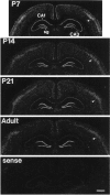
Fig. 1.
Fig. 1.
Expression of HB-EGF mRNA in developing hippocampus. Autoradiograms showing the in situhybridization localization of HB-EGF mRNA in coronal sections through the hippocampus at the indicated postnatal day ages. Note that atP7, labeling is present throughout hippocampal fieldsCA1 and CA3 and dentate gyrus stratum granulosum (sg), whereas in older (P14, P21, Adult) rats, HB-EGF mRNA expression is substantially reduced in the pyramidal cell layer relative to the stratum granulosum. In neocortex, HB-EGF cRNA hybridization is prominent in the middle layers (arrowheads), with levels gradually declining from P7 to adulthood. The bottom panel (sense) demonstrates the lack of hybridization obtained with the control sense cRNA probe in a section through the adult hippocampus. Film exposure, 14 d; section thickness, 14 μm. Scale bar, 1000 μm.
Fig. 2.
Fig. 2.
A, Northern blot analysis of HB-EGF mRNA levels in adult rat hippocampus at various survival times (3–48 hr) after kainate administration. Total RNA (20 μg/lane) was separated on a 1.2% agarose gel, transferred to nylon membrane, and hybridized with an antisense 32P-cRNA HB-EGF probe (top). Membranes were stripped and reprobed with a32P-labeled β actin cRNA to control for equal loading of RNA (bottom). HB-EGF mRNA was detected by autoradiography with an exposure time of 5 d. Levels of the 2.5 kb transcript corresponding to HB-EGF mRNA were elevated 3 hr after kainate treatment and continued to increase through the 24 hr time point. By 48 hr, HB-EGF mRNA levels were declining. Con, Control. B, Densitometric analysis of HB-EGF mRNA levels at various times after kainate administration. HB-EGF mRNA levels were normalized relative to β actin mRNA levels. Data are expressed as mean ± SEM of determinations made in three rats per time point and were analyzed by one-way ANOVA with Duncan post hocanalysis. *p < 0.05 compared with the control (0 hr) value.
Fig. 3.
Fig. 3.
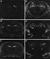
Kainate-induced increase of HB-EGF mRNA expression in hippocampus. Autoradiograms demonstrating the in situhybridization localization of HB-EGF mRNA in coronal sections through the hippocampus in a saline-injected control rat (A) and in rats killed 3 (B), 6 (C), 12 (D), 24 (E), and 48 hr (F) after kainate administration. Expression of HB-EGF mRNA is upregulated in the granule cell layer [stratum granulosum (sg)] of the dentate gyrus 3–6 hr after kainate treatment (B, C) but then declines to control levels by 48 hr (F). Kainate treatment induces prominent hybridization in and around the pyramidal cell layers of hippocampal subfields CA1 and CA3 at the 12 and 24 hr time points (D, E); by 48 hr, labeling is restricted to the CA1 region in the hippocampus (F). In neocortex (nc), HB-EGF mRNA expression increases from 3 to 12 hr after kainate treatment (B–D) and then decreases to control levels by 48 hr (E, F). Note that hybridization signal is also present in and around areas of kainate-induced cell loss at the 24 and 48 hr time points (E, F), including regions of the dorsal lateral thalamus (arrows inF), amygdaloid complex (amg), and piriform cortex (pc). Film exposure, 10 d; section thickness, 10 μm. Note that because of thinner sectioning and shorter exposure time, the control section in A cannot be directly compared with the normal adult section in Figure 1. Scale bar, 1000 μm.
Fig. 4.
Fig. 4.
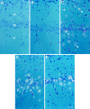
Kainate-induced increase of HB-EGF mRNA in dentate gyrus. Dark-field photomicrographs showing the cellular distribution of HB-EGF mRNA in sections through the dentate gyrus in a saline-injected control rat (A) and in rats killed 3 (B), 6 (C), 24 (D), and 48 hr (E) after kainate administration. Note that kainate treatment initially increased HB-EGF cRNA hybridization in the granule cell layer [stratum granulosum (sg)] at the 3 hr time point (B). However, from 6 to 24 hr after kainate injection, numerous labeled cells (arrowheads indicate several examples) were distributed throughout the hilus (h) and molecular layer (ml) (C, D). By 48 hr, cellular expression of HB-EGF mRNA was barely detectable within the dentate gyrus (E). Scale bar, 50 μm.
Fig. 5.
Fig. 5.
Kainate-induced increase of HB-EGF mRNA in hippocampal region CA1. Dark-field photomicrographs show the autoradiographic localization of HB-EGF cRNA in situhybridization in the CA1 region of hippocampus in a saline-injected control rat (A) and in rats killed 3 (B), 12 (C), 24 (D), and 48 hr (E) after kainate administration. Note that by 12 hr after kainate injection, labeling for HB-EGF mRNA was induced in stratum pyramidale (sp) and particularly within numerous cells scattered throughout the stratum oriens (so) and stratum radiatum (sr) molecular layers (C, arrowheadsindicate examples of labeled somata). By 24 hr, densely labeled cells were situated in close proximity to the pyramidal cell layer (D). At 48 hr after kainate treatment, perikarya expressing HB-EGF mRNA were intermittently dispersed within and along the edges of CA1 stratum pyramidale (E). Scale bar, 50 μm.
Fig. 6.
Fig. 6.
Kainate-induced expression of HB-EGF mRNA in septum and cortex. Autoradiograms demonstrating the regional distribution of HB-EGF cRNA hybridization in coronal sections through the septal nuclei in a saline-injected control rat (A) and in rats killed at 3 (B), 12 (C), 24 (D), and 48 hr (E) after kainate administration. Expression of HB-EGF mRNA is induced in the lateral septal nuclei (ls), neocortex (nc), and piriform cortex (pc) by 3 hr after kainate treatment (B), with peak expression levels found between 12 and 24 hr after kainate treatment (C, D). By 48 hr (E), labeling is restricted to scattered cells within the lateral septum and piriform cortex and to degenerating areas of perirhinal cortex (asterisks). Film exposure, 10 d; section thickness, 10 μm. Scale bar, 1000 μm.
Fig. 7.
Fig. 7.
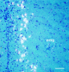
Kainate-induced expression of HB-EGF mRNA in extra-hippocampal regions of neuronal cell loss. Dark-field photomicrograph shows HB-EGF cRNA-hybridizing cells lining the medial edge of the amygdaloid complex (amg) in a rat killed 48 hr after kainate administration. Scale bar, 90 μm.
Fig. 8.
Fig. 8.
Western blot analysis of HB-EGF protein in rat hippocampus (top panel), in a saline-injected control rat (Con), and in rats killed at the indicated time points after kainate administration. Protein (100 μg/lane) was separated by SDS-PAGE, transferred to Hybond (Amersham) membrane, and probed with a polyclonal antibody to HB-EGF (kindly provided by Judith Abraham, Scios Inc.). Proteins were visualized by enhanced chemiluminescence. Recombinant human HB-EGF (10 ng) was used as a positive control. Four bands corresponding to HB-EGF with apparent molecular weights of 27, 24, 17, and 14 kDa are detected in hippocampal extracts. Similar results were obtained in three different rats for each time point. Membranes were also probed with a monoclonal antibody for neurofilament protein (bottom panel) to ensure equivalent gel loading.
Fig. 9.
Fig. 9.
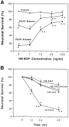
HB-EGF protects against kainate neurotoxicity in hippocampal cell cultures. A, Cultures were pretreated with 1–100 ng/ml human recombinant HB-EGF for 16 hr and were then exposed to either 10 or 50 μm kainate. Neuronal survival was assessed 12 hr after kainate treatment. Values represent the mean ± SEM of determinations made in five separate experiments. Pair-wise comparisons were made by ANOVA with Scheffé’spost hoc analysis. *p < 0.01 compared with the value for vehicle-treated cultures not exposed to HB-EGF; **p < 0.01 compared with values for cultures pretreated with either 0 or 1 ng/ml HB-EGF and then exposed to the same concentration of kainate. B, Cultures were pretreated with 100 ng/ml HB-EGF for 16 hr and then exposed to vehicle or 10 μm kainate. Neuronal survival was assessed at the indicated time points after kainate administration. Values are the mean ± SEM of determinations made in three separate experiments. Pair-wise comparisons were made by ANOVA with Scheffé’spost hoc analysis. *p < 0.01, **p < 0.001 compared with each of the other values at that time point.
Fig. 10.
Fig. 10.
HB-EGF does not affect kainate-induced elevations in [Ca2+]i. Hippocampal cultures were pretreated with 100 ng/ml HB-EGF for 16 hr, and the [Ca2+]i in neurons was then determined (time 0). Cells were then exposed to 50 μm kainate for 5 min, and the [Ca2+]i was again measured in the same neurons. Values are the mean ± SEM of determinations made in four separate cultures (15–22 neurons per culture).

References

    1. Abe K, Xie F-J, Saito H. Epidermal growth factor enhances short-term potentiation and facilitates induction of long-term potentiation in rat hippocampal slices. Brain Res. 1991;547:171–174. - PubMed
    1. Abe K, Ishiyama J, Saito H. Effects of epidermal growth factor and basic fibroblast growth factor on generation of long-term potentiation in the dentate gyrus of fimbria-fornix-lesioned rats. Brain Res. 1992;593:335–338. - PubMed
    1. Abraham JA, Damm D, Bajardi A, Miller J, Klagsbrun M, Ezekowitz AB. Heparin-binding EGF-like growth factor: characterization of rat and mouse cDNA clones, protein domain conservation across species, and transcript expression in tissues. Biochem Biophys Res Commun. 1993;190:125–133. - PubMed
    1. Alexi T, Hefti F. Trophic actions of transforming growth factor-α on mesencephalic dopaminergic neurons developing in culture. Neuroscience. 1993;55:903–918. - PubMed
    1. Barnard JA, Graves-Deal R, Pittelkow MR, DuBois R, Cook P, Ramsey GW, Bishop PR, Damstrup L, Coffey RJ. Auto- and cross-induction within the mammalian epidermal growth factor-related peptide family. J Biol Chem. 1994;269:22817–22822. - PubMed

Publication types