

Functionally dissociating aspects of event memory: the effects of combined perirhinal and postrhinal cortex lesions on object and place memory in the rat
- PMID: 9870977
- PMCID: PMC6782353
- DOI: 10.1523/JNEUROSCI.19-01-00495.1999
Functionally dissociating aspects of event memory: the effects of combined perirhinal and postrhinal cortex lesions on object and place memory in the rat
Abstract
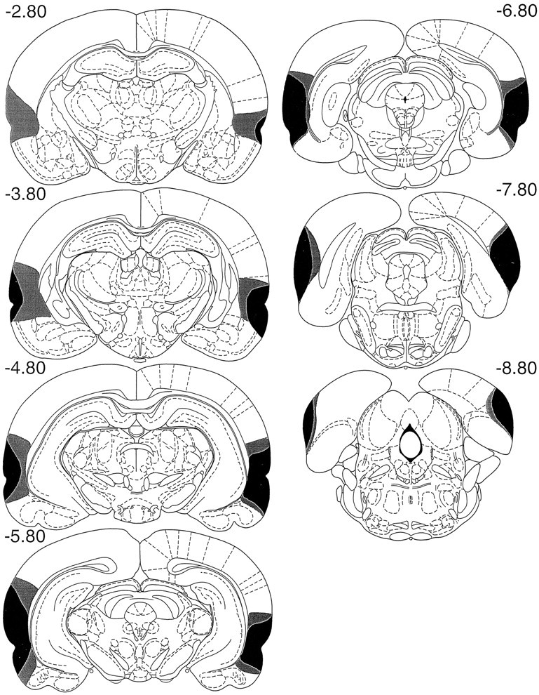
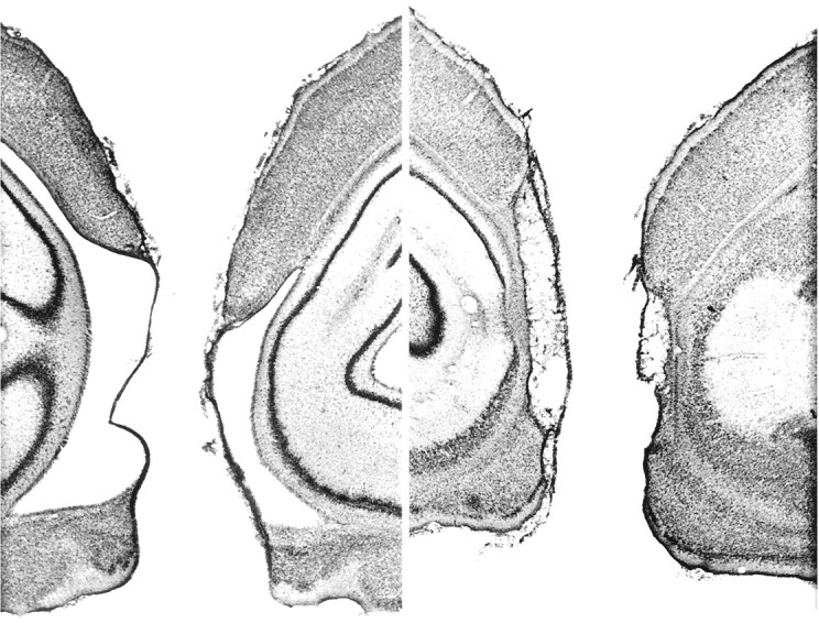
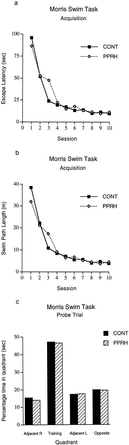
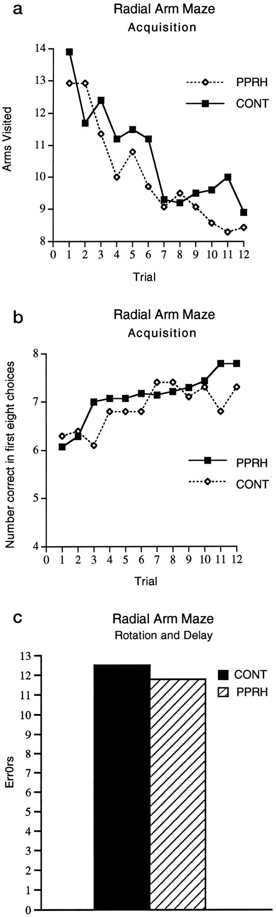
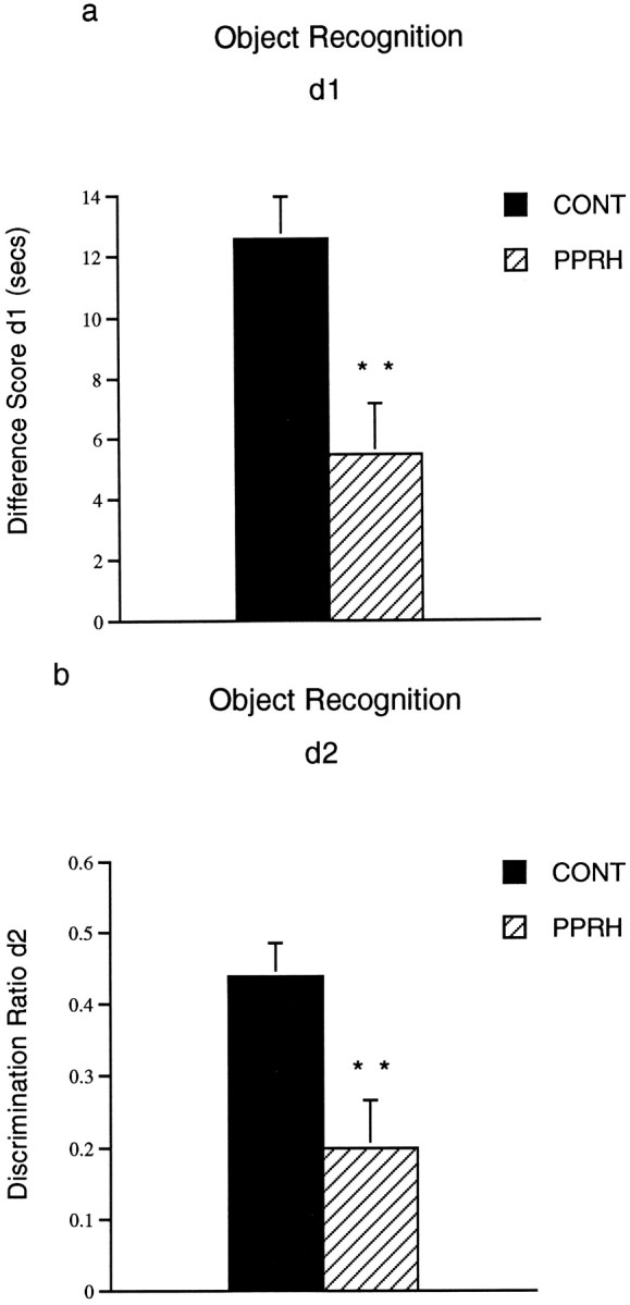
Reciprocal interactions between the hippocampus and the perirhinal and parahippocampal cortices form core components of a proposed temporal lobe memory system. For this reason, the involvement of the hippocampus in event memory is thought to depend on its connections with these cortical areas. Contrary to these predictions, we found that NMDA-induced lesions of the putative rat homologs of these cortical areas (perirhinal plus postrhinal cortices) did not impair performance on two allocentric spatial tasks highly sensitive to hippocampal dysfunction. Remarkably, for one of the tasks there was evidence of a facilitation of performance. The same cortical lesions did, however, disrupt spontaneous object recognition and object discrimination reversal learning but spared initial acquisition of the discrimination. This pattern of results reveals important dissociations between different aspects of memory within the temporal lobe. Furthermore, it shows that the perirhinal-postrhinal cortex is not a necessary route for spatial information reaching the hippocampus and that object familiarity-novelty detection depends on different neural substrates than do other aspects of event memory.
Figures
References
-
- Aggleton JP, Keen S, Warburton EC, Bussey TJ. Extensive cytotoxic lesions involving both the rhinal cortices and area TE impair recognition but spare spatial alternation in the rat. Brain Res Bull. 1997;43:279–287. - PubMed
-
- Brown MW. Why does the cortex have a hippocampus? In: Gabriel M, Moore J, editors. Learning and computational neuroscience: foundations of adaptive networks. MIT; New York: 1990.
-
- Buckley MJ, Gaffan D. Impairment of visual object-discrimination learning after perirhinal cortex ablation. Behav Neurosci. 1997;111:467–475. - PubMed
-
- Burwell RD, Witter MP, Amaral DG. Perirhinal and postrhinal cortices of the rat: a review of the neuroanatomical literature and comparison with findings from the monkey brain. Hippocampus. 1995;5:390–408. - PubMed
Publication types
MeSH terms
Grants and funding
LinkOut - more resources
Full Text Sources
Medical