. 1999 Jan;65(1):169-74.
doi: 10.1128/AEM.65.1.169-174.1999.

In situ population dynamics of bacterial viruses in a terrestrial environment

Affiliations

In situ population dynamics of bacterial viruses in a terrestrial environment

KE Ashelford et al. Appl Environ Microbiol. 1999 Jan.

Abstract

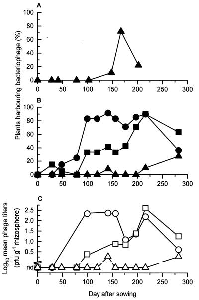
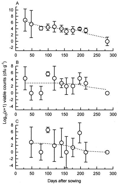
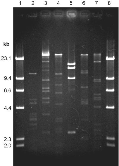
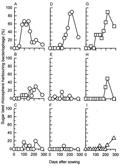
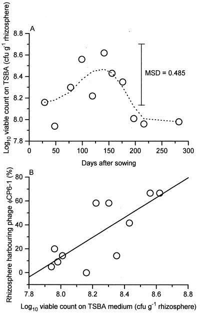
Predation by bacteriophages is thought to control bacterial numbers and facilitate gene transfer among bacteria in the biosphere. A thorough understanding of phage population dynamics is therefore necessary if their significance in natural environments is to be fully appreciated. Here we describe the in situ population dynamics of three separate phage populations predating on separate bacterial species, living on the surface of field-grown sugar beet (Beta vulgaris var. Amethyst), as recorded over a 9-month period. The distributions of the three phage populations were different and fluctuated temporally in 1996 (peak density, approximately 10(3) PFU g-1). One of these populations, predating on the indigenous phytosphere bacterium Serratia liquefaciens CP6, consisted of six genetically distinct DNA phages that varied in relative abundance to the extent that an apparent temporal succession was observed between the two most abundant phages, PhiCP6-1 and PhiCP6-4.


Figures

FIG. 1
FIG. 1
Temporal variation in occurrence of phages isolated from sugar beet during 1994 (A) or 1996 to 1997 (B and C) and antagonistic toward P. fluorescens SBW25 (triangles), P. fluorescens MCP1 (squares), or S. liquefaciens CP6 (circles).
FIG. 2
FIG. 2
Temporal variation during 1996 to 1997 of viable counts of P. fluorescens SBW25 established in the phytosphere microflora of sugar beet rhizosphere (A), leaf (B), and leaf bud (C) samples. Group medians for each sampling occasion are plotted (circles), along with their respective boxplot notches (bars [27]), enabling a pairwise comparison of group medians at the 95% level; overlapping bars indicate medians that are not significantly different. Dotted lines represent the same data after smoothing with the 4253H twice algorithm (27).
FIG. 3
FIG. 3
ClaI restriction digests, run on a 0.7% agarose gel, illustrating the six different phages antagonistic toward S. liquefaciens CP6, isolated from sugar beet during 1996 to 1997. Lanes 1 and 8, HindIII-cut lambda DNA ladder; lanes 2 to 7, phages ΦCP6-1 to ΦCP6-6, respectively.
FIG. 4
FIG. 4
Temporal variation in occurrence of individual phage species in sugar beet rhizosphere samples over the 1996 to 1997 experimental period. Phages ΦCP6-1 to ΦCP6-6 (A to F, respectively) were antagonistic toward S. liquefaciens CP6, while phages ΦMCP1-1 and ΦMCP1-2 (G and H) infected P. fluorescens MCP1. Phages antagonistic toward P. fluorescens SBW25 are also represented (I). Twenty sugar beet plants were sampled on days 29 and 48, 12 were sampled on days 78 to 156 inclusive, 14 were sampled on days 176 and 197, 10 were sampled on day 216, and 11 were sampled on day 282.
FIG. 5
FIG. 5
Temporal variation in total numbers of viable bacteria, isolated on TSBA medium, within the sugar beet rhizosphere during 1996 to 1997. Group means (n = 10 to 20, depending on the sampling day) are plotted against time (A) and phage ΦCP6-1 abundance (B). Smoothed data (––––) was calculated from the means by using the 4253H twice algorithm (27). Least-squares linear regression (9) was used to calculate the regression line (——). Phage abundance (%) = −652.2 + 83.2 × viable count. MSD, minimum significant difference.

Similar articles

Cited by

References

    1. Adams M H. Bacteriophages. New York, N.Y: Interscience Publishers; 1959.
    1. Bailey M J, Lilley A K, Thompson I P, Rainey P B, Ellis R J. Site directed chromosomal marking of a fluorescent pseudomonad isolated from the phytosphere of sugar beet; stability and potential for marker gene transfer. Mol Ecol. 1995;4:755–763. - PubMed
    1. Bergh Ø, Børsheim K Y, Bratbak G, Heldal M. High abundance of viruses found in aquatic environments. Nature. 1989;340:467–468. - PubMed
    1. Bratbak G, Heldal M, Norland S, Thingstad F. Viruses as partners in spring bloom microbial trophodynamics. Appl Environ Microbiol. 1990;56:1400–1405. - PMC - PubMed
    1. Chao L, Levin B R, Stewart F M. A complex community in a simple habitat: an experimental study with bacteria and phage. Ecology. 1977;58:369–378.

LinkOut - more resources