. 1999 Jan 4;189(1):89-102.
doi: 10.1084/jem.189.1.89.

Identification and characterization of novel superantigens from Streptococcus pyogenes

Affiliations

Identification and characterization of novel superantigens from Streptococcus pyogenes

T Proft et al. J Exp Med. .

Abstract

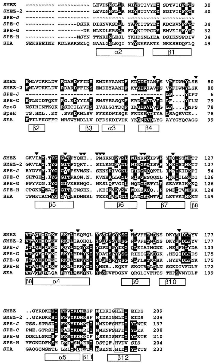
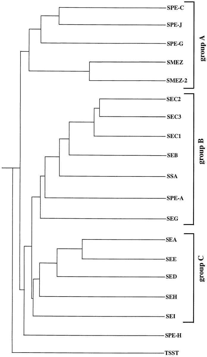
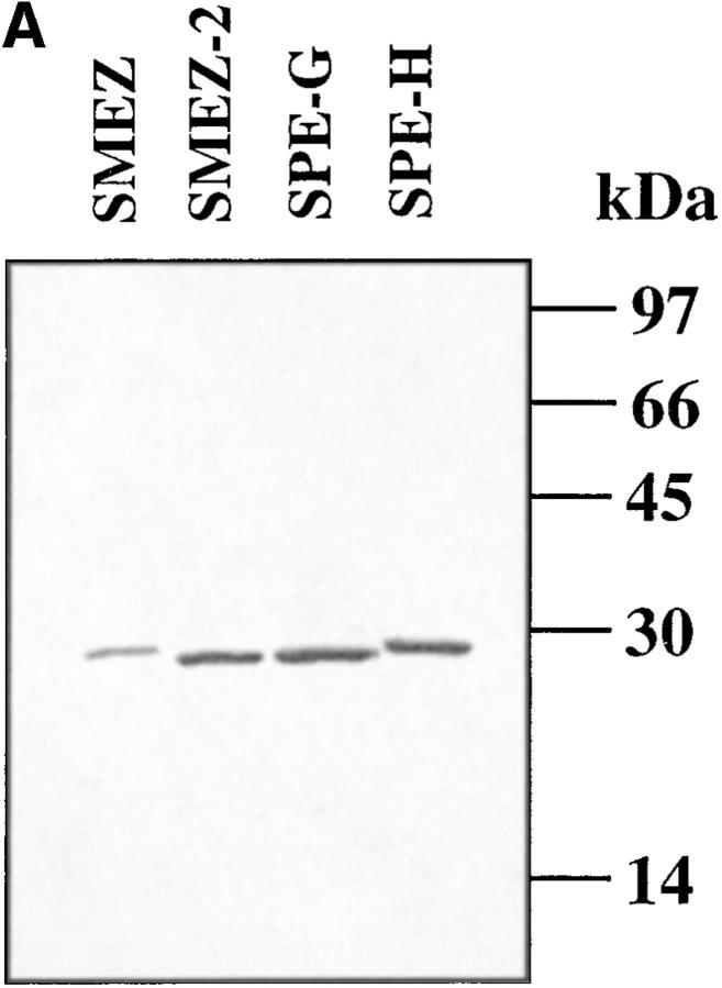
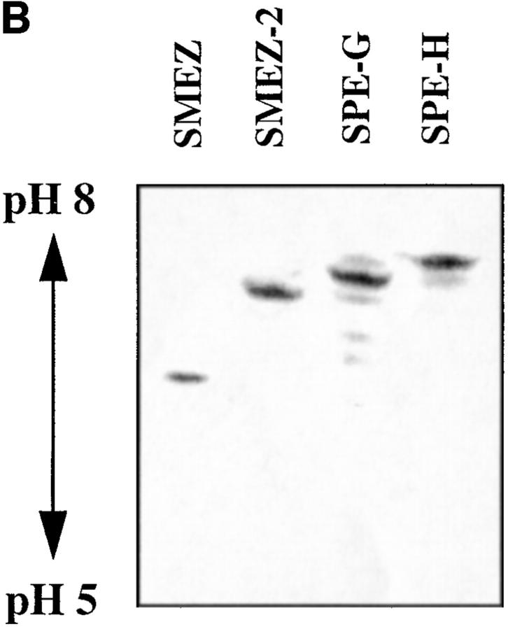
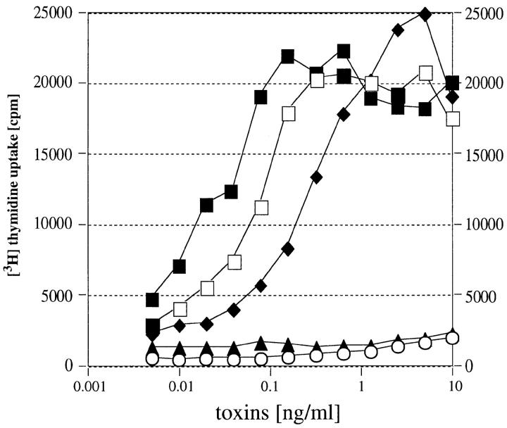
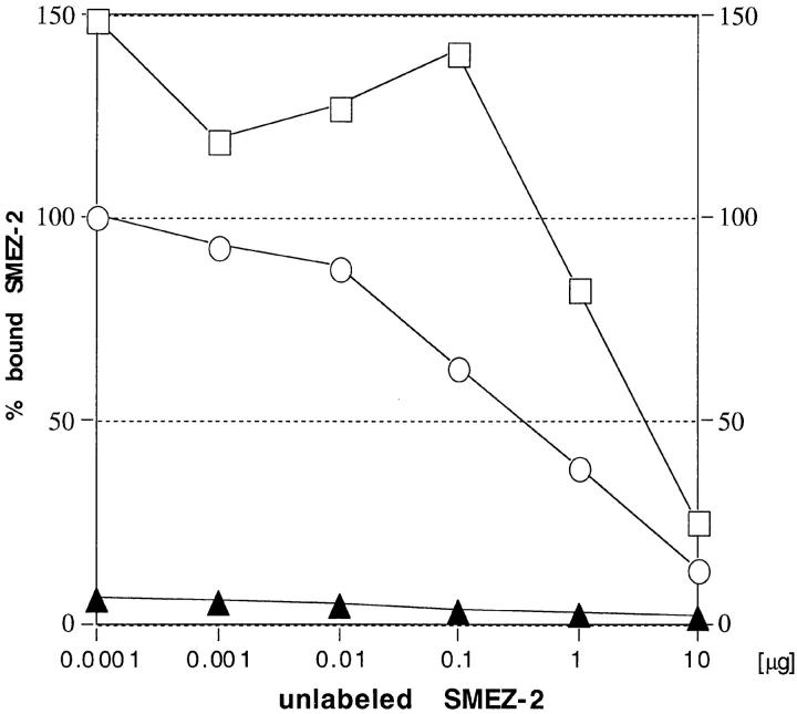
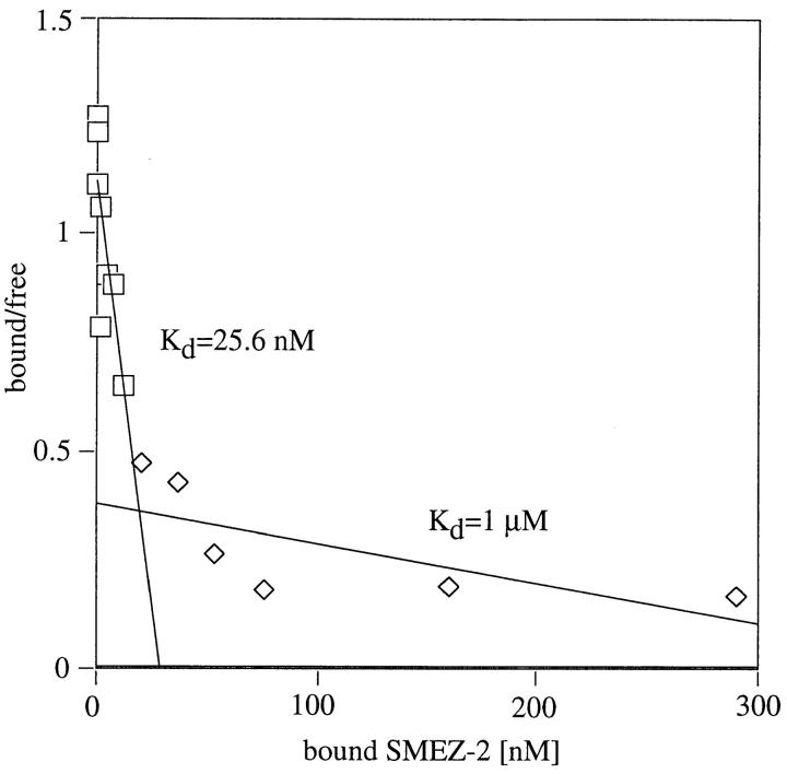
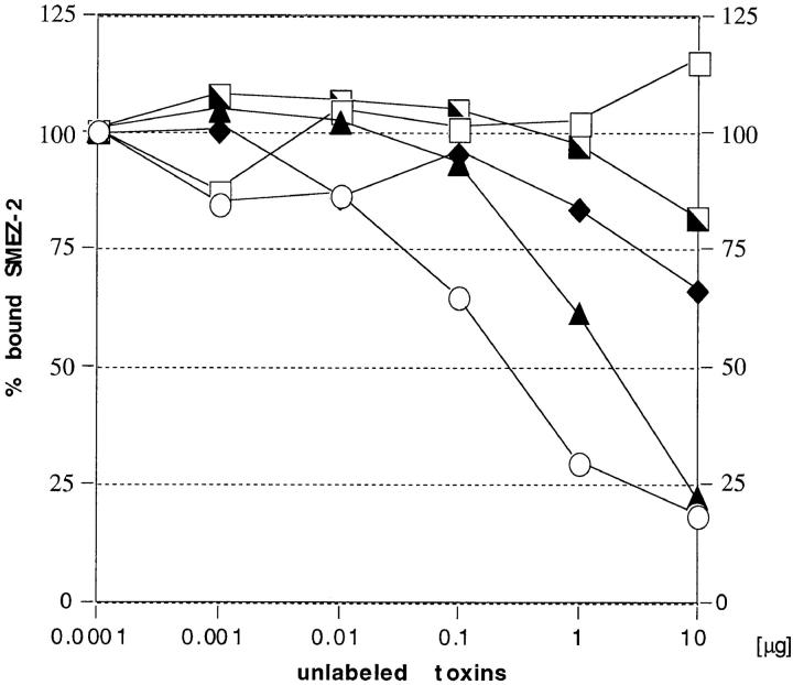
Three novel streptococcal superantigen genes (spe-g, spe-h, and spe-j) were identified from the Streptococcus pyogenes M1 genomic database at the University of Oklahoma. A fourth novel gene (smez-2) was isolated from the S. pyogenes strain 2035, based on sequence homology to the streptococcal mitogenic exotoxin z (smez) gene. SMEZ-2, SPE-G, and SPE-J are most closely related to SMEZ and streptococcal pyrogenic exotoxin (SPE)-C, whereas SPE-H is most similar to the staphylococcal toxins than to any other streptococcal toxin. Recombinant (r)SMEZ, rSMEZ-2, rSPE-G, and rSPE-H were mitogenic for human peripheral blood lymphocytes with half-maximal responses between 0.02 and 50 pg/ml (rSMEZ-2 and rSPE-H, respectively). SMEZ-2 is the most potent superantigen (SAg) discovered thus far. All toxins, except rSPE-G, were active on murine T cells, but with reduced potency. Binding to a human B-lymphoblastoid line was shown to be zinc dependent with high binding affinity of 15-65 nM. Evidence from modeled protein structures and competitive binding experiments suggest that high affinity binding of each toxin is to the major histocompatibility complex class II beta chain. Competition for binding between toxins was varied and revealed overlapping but discrete binding to subsets of class II molecules in the hierarchical order (SMEZ, SPE-C) > SMEZ-2 > SPE-H > SPE-G. The most common targets for the novel SAgs were human Vbeta2.1- and Vbeta4-expressing T cells. This might reflect a specific role for this subset of Vbetas in the immune defense of gram-positive bacteria.


Figures

Figure 1
Figure 1
Multiple alignment of SAg protein sequences. The protein sequences of mature toxins were aligned using the PileUp program on the GCG package. Regions of high sequence identity are in black boxes. The boxes below the sequences indicate the structural elements of SPE-C, as determined from the crystal structure (25). Regions with highest homology correspond to the β4, β5, α4, and α5 regions in SPE-C. The clear box near the COOH terminus represents a primary zinc binding motif, a common feature of all toxins shown here. The arrows on top of the sequence alignment show the regions of sequence diversity between SMEZ and SMEZ-2. The NH2-terminal part of SPE-J is missing, as the corresponding DNA sequence is not yet available in the database.
Figure 2
Figure 2
SAg family tree. The family tree was created using the PileUp program on the GCG package and is based on primary amino acid sequence homology. The SAg family can be divided into three groups (or subfamilies) and two individual branches (SPE-H and TSST). SMEZ, SMEZ-2, SPE-G, and SPE-J build a subfamily together with SPE-C. The complete DNA sequence of the spe-j gene is not yet available, so only the 135 COOH-terminal amino acid residues of SPE-J could be used for the alignment.
Figure 3
Figure 3
Gel electrophoresis of the purified recombinant toxins. (A) Two micrograms of purified recombinant toxin were run on a 12.5% SDS-polyacrylamide gel to show the purity of the preparations. (B) 2 μg of purified recombinant toxin were run on an isoelectric focusing gel (5.5% PAA, pH 5–8). The isoelectric point of rSMEZ-2, rSPE-G, and rSPE-H is similar and was estimated at pH 7–8. The IEP of rSMEZ was estimated at pH 6–6.5.
Figure 3
Figure 3
Gel electrophoresis of the purified recombinant toxins. (A) Two micrograms of purified recombinant toxin were run on a 12.5% SDS-polyacrylamide gel to show the purity of the preparations. (B) 2 μg of purified recombinant toxin were run on an isoelectric focusing gel (5.5% PAA, pH 5–8). The isoelectric point of rSMEZ-2, rSPE-G, and rSPE-H is similar and was estimated at pH 7–8. The IEP of rSMEZ was estimated at pH 6–6.5.
Figure 4
Figure 4
Stimulation of human T cells with recombinant toxins. PBLs were isolated from human blood samples and incubated with varying concentrations of recombinant toxin. After 3 d, 0.1 μCi [3H]thymidine was added and cells were incubated for another 24 h, before being harvested and counted on a gamma counter. ○, unstimulated; ▴, rSMEZ; □, rSMEZ-2; ♦, rSPE-G; ┘, rSPE-H.
Figure 5
Figure 5
Jurkat cell assay. Jurkat cells (bearing a Vβ8 TCR) and LG-2 cells were mixed with varying concentrations of recombinant toxin and incubated for 24 h, before SeI cells were added. After 1 d, 0.1 μCi [3H]thymidine was added and cells were counted after another 24 h. The Vβ8 targeting SEE was used as a positive control. The negative control was SEA. Both SMEZ and SMEZ-2 were potent stimulators of Jurkat cells, indicating their ability to specifically target Vβ8-bearing T cells. ○, unstimulated; ▴, rSEA; □, rSEE; ♦, rSMEZ; ▪, rSMEZ-2.
Figure 6
Figure 6
Computer generated models of protein structures. The models were created on a Silicon Graphic computer using InsightII/Homology software. SEA, SEB, and SPE-C were used as reference proteins to determine structurally conserved regions. The loop regions were generated by random choice. MolScript software (39) was used for displaying the computer generated images. (a) All models show a potential zinc binding site within the β-grasp motif of the toxin structure. Two zinc binding residues are provided by a primary zinc motif (H-X-D) and the third ligand (H or D) comes from either the β9 or β10 strand. The amino acid residues of the β1-β2 loop that corresponds to the HLA-DRI α chain binding site in SEA and SEB are shown on the right side of the models. In all three protein models, this loop region is less hydrophobic than in SEA and SEB, suggesting the lack of the α chain binding site. A, Crystal structures of SPE-C (25); B, SMEZ-2; C, SPE-G; D, SPE-H. (b) SMEZ-2 model, showing the predicted location of the 17 residues that are different between SMEZ and SMEZ-2. The first residue is from SMEZ-2, followed by the position on the primary protein sequence and the corresponding residue on SMEZ.
Figure 6
Figure 6
Computer generated models of protein structures. The models were created on a Silicon Graphic computer using InsightII/Homology software. SEA, SEB, and SPE-C were used as reference proteins to determine structurally conserved regions. The loop regions were generated by random choice. MolScript software (39) was used for displaying the computer generated images. (a) All models show a potential zinc binding site within the β-grasp motif of the toxin structure. Two zinc binding residues are provided by a primary zinc motif (H-X-D) and the third ligand (H or D) comes from either the β9 or β10 strand. The amino acid residues of the β1-β2 loop that corresponds to the HLA-DRI α chain binding site in SEA and SEB are shown on the right side of the models. In all three protein models, this loop region is less hydrophobic than in SEA and SEB, suggesting the lack of the α chain binding site. A, Crystal structures of SPE-C (25); B, SMEZ-2; C, SPE-G; D, SPE-H. (b) SMEZ-2 model, showing the predicted location of the 17 residues that are different between SMEZ and SMEZ-2. The first residue is from SMEZ-2, followed by the position on the primary protein sequence and the corresponding residue on SMEZ.
Figure 7
Figure 7
Zinc dependent binding of SMEZ-2 to LG-2 cells. LG-2 cells were incubated in duplicates with 1 ng of 125I-labeled rSMEZ-2 and increasing amounts of unlabeled toxin at 37°C for 1 h, and then the cells were washed and counted. ○, incubation in media; ▴, incubation in media plus 1 mM EDTA; □, incubation in media plus 1 mM EDTA, 2 mM ZnCl2.
Figure 8
Figure 8
Scatchard analysis of SMEZ-2 binding to LG-2 cells. 1 ng 125I-labeled rSMEZ-2 was incubated in duplicates with LG-2 cells and a twofold dilution series of cold toxin (10 μg to 10 pg). After 1 h, cells were washed and counted. Scatchard plots were performed as described elsewhere (40). ⋄, low affinity binding of SMEZ-2 to LG-2 cells; □, high affinity binding of SMEZ-2 to LG-2 cells.
Figure 10
Figure 10
Summary of competitive binding experiments. Efficiency of each labeled toxin to compete with a 10,000-fold molar excess of any other unlabeled toxin for binding to LG-2 cells. White boxes, no competition; lightest gray boxes, 25% competition; medium gray boxes, 50% competition; dark gray boxes, 75% competition; black boxes, 100% competition. The results within the boxed area at the bottom right have been published elsewhere (29).
Figure 9
Figure 9
Competition binding study with SMEZ-2. LG-2 cells were incubated in duplicates with 1 ng of 125I-labeled rSMEZ-2 and increasing amounts of unlabeled rSMEZ-2, rSEA, rSEB, rTSST, or rSPE-C. After 1 h cells were washed and counted. ○, rSMEZ-2; ▴, rSEA; □, rSEB; ┌, rTSST; ♦, rSPE-C.

References

    1. Marrack P, Kappler J. The staphylococcal enterotoxins and their relatives. Science. 1990;248:705–711. - PubMed
    1. Huber BT, Hsu PN, Sutkowski N. Virus-encoded superantigens. Microbiol Rev. 1996;60:473–482. - PMC - PubMed
    1. Alouf, J.E., H. Knoell, and W. Koehler. 1991. The family of mitogenic, shock-inducing and superantigenic toxins from staphylococci and streptococci. In Sourcebook of Bacterial Protein Toxins. J.E. Alouf and J.H. Freer, editors. Academic Press, San Diego. 367–414.
    1. Betley MJ, Borst DW, Regassa LB. Staphylococcal enterotoxins, toxic shock syndrome toxin and streptococcal exotoxins: a comparative study of their molecular biology. Chem Immunol. 1992;55:1–35. - PubMed
    1. Ren K, Bannan JD, Pancholi V, Cheung AL, Robbins JC, Fischetti VA, Zabriskie JB. Characterization and biological properties of a new staphylococcal exotoxin. J Exp Med. 1994;180:1675–1683. - PMC - PubMed

Publication types

MeSH terms

Associated data