. 1999 Jan 5;96(1):133-8.
doi: 10.1073/pnas.96.1.133.

Alendronate mechanism of action: geranylgeraniol, an intermediate in the mevalonate pathway, prevents inhibition of osteoclast formation, bone resorption, and kinase activation in vitro

Affiliations

Alendronate mechanism of action: geranylgeraniol, an intermediate in the mevalonate pathway, prevents inhibition of osteoclast formation, bone resorption, and kinase activation in vitro

J E Fisher et al. Proc Natl Acad Sci U S A. .

Abstract

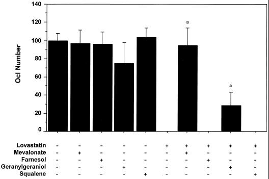
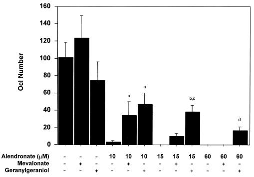
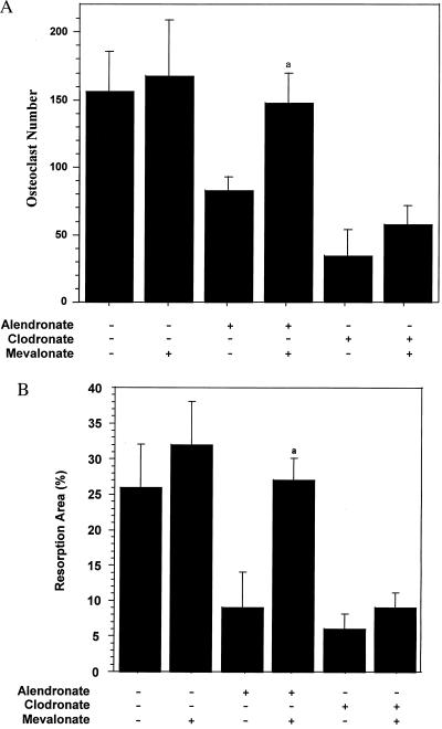
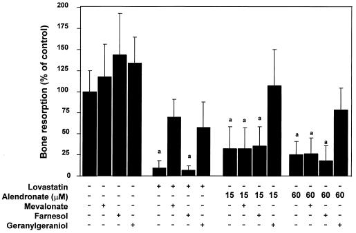
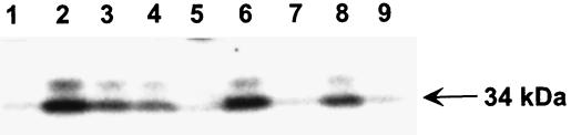
Nitrogen-containing bisphosphonates were shown to cause macrophage apoptosis by inhibiting enzymes in the biosynthetic pathway leading from mevalonate to cholesterol. This study suggests that, in osteoclasts, geranylgeranyl diphosphate, the substrate for prenylation of most GTP binding proteins, is likely to be the crucial intermediate affected by these bisphosphonates. We report that murine osteoclast formation in culture is inhibited by both lovastatin, an inhibitor of hydroxymethylglutaryl CoA reductase, and alendronate. Lovastatin effects are blocked fully by mevalonate and less effectively by geranylgeraniol whereas alendronate effects are blocked partially by mevalonate and more effectively by geranylgeraniol. Alendronate inhibition of bone resorption in mouse calvaria also is blocked by mevalonate whereas clodronate inhibition is not. Furthermore, rabbit osteoclast formation and activity also are inhibited by lovastatin and alendronate. The lovastatin effects are prevented by mevalonate or geranylgeraniol, and alendronate effects are prevented by geranylgeraniol. Farnesol and squalene are without effect. Signaling studies show that lovastatin and alendronate activate in purified osteoclasts a 34-kDa kinase. Lovastatin-mediated activation is blocked by mevalonate and geranylgeraniol whereas alendronate activation is blocked by geranylgeraniol. Together, these findings support the hypothesis that alendronate, acting directly on osteoclasts, inhibits a rate-limiting step in the cholesterol biosynthesis pathway, essential for osteoclast function. This inhibition is prevented by exogenous geranylgeraniol, probably required for prenylation of GTP binding proteins that control cytoskeletal reorganization, vesicular fusion, and apoptosis, processes involved in osteoclast activation and survival.


Figures

Figure 1
Figure 1
Lovastatin inhibits the formation of osteoclasts in vitro but not in the presence of mevalonate or geranylgeraniol. Osteoclastogenesis was assessed by using a coculture of mouse bone marrow cells and MB1.8 osteoblast cells as described in the Materials and Methods. On days 5 and 6 of the coculture, 10 μM LOV was added in the absence (−) or presence (+) of the following cholesterol biosynthesis metabolites: 1 mM MVA, 10 μM FOH, 10 μM GGOH, and 10 μM squalene. Osteoclast number was scored on day 7 by assessing numbers of large (i.e., ≥250 μm) TRAP-positive multinucleate cells (mean ± SD, n ≥ 3). a, statistically significantly different from LOV (P < 0.0001).
Figure 2
Figure 2
Geranylgeraniol and, to a lesser extent, mevalonate blocks alendronate inhibition of osteoclast formation in vitro. Osteoclastogenesis cultures were performed as described in Fig. 1 in the absence or presence of ALN at 10, 15, and 60 μM doses. MVA (1 mM) or GGOH (10 μM) were added to the above cocultures, and TRAP-positive fully mature (i.e., ≥250 μm) multinucleate Ocls were quantitated microscopically (mean ± SD). a, statistically significantly different from ALN (10 μM) (P < 0.0001); b, statistically significantly different from ALN (15 μM) (P < 0.0001); c, statistically significantly different from ALN (15 μM) + MVA (1 mM) (P < 0.0001); d, statistically significantly different from ALN (60 μM) (P < 0.02).
Figure 3
Figure 3
Mevalonate blocks alendronate, but not clodronate, inhibition of osteoclast number and bone resorption in mouse calvaria. Calvariae from 1-day-old mice were halved and cultured for 24 hr in media containing parathyroid hormone and either ALN (10 μM) or CLOD (100 μM) in the presence or absence of MVA (1 mM) as described in Materials and Methods. Osteoclasts were scored over the entire area of the parietal bones after staining with nitroblue tetrazolium (A), and bone resorption was scored by increased lucency within the central region of the parietal bones (B) (mean ± SD, n ≥ 3). a, statistically significantly different from ALN (P < 0.01).
Figure 4
Figure 4
Effects of cholesterol biosynthesis metabolites on inhibition of rabbit osteoclast-mediated bone resorption in vitro. Rabbit osteoclasts were isolated from the tibiae and femora of 10-day-old New Zealand White rabbits and then were plated onto bovine bone slices (6-mm diameter × 200-μm thickness). LOV (10 μM) or ALN (15 or 60 μM) were added in the absence or presence of the following: MVA (1 mM), FOH (10 μM), and GGOH (10 μM). After 3 days incubation, collagen fragments released into the medium were measured by the CROSSLAPS ELISA assay as described in the Materials and Methods. (mean ± SD, n = 3) a, statistically significantly different from control (P < 0.0001).
Figure 5
Figure 5
Activation of a 34-kDa kinase by alendronate and lovastatin is blocked by geranylgeraniol and, to a lesser extent, mevalonate. Ocls were purified from cocultures (see Fig. 1) by sequential treatment of culture dishes with collagenase and then EDTA, as described in Materials and Methods. Ocls then were left untreated (lane 1) or were incubated with ALN (30 μM; lanes 2–5) or LOV (10 μM; lanes 6–9) in the presence of MVA (1 mM; lanes 3 and 7), FOH (10 μM; lanes 4 and 8), or GGOH (10 μM; lanes 5 and 9) for 17 hr. Cell lysates were analyzed by in-gel kinase assay using myelin basic protein as a substrate. Activity of 34-kDa (arrow) and 36-kDa kinases was visualized by autoradiography. Data is representative of three independent experiments.

References

    1. Rodan G A, Fleisch H A. J Clin Invest. 1996;97:2692–2696. - PMC - PubMed
    1. Sahni M, Guenther H L, Fleisch H, Collin P, Martin T J. J Clin Invest. 1993;91:2004–2011. - PMC - PubMed
    1. Hughes D E, Wright K R, Uy H L, Sasaki A, Yoneda T, Roodman G D, Mundy G R, Boyce B F. J Bone Miner Res. 1995;10:1478–1487. - PubMed
    1. van Beek E, Hoekstra M, van der Ruit M, Lowik C, Papapoulos S. J Bone Miner Res. 1994;9:1875–1882. - PubMed
    1. Rogers M J, Xiong X, Brown R J, Watts D J, Russell R G G, Bayless A V, Ebetino F H. Mol Pharmacol. 1995;47:398–402. - PubMed

MeSH terms

LinkOut - more resources