. 1999 Jan 5;96(1):139-44.
doi: 10.1073/pnas.96.1.139.

Nuclear endpoint of Wnt signaling: neoplastic transformation induced by transactivating lymphoid-enhancing factor 1

Affiliations

Nuclear endpoint of Wnt signaling: neoplastic transformation induced by transactivating lymphoid-enhancing factor 1

M Aoki et al. Proc Natl Acad Sci U S A. .

Abstract

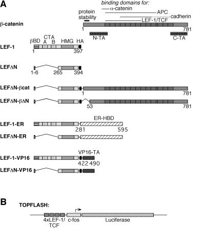
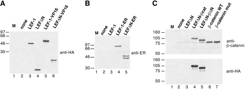
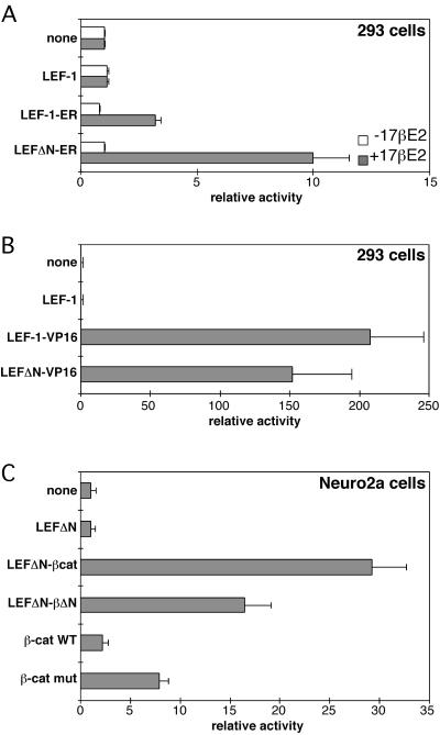
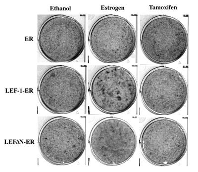
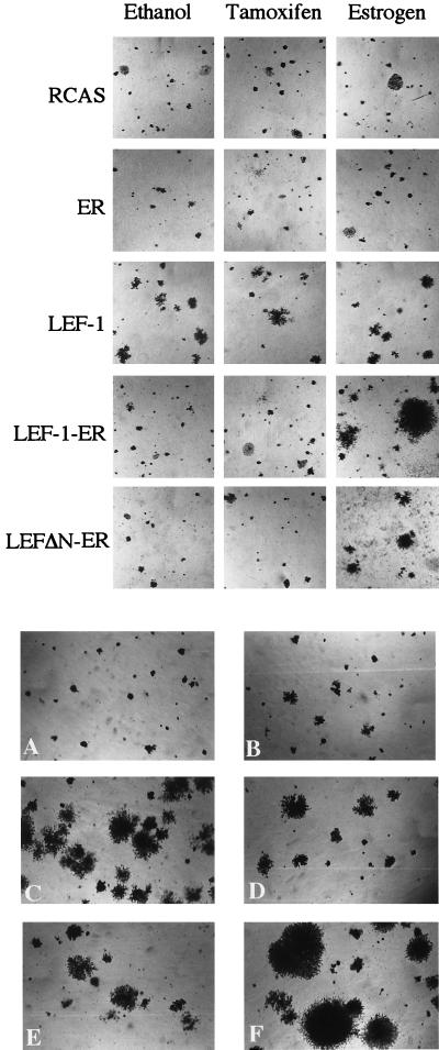
The interaction between beta-catenin and LEF-1/TCF transcription factors plays a pivotal role in the Wnt-1 signaling pathway. The level of beta-catenin is regulated by partner proteins, including glycogen synthase kinase-3beta (GSK-3beta) and the adenomatous polyposis coli (APC) tumor suppressor protein. Genetic defects in APC are responsible for a heritable predisposition to colon cancer. APC protein and GSK-3beta bind beta-catenin, retain it in the cytoplasm, and facilitate the proteolytic degradation of beta-catenin. Abrogation of this negative regulation allows beta-catenin to translocate to the nucleus and to form a transcriptional activator complex with the DNA-binding protein lymphoid-enhancing factor 1 (LEF-1). This complex is thought to be involved in tumorigenesis. Here we show that covalent linkage of LEF-1 to beta-catenin and to transcriptional activation domains derived from the estrogen receptor or the herpes simplex virus protein VP16 generates transcriptional regulators that induce oncogenic transformation of chicken embryo fibroblasts. The chimeras between LEF-1 and beta-catenin or VP16 are constitutively active, whereas fusions of LEF-1 to the estrogen receptor are regulatable by estrogen. These experiments document the oncogenicity of transactivating LEF-1 and show that the transactivation domain normally provided by beta-catenin can be replaced by heterologous activation domains. These results suggest that the transactivating function of the LEF-1/beta-catenin complex is critical for tumorigenesis and that this complex transforms cells by activating specific LEF-1 target genes.


Figures

Figure 1
Figure 1
Structure of LEF-1 fusion proteins. (A) Schematic representation of β-catenin, LEF-1, and LEF-1-fusions. On the β-catenin molecule, several specific domains are marked: an amino-terminal region responsible for the instability of the protein and two transactivating domains, N-TA and C-TA. The armadillo repeats are represented as dark boxes. On the LEF-1 molecule, the β-catenin-binding domain (βBD), two context-dependent transactivation domains (CTA-A and CTA-B), and the HMG-box mediating DNA binding are marked. The HA-epitope tag on all constructs is represented by a black box. The hormone-binding domain (HBD) of the human ER and the carboxyl-terminal transactivation (TA) domain of the herpes simplex virus VP16 protein are shown as hatched boxes. Amino acid positions of the termini of the various protein domains are given. (B) Schematic representation of the Topflash reporter construct. Expression of the luciferase gene (hatched bar) in this plasmid is driven by the c-fos minimal promoter (gray box) and four copies of a LEF-1/TCF consensus binding motif (dark gray boxes).
Figure 2
Figure 2
(A) Expression of chimeric LEF-1 constructs. Human 293 cells were transfected with empty expression vector (lane 2) or expression vectors for LEF-1, LEFΔN, LEF-1-VP16, or LEFΔN-VP16. Whole cell lysates were prepared and resolved by electrophoresis in an SDS/10% polyacrylamide gel. Proteins were transferred onto nitrocellulose and probed with a monoclonal antibody recognizing the HA tag. (B) Cellular lysates from 293 cells transfected with empty expression vector (lane 2) or expression vectors for LEF-1, LEF-1-ER, or LEFΔN-ER were resolved by electrophoresis in an SDS/10% polyacrylamide gel and transferred onto nitrocellulose. The blot was probed with a polyclonal antibody directed against the hormone-binding domain of the human ER. (C) Cellular lysates from Neuro2a cells transfected with empty expression vector (lane 2) or expression vectors for LEFΔN, LEFΔNβ-cat, LEFΔN-βΔN, or wild-type (WT) and mutant β-catenin, respectively, as indicated were resolved by electrophoresis in SDS/7.5% polyacrylamide gels and transferred onto nitrocellulose. The upper blot was probed with a monoclonal antibody directed against β-catenin. The faint band visible in lanes 2–5 that migrates at the same position as the transfected β-catenin (lanes 6 and 7) represents endogenous protein of Neuro2a cells. The lower blot was probed with a monoclonal antibody against the HA tag. M lanes contain molecular size markers. The mass of the marker proteins in kDa is shown on the left.
Figure 3
Figure 3
Activation of the Topflash reporter by LEF-1-fusion constructs. (A) Human 293 cells were transfected with the Topflash reporter plasmid and expression plasmids for LEF-1, LEF-1-ER, or LEFΔN-ER. Cells were treated with 10−6 M 17β-estradiol (+17βE2) in ethanol or solvent only (−17βE2) for 24 hr. Luciferase activities were determined as described in Materials and Methods and are expressed as relative activity. Reporter gene activity in control cells transfected with Topflash and empty expression vector were arbitrarily set as 1. Values given are derived from at least three independent experiments. (B) Human 293 cells were transfected with the Topflash reporter plasmid and expression plasmids for LEF-1, LEF-1-VP16, or LEFΔN-VP16 as indicated. Luciferase activities were determined as in A. (C) Mouse Neuro2a cells were transfected with the Topflash reporter plasmid and expression plasmids for LEFΔN, LEFΔN-βcat, LEFΔN-βΔN, and wild-type (WT) or mutant β-catenin as indicated. Luciferase activities were determined as in A. Note the different scales in A, B, and C.
Figure 4
Figure 4
Estrogen-dependent formation of transformed cell foci by LEF-1-ER fusion products. CEF were infected with the constructs indicated on the left and then overlaid with nutrient agar containing estrogen (2 × 10⁻⁶ M), tamoxifen (1 × 10⁻⁷ M), or ethanol (vehicle). On day 20 after infection the cells were fixed and stained with crystal violet and photographed.
Figure 5
Figure 5
(Upper) Estrogen-dependent agar colony formation by LEF-1-ER fusion products. CEF transfected with the indicated constructs were seeded in soft nutrient agar containing estrogen (2 × 10⁻⁶ M), tamoxifen (1 × 10⁻⁷ M), or ethanol (vehicle). Photographs of the agar colonies were taken on day 17 after infection. (Lower) Agar colony formation by various LEF-1 constructs. CEF transfected with RCAS (A), LEF-1 (B), LEF-1-VP16 (C), LEFΔN-VP16 (D), LEFΔN-βcat (E), and LEFΔN-βΔN (F) were seeded in soft nutrient agar. Photographed 17 days after infection.

Similar articles

Cited by

References

    1. Cadigan K M, Nusse R. Genes Dev. 1997;11:3286–3305. - PubMed
    1. Morin P J, Sparks A B, Korinek V, Barker N, Clevers H, Vogelstein B, Kinzler K W. Science. 1997;275:1787–1790. - PubMed
    1. Rubinfeld B, Robbins P, El-Gamil M, Albert I, Porfiri E, Polakis P. Science. 1997;275:1790–1792. - PubMed
    1. Nusse R, Varmus H E. Cell. 1982;31:99–109. - PubMed
    1. Nusse R, van Ooyen A, Cox D, Fung Y K, Varmus H. Nature (London) 1984;307:131–136. - PubMed

Publication types

MeSH terms

LinkOut - more resources