. 1999 Jan 5;96(1):179-84.
doi: 10.1073/pnas.96.1.179.

Polyglutamine-mediated dysfunction and apoptotic death of a Caenorhabditis elegans sensory neuron

Affiliations

Polyglutamine-mediated dysfunction and apoptotic death of a Caenorhabditis elegans sensory neuron

P W Faber et al. Proc Natl Acad Sci U S A. .

Abstract

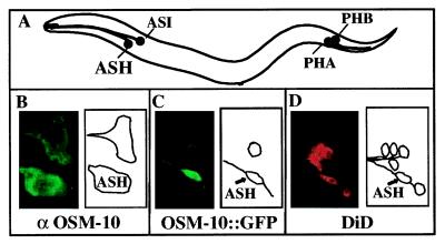
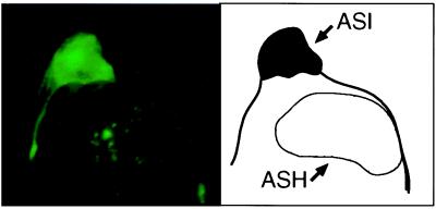
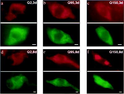
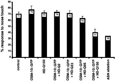
The effect of expressing human huntingtin fragments containing polyglutamine (polyQ) tracts of varying lengths was assessed in Caenorhabditis elegans ASH sensory neurons in young and old animals. Expression of a huntingtin fragment containing a polyQ tract of 150 residues (Htn-Q150) led to progressive ASH neurodegeneration but did not cause cell death. Progressive cell death and enhanced neurodegeneration were observed in ASH neurons that coexpressed Htn-Q150 and a subthreshold dose of a toxic OSM-10::green fluorescent protein (OSM-10::GFP) fusion protein. Htn-Q150 huntingtin protein fragments formed protein aggregates in ASH neurons, and the number of ASH neurons containing aggregates increased as animals aged. ASH neuronal cell death required ced-3 caspase function, indicating that the observed cell death is apoptotic. Of interest, ced-3 played a critical role in Htn-Q150-mediated neurodegeneration but not in OSM10::GFP-mediated ASH neurodegeneration. ced-3 function was important but not essential for the formation of protein aggregates. Finally, behavioral assays indicated that ASH neurons, coexpressing Htn-Q150 and OSM10::GFP, were functionally impaired at 3 days before the detection of neurodegeneration, cell death, and protein aggregates.


Figures

Figure 1
Figure 1
Overview of the system. (A) A schematic presentation of a nematode, indicating the positions of neurons that express the osm-10 gene. Neurons are referred to by their class designation, e.g., ASHL and ASHR as ASH. (B) (Left) ASH and ASI visualized by using the OSM-10 antiserum. (Right) Diagrammed presentation of the stained neurons. (C) (Left) ASH and ASI visualized in transgenic animals expressing OSM-10∷GFP. (Right) Diagrammed presentation of the stained neurons. (D) (Left) DiD stains six classes of neurons, including the ASH, in the head. (Right) Diagrammed presentation of the stained neurons.
Figure 2
Figure 2
Htn-Q150/OSM-10∷GFP expression led to morphological changes of the ASH in aged animals. (Left) An Htn-Q150/ OSM-10∷GFP-expressing ASH neuron from an 8-day-old animal with a speckled bag phenotype, visualized by GFP fluorescence. Multiple serial section confocal planes were combined for this image. Htn-Q150/OSM-10∷GFP expression does not affect the size and morphology of the ASI sensory neurons (as illustrated), and the GFP marker in the ASI neuron shows the normal subcellular expression pattern (A.C.H., J. Kaas, J. E. Shapiro, and J. M. Kaplan, unpublished work). In contrast, a subset of ASH sensory neurons are severely affected, displaying a speckled bag phenotype, swelling to 2–3 times the size of a normal ASH neuron, and lacking intracellular morphology. The subcellular structure, if any, that contains the faint GFP-positive dots is unclear. The size of the ASH and ASI sensory neurons are comparable in wild-type animals. osm-10 expression in ASH neurons is significantly higher than expression in ASI neurons (A.C.H., J. Kaas, J. E. Shapiro, and J. M. Kaplan, unpublished work), and, consequently, polyQ induced effects are less common in ASI neurons. (Right) A schematic representation of the upper panel. The ASI and ASH neurons are outlined.
Figure 3
Figure 3
Htn-Q150 expression led to time-dependent protein aggregation in sensitized ASH neurons. (ac Upper) Visualization of Htn-Q2, Htn-Q95, and Htn-Q150 expression in sensitized ASH sensory neurons in 3-day-old transgenic animals by using the antihuntingtin HP1 (35) antiserum in red. All huntingtin fragments were expressed at similar levels and were found in the cytoplasm and processes without accumulation in obvious intracellular structures. For Htn-Q150, the huntingtin fragments accumulated into discrete foci or aggregates in a very small percentage of the Htn-Q150 expressing ASH sensory neurons (aggregates were found in random locations in the cytoplasm and processes but not in the nucleus, based on visual examination). (ac Lower) The ASH sensory neuron shown in the upper panels visualized with the GFP marker in green. (df Upper) Visualization of Htn-Q2, Htn-Q95, and Htn-Q150 expression in sensitized ASH sensory neurons in 8-day-old transgenic animals by using the antihuntingtin HP1 (35) antiserum in red. Localization of Htn-Q2 and Htn-Q95 fragments was still broadly cytoplasmic. For Htn-Q150, the huntingtin fragments were detected in large and small cytoplasmic aggregates in over half of the Htn-Q150-expressing ASH neurons. (df Lower) The ASH sensory neuron shown in the upper panels visualized with the GFP marker in green. (Bar = 3 μm.)
Figure 4
Figure 4
Htn-Q150 expression in sensitized ASH neurons leads to a severe defect in the nose touch response of transgenic animals. Each bar represents the percentage of trials in which transgenic or control animals (genotype indicated below bar) responded to nose touch by stopping forward movement or reversing. Error bars indicate the SEM). The data for ASH-ablated animals is from Kaplan and Horvitz (29).

References

    1. Huntington’s Disease Collaborative Research Group. Cell. 1993;72:971–983. - PubMed
    1. Strong T V, Tagle D A, Valdes J M, Elmer L W, Boehm K, Swaroop M, Kaatz K W, Collins F S, Albin R L. Nat Genet. 1993;5:259–265. - PubMed
    1. Martin J B, Gusella J F. N Engl J Med. 1986;315:1267–1276. - PubMed
    1. Vonsattel J P, Myers R H, Stevens T J, Ferrante R J, Bird E D, Richardson E P., Jr J Neuropathol Exp Neurol. 1985;44:559–577. - PubMed
    1. Gusella J F, MacDonald M E. Curr Opin Neurobiol. 1995;5:656–662. - PubMed

Publication types

MeSH terms