. 1999 Jan 5;96(1):232-7.
doi: 10.1073/pnas.96.1.232.

Virus-specific immunity after gene therapy in a murine model of severe combined immunodeficiency

Affiliations

Virus-specific immunity after gene therapy in a murine model of severe combined immunodeficiency

K D Bunting et al. Proc Natl Acad Sci U S A. .

Abstract

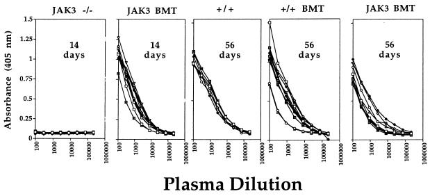
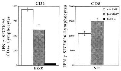
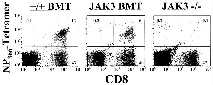
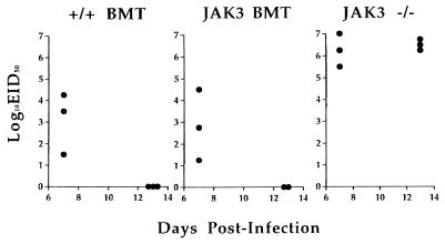
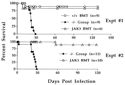
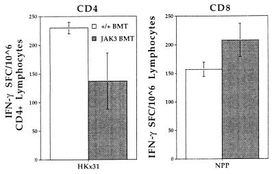
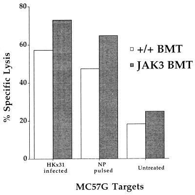
Human severe combined immunodeficiency (SCID) can be caused by defects in Janus kinase 3 (JAK3)-dependent cytokine signaling pathways. As a result, patients are at high risk of life-threatening infection. A JAK3 -/- SCID mouse model for the human disease has been used to test whether transplant with retrovirally transduced bone marrow (BM) cells (JAK3 BMT) could restore immunity to an influenza A virus. The immune responses also were compared directly with those for mice transplanted with wild-type BM (+/+ BMT). After infection, approximately 90% of the JAK3 BMT or +/+ BMT mice survived, whereas all of the JAK3 -/- mice died within 29 days. Normal levels of influenza-specific IgG were present in plasma from JAK3 BMT mice at 14 days after respiratory challenge, indicating restoration of B cell function. Influenza-specific CD4(+) and CD8(+) T cells were detected in the spleen and lymph nodes, and virus-specific CD8(+) effectors localized to the lungs of the JAK3 BMT mice. The kinetics of the specific host response correlated with complete clearance of the virus within 2 weeks of the initial exposure. By contrast, the JAK3 -/- mice did not show any evidence of viral immunity and were unable to control this viral pneumonia. Retroviral-mediated JAK3 gene transfer thus restores diverse aspects of cellular and humoral immunity and has obvious potential for human autologous BMT.


Figures

Figure 1
Figure 1
Levels of influenza-specific IgG in plasma from JAK3 BMT mice 14 and 56 days after challenge. Mice were infected intranasally with 106.8 EID50 of the HKx31 influenza virus, and plasma was collected from the peripheral blood at various time points. Antibody titers were determined by ELISA using the absorbance at 405 nm. No antibody response was detectable for the JAK3 −/− mice 14 days after infection. By contrast, all JAK3 BMT mice showed normal titers of specific antibody at this early time point. Anti-influenza IgG levels also were determined in plasma from JAK3 BMT mice 56 days after challenge. The +/+ BMT and JAK3 BMT mice showed normal levels of circulating virus-specific Ig when compared with untransplanted wild-type mice (+/+). The titers in JAK3 BMT and +/+ BMT mice did not differ significantly between days 14 and 56. Each line represents the antibody titer from an individual mouse.
Figure 2
Figure 2
ELISpot assay to quantitate the cellular immune response in spleen and lymph nodes of JAK3 BMT mice 13 days after influenza challenge. Spleen and lymph nodes were collected from infected mice and pooled for analysis. The cells were cocultured with antigen-presenting cells that were either HKx31-infected (CD4) or NP366-pulsed (CD8). Uninfected or nonpulsed samples served as negative controls and the background level of IFN-γ production was less than 38 SFC/106 cells. Numbers of IFN-γ producing cells were determined by ELISpot assay in 96-well plates. The JAK3 BMT (gray bars) and +/+ BMT (white bars) mice showed a similar response for both CD4+ and CD8+ T cells. However, no significant response was detectable in the JAK3 −/− mice (black bars). Results are expressed as the number of SFCs and represent data from three mice assayed as a pool.
Figure 3
Figure 3
Influenza-specific CD8+ T cells in the lungs of JAK3 BMT mice 13 days after influenza challenge. The BAL population was stained for influenza virus-specific CD8+ T cells by using the NPP tetramer (NP366 + H-2Db)-phycoerythrin and CD8- fluorescein isothiocyanate, followed by flow cytometric analysis. Significant populations of CD8+NPP+ cells were found by flow cytometric analysis of BAL cells from the +/+ BMT and JAK3 BMT mice, demonstrating the presence of infiltrating CD8+ T cells specific for influenza virus. No significant numbers could be detected in JAK3 −/− mice.
Figure 4
Figure 4
Clearance of influenza virus from the JAK3 BMT mice. Lungs were collected from infected mice 7 and 13 days after intranasal challenge with 106.8 EID50 of the HKx31 influenza A virus. Virus was detectable after inoculation in embryonated hen’s eggs for all three groups on day 7, but the titers in the +/+ BMT and JAK3 BMT mice were much lower than those in JAK3 −/− mice. By day 13, the +/+ BMT and JAK3 BMT mice had cleared the infection from the lungs, but titers remained high in the JAK3 −/− mice. Each symbol represents a single mouse.
Figure 5
Figure 5
Protection against influenza virus in JAK3 BMT mice. The HKx31-infected mice were followed long term to determine the effects of JAK3 gene transfer on survival. All mice from the JAK3 −/− group (includes mock BMT mice and untransplanted JAK3 −/− mice) became progressively ill and died by day 29 after infection. By contrast, roughly 90% of JAK3 BMT mice survived the challenge and lived long term. The relative survival of JAK3 BMT mice was the same as that seen after transplant of normal wild-type BM (+/+ BMT, Expt #1).
Figure 6
Figure 6
Memory T cells in JAK3 BMT mice. Spleen and lymph nodes were collected from HKx31-infected surviving mice at 130 days after challenge. ELISpot assay of pooled lymphocyte populations for IFN-γ SFC demonstrated the presence of substantial numbers of memory CD4+ and CD8+ T cells from both +/+ BMT (open bars) and JAK3 BMT (filled bars) mice. The background level of IFN-γ production after incubation with untreated stimulators was <33 SFC/106 cells.
Figure 7
Figure 7
Influenza-specific cell-mediated cytotoxicity after in vitro stimulation of memory CD8+ T cells. Spleen and lymph nodes were collected and pooled from +/+ BMT (open bars) or JAK3 BMT (filled bars) mice that had been infected 130 days previously with the HKx31 virus. After 5 days of in vitro stimulation in the presence of irradiated stimulator cells that were HKx31-infected or untreated, effectors were harvested and cytotoxic T cell activity measured in 51Cr release assay against HKx31-infected, NP-pulsed, or untreated MC57G (H-2b) target cells. This result shows lysis of targets at an effector-to-target cell ratio of 3.8:1. The background level of specific lysis was <2% for lymphocytes cultured with uninfected stimulator cells.

Similar articles

Cited by

References

    1. Gurniak C B, Berg L J. Blood. 1996;87:3151–3160. - PubMed
    1. Chen M, Cheng A, Chen Y Q, Hymel A, Hanson E P, Kimmel L, Minami Y, Taniguchi T, Changelian P S, O’Shea J J. Proc Nat Acad Sci USA. 1997;94:6910–6915. - PMC - PubMed
    1. Ihle J N. Philos Trans R Soc London B. 1996;351:159–166. - PubMed
    1. Oakes S A, Candotti F, Johnston J A, Chen Y Q, Ryan J J, Taylor N, Liu X, Hennighausen L, Notarangelo L D, Paul W E, et al. Immunity. 1996;5:605–615. - PubMed
    1. Teglund S, McKay C, Schuetz E, van Deursen J M, Stravopodis D, Wang D, Brown M, Bodner S, Grosveld G, Ihle J N. Cell. 1998;93:841–850. - PubMed

Publication types