Comparative Study
. 1999 Jan 5;96(1):260-5.
doi: 10.1073/pnas.96.1.260.

Neurons in the dorsal column white matter of the spinal cord: complex neuropil in an unexpected location

Affiliations
Comparative Study

Neurons in the dorsal column white matter of the spinal cord: complex neuropil in an unexpected location

C Abbadie et al. Proc Natl Acad Sci U S A. .

Abstract

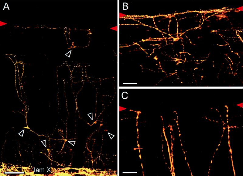
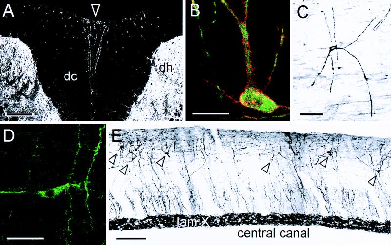
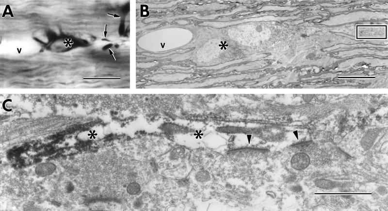
It is common to think of gray matter as the site of integration in neural circuits and white matter as the wires that connect different groups of neurons. The dorsal column (DC) white matter, for example, is the spinal cord axonal pathway through which a topographic map of the body is conveyed to the somatosensory cortex. We now describe a network of neurons located along the midline of the DCs. The neurons are present in several mammals, including primates and birds, and have a profuse dendritic arbor that expresses both the neuron-specific marker, microtubule-associated protein-2, and the neurokinin-1 receptor, a target of the neuropeptide, substance P. Electron microscopy and double immunostaining for synaptophysin and a marker of gamma-aminobutyric acid-ergic terminals documented a rich synaptic input to these neurons. Finally, injection of a gamma-aminobutyric acid type A receptor antagonist or of substance P into the cerebrospinal fluid of the rat spinal cord induced Fos expression and internalization of the neurokinin-1 receptor in these neurons, respectively, indicating that the DC neurons are under tonic inhibitory control and can respond to neurotransmitters that circulate in the cerebrospinal fluid.


Figures

Figure 1
Figure 1
(A) NK-1 receptor immunoreactive neurons (white arrowheads) are scattered through this 30-μm midsagittal section of the lumbar spinal cord of the adult rat. The dense NK-1 receptor immunoreactivity in lamina X (lam X) distinguishes the gray matter from the white matter of the DCs. (B and C) The neurons are also MAP-2 positive and have highly varicose dendrites that arborize extensively, often contacting the dorsal surface of the cord (red arrowheads). (Bars: 100 μm, A; 50 μm, B; and 25 μm, C.)
Figure 2
Figure 2
(A) Although the neurons in the DCs (dc) are best viewed in sagittal section, MAP-2 immunoreactive dendrites occasionally can be seen along the midline (arrowhead) in transverse sections. The dendrites are located at a distance from the heavily labeled dorsal horn gray matter (dh). (B) Superimposition of confocal images for MAP-2 (green) and the NK-1 receptor (red) reveals that every DC neuron labels for both molecules. Where there is overlap, the image appears yellow. C and D illustrate DC neurons from midsagittal sections of the cervical spinal cord in monkey and bird (Zebra finch), respectively. Both are stained for MAP-2 by using an immunoperoxidase (C) or an immunofluorescence protocol (D). (E) This midsagittal montage of sections from a 13-day-old rat spinal cord illustrates the widespread distribution of the DC neurons (labeled with MAP-2; arrowheads); many are located close to the surface of the cord, distant from the gray matter (lamina X) around the central canal. A, B, and D are confocal images; C and E were digitized from slides and prepared with Adobe Photoshop. (Bars: 200 μm, A; 20 μm, B; 50 μm, C; 4 μm, D; and 50 μm, E.)
Figure 3
Figure 3
Sagittal sections of the rat lumbar spinal cord provide information on the functional nature of the neurons of the DC white matter. (A) Labeling for synaptophysin (green) reveals a dense somatic and dendritic synaptic input to the DC neurons (stained for NK-1 receptor; red). (B) A large proportion of the synaptic input to the neurons (stained for MAP-2; red) are GABAergic as they express the GABA vesicle transporter (green). (C and D) Direct CSF injection of the GABAA receptor antagonist, bicuculline, induces Fos expression (black nucleus; arrowheads in C) in approximately 50% of the DC neurons (identified by the brown MAP-2 immunoreaction product). (E) Direct CSF injection of SP induces internalization of the NK-1 receptor in approximately one-third of the DC neurons. The receptor is concentrated in cytoplasmic endosomes. (F) Approximately 20% of the neurons (stained for MAP-2; red) express neuronal nitric oxide synthase (green). In A, B, and F, superimposition of the confocal images produces a yellow image where the immunostaining of two antigens overlaps. (Bars: 25 μm, A; and 50 μm, BF.)
Figure 4
Figure 4
(A) Light micrograph of MAP-2-labeled neuron (∗) in 50-μm thick plastic embedded section of the DCs showing adjacent landmark blood vessel (v) and labeled dendrites (arrows) near the surface of the section. (B) Low-magnification electron micrograph of this region shows the selected neuronal elements with greater resolution. Using the light micrograph as a map permits identification of individual dendrites with accuracy at the EM level. (C) This high-magnification image of the box in B illustrates axonal boutons that make asymmetric synapses (arrowheads) upon a MAP-2-immunoreactive dendrite (∗). (Bars: 5 μm, A; 10 μm, B; and 1 μm, C.)

References

    1. Brodal A, Rexed J. J Comp Neurol. 1953;98:179–211. - PubMed
    1. Gwyn D G, Waldron H A. Brain Res. 1968;10:342–351. - PubMed
    1. Willis W D, Coggeshall R E. Sensory Mechanisms of the Spinal Cord. New York: Plenum; 1991.
    1. Mantyh P W, Demaster E, Malhotra A, Ghilardi J R, Rogers S, Mantyh C R, Liu H, Basbaum A I, Vigna S R, Maggio J E, Simone D. Science. 1995;268:1629–1632. - PubMed
    1. McIntire S L, Reimer R J, Schuske K, Edwards R H, Jorgensen E M. Nature (London) 1997;389:870–876. - PubMed

Publication types

MeSH terms

Substances

LinkOut - more resources