. 1999 Jan;10(1):9-22.
doi: 10.1091/mbc.10.1.9.

Regulation of cadherin function by Rho and Rac: modulation by junction maturation and cellular context

Affiliations
Free PMC article

Regulation of cadherin function by Rho and Rac: modulation by junction maturation and cellular context

V M Braga et al. Mol Biol Cell. 1999 Jan.
Free PMC article

Abstract

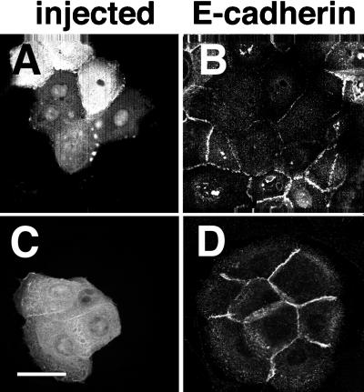
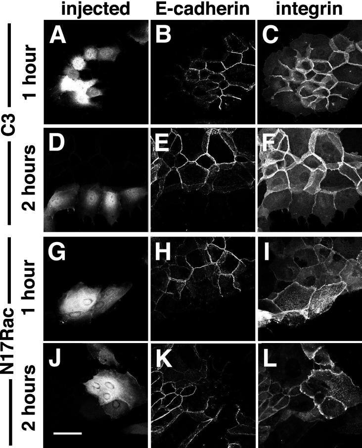
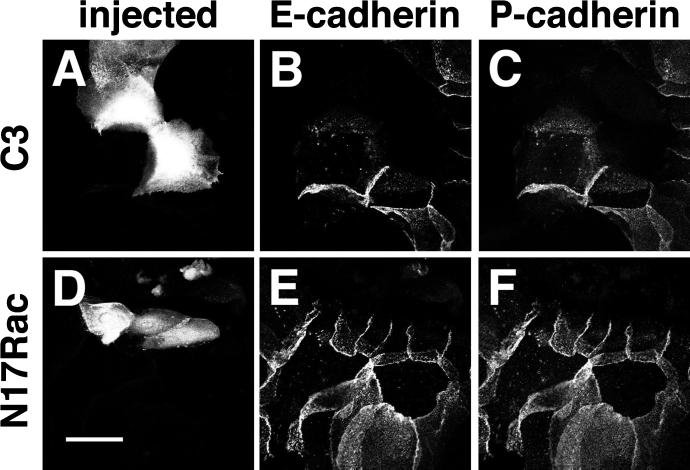
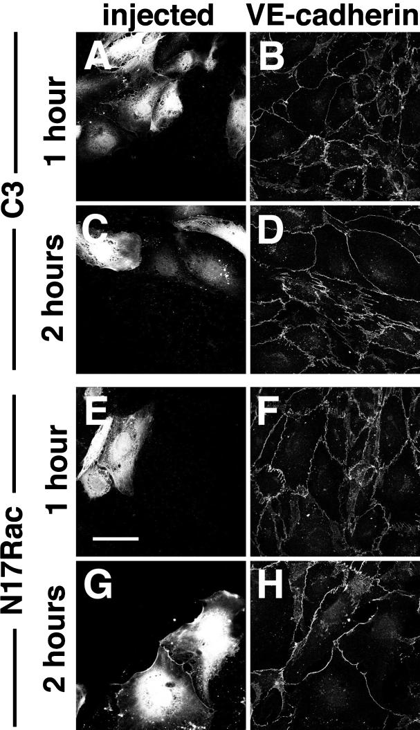
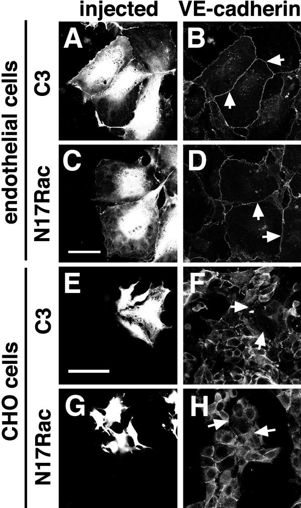
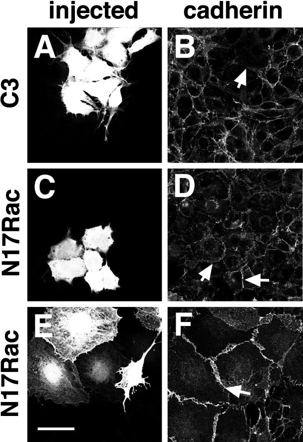
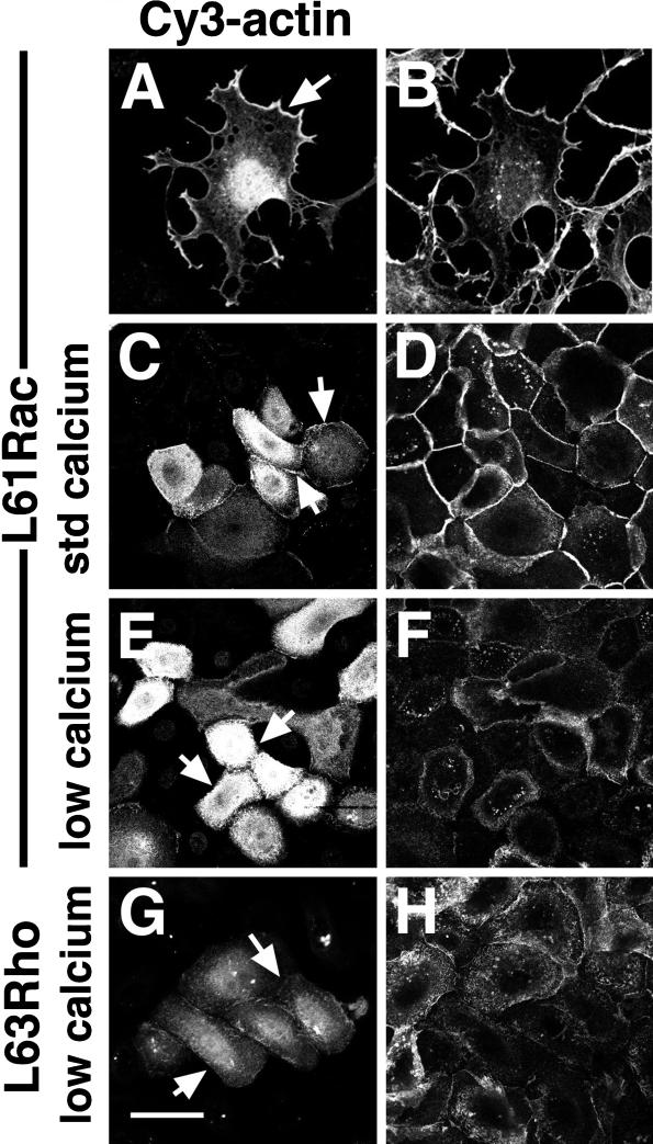
Cadherins are cell-cell adhesion receptors whose adhesive function requires their association with the actin cytoskeleton via proteins called catenins. The small guanosine triphosphatases (GTPases), Rho and Rac, are intracellular proteins that regulate the formation of distinct actin structures in different cell types. In keratinocytes and in other epithelial cells, Rho and Rac activities are required for E-cadherin function. Here we show that the regulation of cadherin adhesiveness by the small GTPases is influenced by the maturation status of the junction and the cellular context. E-cadherin localization was disrupted in mature keratinocyte junctions after inhibition of Rho and Rac. However, an incubation of 2 h was required after GTPase inhibition, when compared with newly established E-cadherin contacts (30 min). Regarding other cadherin receptors, P-cadherin was effectively removed from mature keratinocytes junctions by blocking Rho or Rac. In contrast, VE-cadherin localization at endothelial junctions was independent of Rho/Rac activity. We demontrate that the insensitivity of VE-cadherin to inhibition of Rho and Rac was not due to the maturation status of endothelial junction, but rather the cellular background: when transfected into CHO cells, the localization of VE-cadherin was perturbed by inhibition of Rho proteins. Our results suggest that the same stimuli may have different activity in regulating the paracellular activity in endothelial and epithelial cells. In addition, we uncovered possible roles for the small GTPases during the establishment of E-cadherin-dependent contacts. In keratinocytes, Rac activation by itself cannot promote accumulation of actin at the cell periphery in the absence of cadherin-dependent contacts. Moreover, neither Rho nor Rac activation was sufficient to redistribute cadherin molecules to cell borders, indicating that redistribution results mostly from the homophilic binding of the receptors. Our results point out the complexity of the regulation of cadherin-mediated adhesion by the small GTPases, Rho and Rac.


Figures

Figure 1
Figure 1
The effects of Rho and Rac on cadherin receptors is influenced by the maturation of the intercellular contacts. Keratinocytes grown in low calcium medium were transferred to standard medium to induce calcium-dependent cell–cell contacts for 3 h (newly formed junctions) (A and B) or alternatively, cells were grown in standard medium (mature junctions) (C and D). After microinjection of a dominant negative form of Rac (N17Rac) to block endogenous Rac, cells were incubated for further 30 min. E-cadherin was visualized by staining with HECD-1 followed by FITC-conjugated anti-mouse (B and D); injected cells are seen in panels A and C. Bar, 50 μm.
Figure 2
Figure 2
Mature keratinocyte cadherin junctions are disrupted by inhibition of endogenous Rho or Rac. Keratinocytes grown in the presence of cell–cell contacts (standard medium) were microinjected with C3 transferase to inhibit Rho (C3, A–F) or N17Rac (N17Rac, G–L) and subsequently incubated for 1 h (A–C and G–I) and 2 h (D–F and J–L). Cells were fixed, permeabilized, and double labeled with an antibody against E-cadherin (ECCD-2) followed by FITC-conjugated anti-rat IgG (B, E, H, and K), and an antibody against β1 integrins (P5D2) followed by Cy5-conjugated anti-mouse IgG (C, F, I, and L). Microinjected cells were visualized by coinjection of Dextran-Texas Red (A, D, G, and J). Bar, 50 μm.
Figure 3
Figure 3
Rho and Rac function is required for the maintenance of both E- and P-cadherin–mediated cell–cell adhesion. Keratinocytes grown in standard calcium medium were injected with C3 transferase (A–C) or N17Rac (D–F). After 2 h incubation, cells were double stained for E-cadherin (ECCD-2) followed by FITC-conjugated anti-rat IgG (B and E) and P-cadherin (NCC-CAD-299) followed by Cy5-conjugated anti-mouse IgG (C and F). Microinjected cells were observed by coinjection of Dextran-Texas Red (A and D). Bar, 50 μm
Figure 4
Figure 4
VE-cadherin–dependent cell–cell contacts are not perturbed by inhibiting Rho or Rac activity. Confluent endothelial cells were injected with C3 transferase to block endogenous Rho (A–D) or N17Rac to inhibit endogenous Rac (E–H) and subsequently incubated for 1 h (A, B, E, and F) or 2 h (C, D, G, and H). Cells were labeled with anti–VE-cadherin monoclonal antibody (TEA-1) followed by anti-mouse FITC. Injected cells are shown in panels A, C, E, and G, and VE-cadherin staining is shown in panels B, D, F, and H. Bar, 50 μm.
Figure 5
Figure 5
Formation of new VE-cadherin junctions in endothelial cells does not require the activity of Rho or Rac. Confluent endothelial cells (A–D) were incubated in medium without serum or complements and containing 5 mM EGTA to disrupt cadherin-dependent contacts for 5–20 min. After microinjection of C3 (A and B) or N17Rac (C and D), cells were washed to remove the EGTA and incubated in medium with serum and complements for 1 h to induce cell–cell contacts. Subconfluent CHO cells transfected with full-length VE-cadherin were also microinjected with C3 (E and F) or N17Rac (G and H) and incubated for up to 2 h. Staining of VE-cadherin was performed and visualized with anti-mouse FITC (B, D, F, and H); injected cells were identified with Dextran-Texas Red (A, C, E, and G). Arrows in panels B and D show the presence of VE-cadherin–dependent contacts; arrows in panels F and H show the dismantling of VE-cadherin contacts in CHO cells. Bar, 50 μm.
Figure 6
Figure 6
N17Rac is unable to disrupt cadherin-mediated adhesion in fibroblasts. L-cells expressing full-length E-cadherin were microinjected with C3 (A and B) or a dominant negative form of Rac (N17Rac; C and D), incubated for 1 or 2 h (A–D and our unpublished observations) and stained for E-cadherin (B and D). Quiescent Swiss 3T3 fibroblasts were also injected with N17Rac and incubated for up to 2 h (E and F and our unpublished observations) and stained with an anti–pan-cadherin antibody (F). Similar results were obtained after 1 or 2 h of incubation with either cell type. Injected cells are shown in panels A, C, and E. Arrows point to the absence (B) or presence (D and F) of cadherin at sites of cell–cell contacts. Bar, 50 μm.
Figure 7
Figure 7
Activation of Rho or Rac is not sufficient to redistribute cadherin receptors and Cy3-labeled actin to the periphery of keratinocyte cells. Starved Swiss 3T3 fibroblasts were microinjected with L61Rac and Cy3-labeled actin (A) and after 20 min incubation, cells were fixed and stained with FITC-phalloidin (B). Keratinocytes grown in the absence of cell–cell contacts were injected with Cy3-actin and activated Rac (L61Rac, panels C–F) or activated Rho (L63Rho, panels G and H). Cells were incubated for 20 min in the same medium (low calcium, panels E–H) or immediately transferred to standard calcium medium to induce intercellular contacts (std calcium, panels C and D). Staining for E-cadherin receptors was performed (D, F, and H) and Cy3-actin pattern in injected cells was shown (A, C, E, and G). Arrows in panels A and C indicate accumulation of Cy3-actin at the cell periphery; arrows in E and G show diffuse localization of labeled actin. Bar, 50 μm.

References

    1. Bixby JL, Zhang R. Purified N-cadherin is a potent substrate for the rapid induction of neurite outgrowth. J Cell Biol. 1990;110:1253–1260. - PMC - PubMed
    1. Brady-Kalnay S, Tonks NK. Protein tyrosine phosphatases as cell adhesion receptors. Curr Biol. 1995;7:650–657. - PubMed
    1. Braga VMM, Hodivala KJ, Watt FM. Calcium-induced changes in distribution and solubility of cadherins and their associated cytoplasmic proteins in human keratinocytes. Cell Adhesion Com. 1995;3:201–215. - PubMed
    1. Braga VMM, Machesky LM, Hall A, Hotchin NA. The small GTPases Rho and Rac are required for the establishment of cadherin-dependent cell-cell conacts. J Cell Biol. 1997;137:1421–1431. - PMC - PubMed
    1. Braga VMM, Najabagheri N, Watt FM. Calcium-induced intercellular adhesion of keratinocytes does not involve accumulation of β1 integrins at cell-cell contact sites and does not involve changes in the levels or phosphorylation of the catenins. Cell Adhesion Com. 1998;5:137–149. - PubMed

Publication types

LinkOut - more resources