. 1999 Jan;10(1):47-61.
doi: 10.1091/mbc.10.1.47.

Association of Rab25 and Rab11a with the apical recycling system of polarized Madin-Darby canine kidney cells

Affiliations
Free PMC article

Association of Rab25 and Rab11a with the apical recycling system of polarized Madin-Darby canine kidney cells

J E Casanova et al. Mol Biol Cell. 1999 Jan.
Free PMC article

Abstract

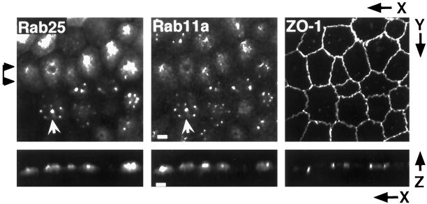
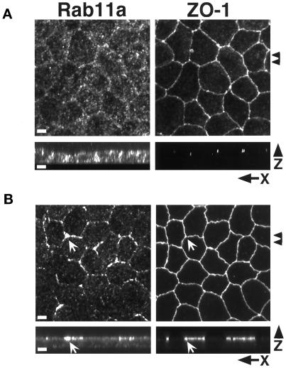
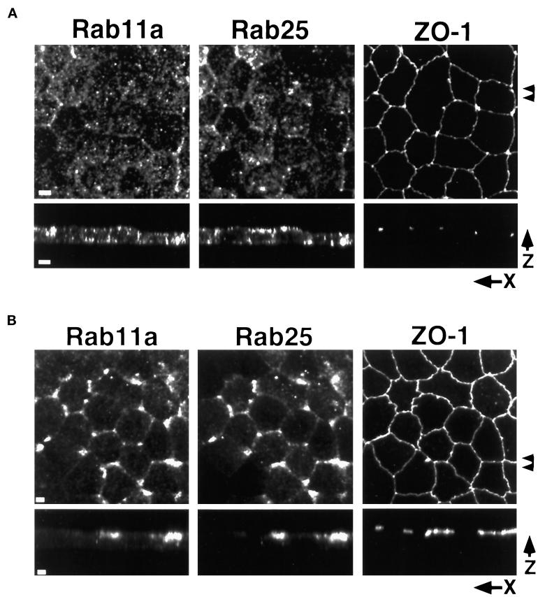
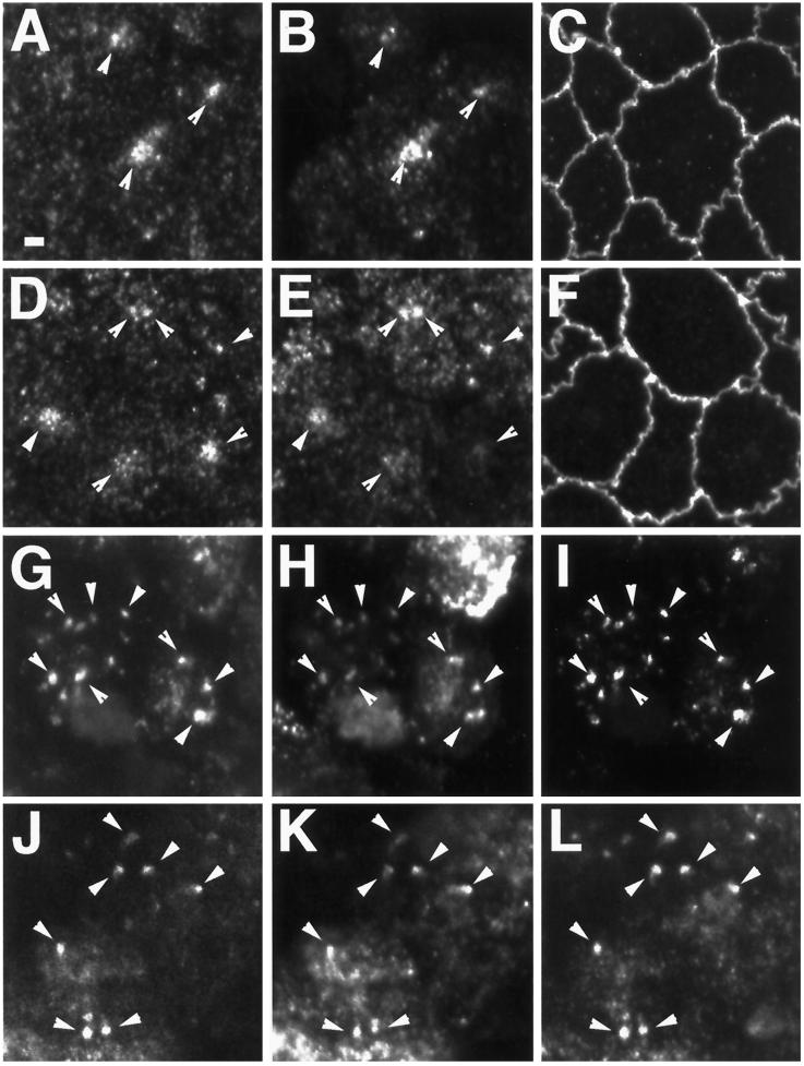
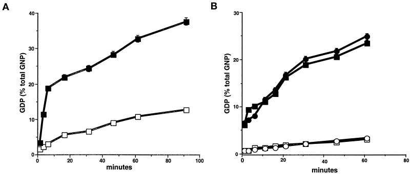
Recent evidence suggests that apical and basolateral endocytic pathways in epithelia converge in an apically located, pericentriolar endosomal compartment termed the apical recycling endosome. In this compartment, apically and basolaterally internalized membrane constituents are thought to be sorted for recycling back to their site of origin or for transcytosis to the opposite plasma membrane domain. We report here that in the epithelial cell line Madin-Darby Canine Kidney (MDCK), antibodies to Rab11a label an apical pericentriolar endosomal compartment that is dependent on intact microtubules for its integrity. Furthermore, this compartment is accessible to a membrane-bound marker (dimeric immunoglobulin A [IgA]) internalized from either the apical or basolateral pole, functionally defining it as the apical recycling endosome. We have also examined the role of a closely related epithelial-specific Rab, Rab25, in the regulation of membrane recycling and transcytosis in MDCK cells. When cDNA encoding Rab25 was transfected into MDCK cells, the protein colocalized with Rab11a in subapical vesicles. Rab25 transfection also altered the distribution of Rab11a, causing the coalescence of immunoreactivity into multiple denser vesicular structures not associated with the centrosome. Nevertheless, nocodazole still dispersed these vesicles, and dimeric IgA internalized from either the apical or basolateral membrane was detected in endosomes labeled with antibodies to both Rab11a and Rab25. Overexpression of Rab25 decreased the rate of IgA transcytosis and of apical, but not basolateral, recycling of internalized ligand. Conversely, expression of the dominant-negative Rab25T26N did not alter either apical recycling or transcytosis. These results indicate that both Rab11a and Rab25 associate with the apical recycling system of epithelial cells and suggest that Rab25 may selectively regulate the apical recycling and/or transcytotic pathways.


Figures

Figure 1
Figure 1
Localization of Rab11a in MDCK cells. MDCK cells were grown on permeable supports at confluence for 3 d and then fixed in 4% paraformaldehyde. (A) Polarized MDCK cells were triple-stained for γ-tubulin, Rab11a, and ZO-1; specific labeling was identified with secondary antibodies conjugated with Cy5, Cy2, and Cy3, respectively. Top panels show X–Y projections of 40 optical confocal fluorescence sections (0.3 μm each). Arrowheads at right indicate the region used to construct the X–Z projections shown in the bottom panels. Rab11a antibodies labeled vesicular aggregates located adjacent to the centrosome (arrows). Bar, 1 μm.
Figure 2
Figure 2
Rab25 and Rab11a colocalize in Rab25-transfected cells. MDCK cells stably expressing Rab25 were cultured on permeable supports. Fixed cells were triple-stained with antibodies against Rab11a, Rab25, and ZO-1 with detection by secondary antibodies conjugated with Cy5, Cy2, and Cy3, respectively. Maximum intensity projections were constructed from 40 confocal optical sections (0.3 μm each). Rab25 staining overlapped with Rab11a staining in most cells (arrows). In many cells, instead of a single nidus of immunoreactivity for Rab11a/Rab25, several brightly staining foci were observed in the apical region of the cell. Bar, 4 μm.
Figure 3
Figure 3
Effects of microtubule integrity on Rab11a distribution. (A) MDCK cells cultured on permeable supports were treated with 33 μM nocodazole for 2 h and then fixed in 4% paraformaldehyde. Cells were dual-stained with antibodies against Rab11a and ZO-1 with detection by secondary antibodies conjugated with Cy5 and Cy2, respectively. The top two panels show confocal projections of 40 X–Y optical sections (0.3 μm each) from the apical regions of the cells. Arrowheads at the right mark the region used to construct the X–Z projections shown in the bottom two panels. Punctate Rab11a immunoreactivity is dispersed throughout the cell with some concentration at the lateral margins. (B) Polarized MDCK cells cultured on permeable supports were treated with 5 μM taxol for 4 h at 37°C. Cells were dual-stained with antibodies against Rab11a and ZO-1. Top two panels show maximum intensity projection reconstructions of 40 confocal X–Y optical sections (0.3 μm each). Arrowheads at left indicate the region used to construct X–Z projections shown in bottom panels. Taxol caused the redistribution of Rab11a immunoreactivity to the region of the tight junctions (arrows) as well as a diffuse subapical staining pattern. Bar, 4 μm.
Figure 4
Figure 4
Effects of microtubule integrity on Rab11a and Rab25 distribution in Rab25-overexpressing MDCK cells. MDCK cells stably overexpressing Rab25 were grown on permeable filters and treated with either (A) 33 μM nocodazole for 2 h at 4°C or (B) 5 μM taxol for 4 h at 37°C. Cells were fixed in 4% paraformaldehyde and triple-stained with antibodies against Rab11a, Rab25, and ZO-1. Top panels in both A and B show maximum intensity projection reconstructions of 40 (0.3 μm each) confocal X–Y optical sections. Arrowheads at right indicate the region used to construct X–Z projections shown in the bottom panels. In A, nocodazole treatment dispersed both the Rab11a and Rab25 staining vesicles throughout the cytoplasm with some concentration at the lateral and apical surfaces. In B, taxol caused the redistribution of both Rab11a and Rab25 immunoreactivity to the region of the tight junctions. Rab11a and Rab25 immunoreactivity codistributed in taxol-treated cells. Bar, 4 μm.
Figure 5
Figure 5
Trafficking of dimeric IgA into the Rab11a- and Rab25-positive endosomal compartment. MDCK cells stably expressing the pIgR alone (A–F) or stably expressing both pIgR and Rab25 (G–L) were cultured on permeable supports and loaded with dimeric IgA for 30 min at 37°C either from the apical (A–C, G–I) or basal media (D–F, J–L). Cells were-triple labeled with anti-Rab11a (A, D, G, J), FITC-conjugated anti-IgA (B, E, H, K) and either anti-ZO-1 (C, F) or anti-Rab25 (I, L). Ten (0.3 μm) X–Y confocal optical sections from the apical regions were used to construct maximum intensity projections. In cells expressing only pIgR (A–F), under both loading conditions, IgA was observed in Rab11a-positive endosomal populations (arrowheads) in the apical region of the cells. In cells expressing both pIgR and Rab25 (G–L), IgA internalized from either pole of the cell could be observed in endosomes that stained for both Rab11a and Rab25 (arrowheads) in the apical region of the cells. Bar, 2 μm.
Figure 6
Figure 6
GTPase activity in Rab11 family members. The GTPase activities of (A) recombinant Rab25 and (B) recombinant Rab11a and Rab11aQ70L were assessed in vitro. (A) GTPase activity was assessed as described in MATERIALS AND METHODS in 1 μg of recombinant GST-Rab25 in the absence (□) or presence (▪) of gastric cytosol as a GAP donor. (B) GTPase activity was assessed in recombinant His-tagged Rab11a (circles) and His-tagged Rab11aQ70L (squares) in the absence (open symbols) or presence (closed symbols) of gastric cytosol as a GAP donor. All results represent the mean + SEM of three separate experiments each with duplicate samples. The results indicate that Rab25, Rab11a, and Rab11aQ70L are all active GTPases.
Figure 7
Figure 7
Rab25 inhibits transcytosis and apical recycling in adenovirus-transfected MDCK cells. (A) MDCK cells stably expressing the pIgR and the tetracycline-regulated transactivator were either mock-infected (triangles) or infected with recombinant adenovirus (squares and circles) encoding either wild-type Rab25 (A–C) or the Rab25T26N (D–F) for 18 h at 37°C. Expression of the recombinant proteins was confirmed by Western blot (B and E, insets). Infections were performed either in the absence of doxycycline (circles) or the presence of 20 ng/ml doxycycline (squares) to repress Rab25 synthesis. Cells were loaded with [125I]-dimeric IgA from the basolateral medium for 10 min at 37°C. Filters were then washed, and fresh medium was added to apical and basolateral chambers and incubated for the times indicated. Apical media (containing transcytosed ligand, filled symbols) and basolateral media (recycled ligand, open symbols) were harvested at each time point, and cells were collected after the last time point. Data are the average of two determinations and are expressed as a percentage of total internalized cpm. Triangles indicate mock-infected; circles, − doxycycline; squares, + doxycycline. (B and E) Fraction of internalized IgA remaining inside the cells at the end of the experiment shown in A and D, respectively. (C and F) Apical recycling. Ligand was internalized from the apical surface for 30 min at 37°C. Cells were then cooled to 4°C and washed, and surface-bound ligand was removed by incubation with trypsin (10 μg/ml) for 1 h at 4°C. Trypsinization was stopped by washing with cold medium containing soybean trypsin inhibitor (50 μg/ml). Filters were then placed in warm medium, and ligand was recovered from the apical (recycled) or basolateral (transcytosed) medium at the times indicated. Apical recycling was assayed in both (C) Rab25-infected and (E) Rab25T26N-infected cells. Filled symbols indicate apical recycling; open symbols, apical-to-basolateral transcytosis: triangles, mock-infected; circles, − doxycycline; squares, + doxycycline. All data represent trichloroacetic acid-precipitable counts (typically 97% of internalized ligand was trichloroacetic acid-precipitable) and are representative of at least three separate experiments.
Figure 8
Figure 8
Rab25 does not alter basolateral recycling in MDCK cells. (A) Cells were loaded with [125I]-labeled canine transferrin for 30 min at 37°C, washed, and then placed in fresh medium for the times indicated. Apical and basolateral media were harvested at each time point, and (B) the fraction of ligand remaining inside the cells was determined after the last time point. Recycling was measured in mock-infected cells (○) and in cells infected with Rab25 in the presence of doxycycline (□), with Rab25 in the absence of doxycycline (▵), and with Rab25T26N in the absence of doxycycline (⋄). All data are representative of three separate experiments.

References

    1. Adari H, Lowy DR, Willumsen BM, Der CJ, McCormick F. Guanosine triphosphatase activating protein (GAP) interacts with the p21 ras effector binding domain. Science. 1988;240:518–521. - PubMed
    1. Albright CF, Giddings BW, Liu J, Vita M, Weinberg RA. Characterization of a guanine nucleotide dissociation stimulator for a Ras related GTPase. EMBO J. 1993;12:339–347. - PMC - PubMed
    1. Apodaca G, Katz LA, Mostov KE. Receptor-mediated transcytosis of IgA in MDCK cells is via apical recycling endosomes. J Cell Biol. 1994;125:67–86. - PMC - PubMed
    1. Barroso M, Sztul E. Basolateral to apical transcytosis in polarized cells is indirect and involves BFA and trimeric G protein sensitive passage through the apical endosome. J Cell Biol. 1994;124:83–100. - PMC - PubMed
    1. Barth AI, Pollack AL, Altschuler Y, Mostov KE, Nelson WJ. NH2-terminal deletion of beta-catenin results in stable colocalization of mutant beta-catenin with adenomatous polyposis coli protein and altered MDCK adhesion. J Cell Biol. 1997;136:693–706. - PMC - PubMed

Publication types

MeSH terms

LinkOut - more resources