. 1999 Jan;10(1):77-90.
doi: 10.1091/mbc.10.1.77.

Association of nonribosomal nucleolar proteins in ribonucleoprotein complexes during interphase and mitosis

Affiliations
Free PMC article

Association of nonribosomal nucleolar proteins in ribonucleoprotein complexes during interphase and mitosis

S Piñol-Roma. Mol Biol Cell. 1999 Jan.
Free PMC article

Abstract

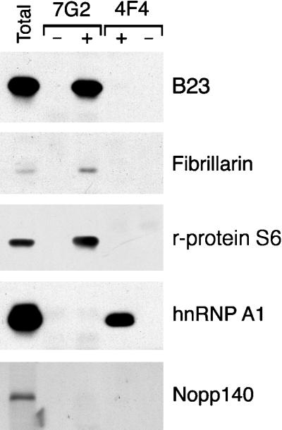
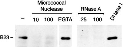
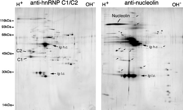
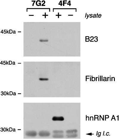
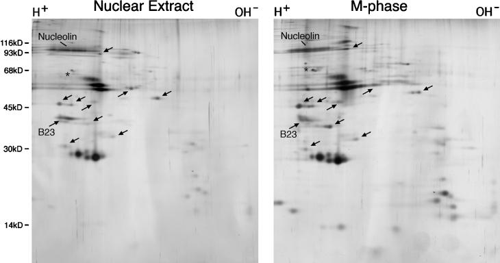
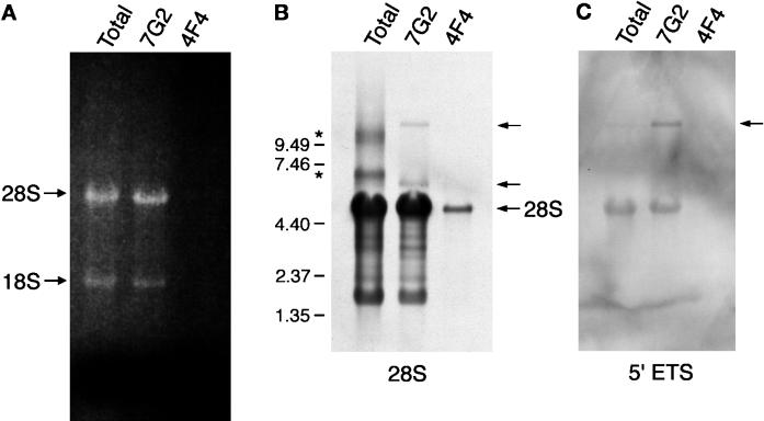
rRNA precursors are bound throughout their length by specific proteins, as the pre-rRNAs emerge from the transcription machinery. The association of pre-rRNA with proteins as ribonucleoprotein (RNP) complexes persists during maturation of 18S, 5.8S, and 28S rRNA, and through assembly of ribosomal subunits in the nucleolus. Preribosomal RNP complexes contain, in addition to ribosomal proteins, an unknown number of nonribosomal nucleolar proteins, as well as small nucleolar RNA-ribonucleoproteins (sno-RNPs). This report describes the use of a specific, rapid, and mild immunopurification approach to isolate and analyze human RNP complexes that contain nonribosomal nucleolar proteins, as well as ribosomal proteins and rRNA. Complexes immunopurified with antibodies to nucleolin-a major nucleolar RNA-binding protein-contain several distinct specific polypeptides that include, in addition to nucleolin, the previously identified nucleolar proteins B23 and fibrillarin, proteins with electrophoretic mobilities characteristic of ribosomal proteins including ribosomal protein S6, and a number of additional unidentified proteins. The physical association of these proteins with one another is mediated largely by RNA, in that the complexes dissociate upon digestion with RNase. Complexes isolated from M-phase cells are similar in protein composition to those isolated from interphase cell nuclear extracts. Therefore, the predominant proteins that associate with nucleolin in interphase remain in RNP complexes during mitosis, despite the cessation of rRNA synthesis and processing in M-phase. In addition, precursor rRNA, as well as processed 18S and 28S rRNA and candidate rRNA processing intermediates, is found associated with the immunopurified complexes. The characteristics of the rRNP complexes described here, therefore, indicate that they represent bona fide precursors of mature cytoplasmic ribosomal subunits.


Figures

Figure 1
Figure 1
Distribution of nucleolin in interphase and M-phase HeLa cells. (A) Asynchronously growing HeLa cells were fixed, permeabilized, and immunostained with the anti-nucleolin monoclonal antibody 7G2, followed by detection with FITC-conjugated secondary antibody. The position of the nuclei and nucleoli, as well as of cells in mitotic prophase and metaphase (as judged by phase microscopy, not shown here), is indicated. (B) Immunoblot analysis of total HeLa cell proteins with the anti-nucleolin monoclonal antibody 7G2. The position of molecular weight standards run in the same gel is indicated on the left (molecular masses are indicated in kilodaltons).
Figure 2
Figure 2
Immunopurification of nucleolin from HeLa cell nuclear extracts. Immunopurifications were carried out from interphase cell nuclear extracts using anti-nucleolin (7G2), anti-hnRNP C1/C2 (4F4), and nonimmune (SP2/0) monoclonal antibodies, in the presence of either 0.5% Triton X-100 (lanes T) or 1% Empigen BB (lanes E). The immunopurified complexes were resolved by SDS-PAGE, and proteins were visualized by silver staining. Mock immunopurifications in which nuclear extract was omitted were run in parallel to allow identification of bands that resulted from the antibody used in the isolation (lanes −). Examples of bands specific to 7G2-immunopurified complexes are indicated with •, as discussed in the text. Ig h.c. and Ig l.c. refer to the Ig heavy and light chains, respectively.
Figure 3
Figure 3
Immunoblot analysis of RNP complexes immunopurified from nuclear extracts. Proteins from complexes immunopurified with 7G2 (lanes 7G2) or with 4F4 (lanes 4F4) monoclonal antibodies (to nucleolin and hnRNP C1/C2 respectively) were resolved by SDS-PAGE, transferred to nitrocellulose, and immunoblotted with antibodies to B23, fibrillarin, ribosomal protein S6 (r-protein S6), hnRNP A1, or Nopp140 (lanes +), as indicated on the right. As a control for reactivity with the antibodies used in the immunopurification, mock immunopurifications were also included in which HeLa cell proteins were omitted (lanes -).
Figure 4
Figure 4
Sensitivity of nucleolin complexes to RNase. Nuclear extracts were incubated with micrococcal nuclease, RNase A, or DNase I, as indicated, and immunopurifications were then carried out with the 7G2 monoclonal antibody in RSB-100 containing 0.5% Triton X-100. The proteins in the immunopurified complexes were resolved by SDS-PAGE, transferred to nitrocellulose, and probed with the antiB23 monoclonal antibody. Micrococcal nuclease digestions were carried out using either 10 or 100 units/ml, as indicated, in the presence of 2 mM CaCl2. lane EGTA: incubation with 100 u/ml micrococcal nuclease in the presence of both CaCl2 and 5 mM EGTA. RNase A digestions were carried out using either 25 or 100 μg/ml, as indicated. DNase I incubations were carried out in the presence of 100 units RNase-free DNase I/ml. lane -: complexes immunopurified from nuclear extract incubated similarly in the absence of added nuclease.
Figure 5
Figure 5
Immunopurification of nucleolin and hnRNP C proteins from M-phase cells. Whole cell lysates were prepared from metaphase-arrested HeLa cells in RSB-100 containing 0.5% Triton X-100. Immunopurifications were then carried out from these lysates using antinucleolin (right panel) or antihnRNP C (left panel) monoclonal antibodies. The antibody-antigen complexes were eluted with NEPHGE sample buffer, resolved by two-dimensional gel electrophoresis (NEPHGE in the first dimension, left to right, followed by SDS-PAGE in the second dimension; top to bottom), and proteins visualized by silver staining. Analysis of immunoglobulins incubated similarly but in the absence of cell lysate was carried out in parallel gels (not shown) to identify those polypeptides resulting from the antibody and not from the HeLa cells. Arrows indicate some of the proteins specifically associated in complexes with nucleolin. Ig h.c. and Ig l.c. refer to the Ig heavy and light chains respectively.
Figure 6
Figure 6
Immunoblot analysis of RNP complexes immunopurified from metaphase-arrested HeLa cells. RNP complexes were immunopurified with either monoclonal antibody 4F4 or 7G2 from metaphase-arrested HeLa cells (lanes +). The proteins in the respective complexes were resolved by SDS-PAGE and blotted onto nitrocellulose. Identical filters were probed with antiserum to fibrillarin, a monoclonal antibody to B23, or 4B10 monoclonal antibody to hnRNP A1, as indicated. Lanes - contain mock immunopurifications carried out without HeLa cell lysate, to ascertain whether any of the signal observed is due to the antibodies used in the immunopurification. Ig l.c. refers to the Ig light chains originating from the antibody used for immunopurification, and which are detected by the secondary antibody used in the immunoblot.
Figure 7
Figure 7
Two-dimensional gel electrophoresis comparison of complexes immunopurified from interphase cell nuclear extracts and from M-phase cells. RNP complexes were immunopurified with the 7G2 monoclonal antibody from HeLa cell nuclear extracts (left panel) or from whole nocodazole-arrested cell lysates (right panel) as described for Figures 2 and 5, respectively. Proteins in each complex were resolved by two-dimensional gel electrophoresis (see legend to Figure 5) and visualized by silver staining. Arrows point to candidate nonribosomal proteins, as discussed in the text.
Figure 8
Figure 8
Analysis of RNA immunopurified with antibodies to nucleolin. (A) RNP complexes were immunopurified from metaphase-arrested HeLa cells using monoclonal antibodies to nucleolin (7G2) or to hnRNP C1/C2 (4F4). RNA was eluted from the complexes as described in the main text and resolved in a 1% agarose gel containing formaldehyde. For comparison, total RNA isolated from whole cell lysates was also included (lane Total). The RNA was visualized by staining with ethidium bromide. The position of 18S and 28S rRNA is indicated on the left. (B) Northern blot analysis of the RNA shown in panel A. The RNA was transferred to a nitrocellulose membrane and probed with biotinylated oligonucleotide probes complementary to 28S rRNA. Detection of hybridized oligonucleotides was carried out with HRP-conjugated avidin, followed by ECL detection. The arrows point to the position of the mature 28S rRNA (bottom) and of precursor (top) and candidate 32S intermediate (middle) rRNA, as described in the text. Asterisks on the left indicate the position of likely RNA aggregates. The position of RNAs of known length run on the same gel is also indicated on the left. (C) Northern blot analysis carried out under identical conditions to those shown in panel B, but using an oligonucleotide complementary to the 5′-pre-rRNA external transcribed spacer. The arrow points to the precursor rRNA.

References

    1. Auger-Buendia M-A, Longuet M. Characterization of proteins from nucleolar preribosomes of mouse leukemia cells by two-dimensional polyacrylamide gel electrophoresis. Eur J Biochem. 1978;85:105–114. - PubMed
    1. Belenguer P, Caizergues-Ferrer M, Labbé J-C, Dorée M, Amalric F. Mitosis-specific phosphorylation of nucleolin by p34cdc2 protein kinase. Mol Cell Biol. 1990;10:3607–3618. - PMC - PubMed
    1. Borer RA, Lehner CF, Eppenberger HM, Nigg EA. Major nucleolar proteins shuttle between nucleus and cytoplasm. Cell. 1989;56:379–390. - PubMed
    1. Bostock CJ, Prescott DM, Kirkpatrick JB. An evaluation of the double thymidine block for synchronizing mammalian cells at the G1-S border. Exp Cell Res. 1971;68:163–168. - PubMed
    1. Bugler B, Bourbon H, Lapeyre B, Wallace MO, Chang J-H, Amalric F, Olson MOJ. RNA binding fragments from nucleolin contain the ribonucleoprotein consensus sequence. J Biol Chem. 1987;262:10922–10925. - PubMed

Publication types

MeSH terms

LinkOut - more resources