

Ultrastructural analysis of transcription and splicing in the cell nucleus after bromo-UTP microinjection
- PMID: 9880337
- PMCID: PMC25164
- DOI: 10.1091/mbc.10.1.211
Ultrastructural analysis of transcription and splicing in the cell nucleus after bromo-UTP microinjection
Abstract
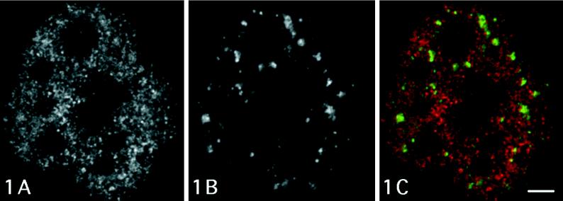
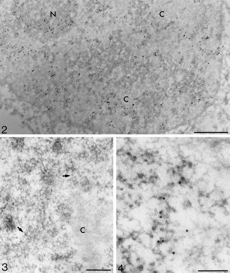
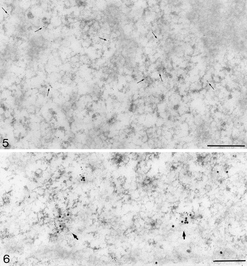
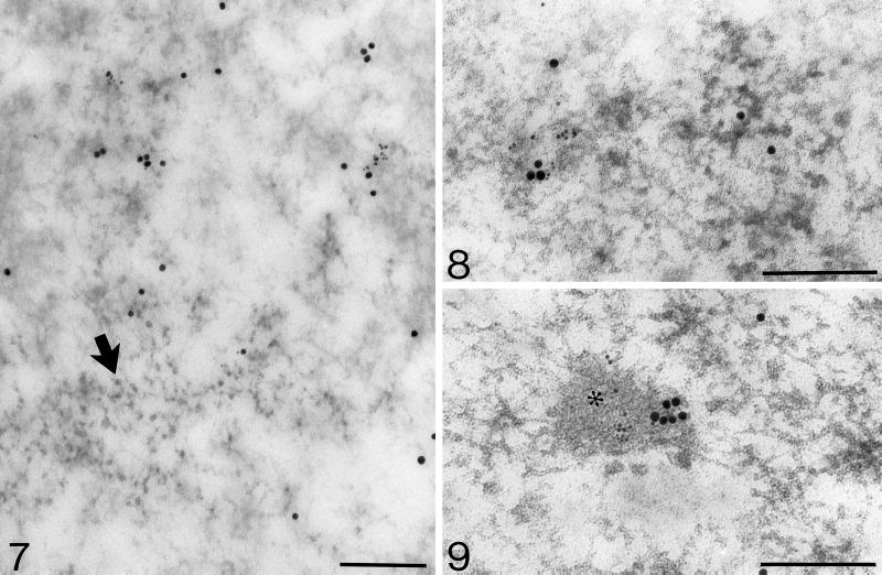
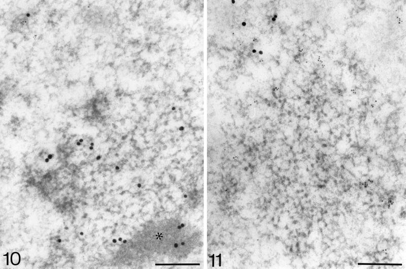
In this study we demonstrate, at an ultrastructural level, the in situ distribution of heterogeneous nuclear RNA transcription sites after microinjection of 5-bromo-UTP (BrUTP) into the cytoplasm of living cells and subsequent postembedding immunoelectron microscopic visualization after different labeling periods. Moreover, immunocytochemical localization of several pre-mRNA transcription and processing factors has been carried out in the same cells. This high-resolution approach allowed us to reveal perichromatin regions as the most important sites of nucleoplasmic RNA transcription and the perichromatin fibrils (PFs) as in situ forms of nascent transcripts. Furthermore, we show that transcription takes place in a rather diffuse pattern, without notable local accumulation of transcription sites. RNA polymerase II, heterogeneous nuclear ribonucleoprotein (hnRNP) core proteins, general transcription factor TFIIH, poly(A) polymerase, splicing factor SC-35, and Sm complex of small nuclear ribonucleoproteins (snRNPs) are associated with PFs. This strongly supports the idea that PFs are also sites of major pre-mRNA processing events. The absence of nascent transcripts, RNA polymerase II, poly(A) polymerase, and hnRNPs within the clusters of interchromatin granules rules out the possibility that this domain plays a role in pre-mRNA transcription and polyadenylation; however, interchromatin granule-associated zones contain RNA polymerase II, TFIIH, and Sm complex of snRNPs and, after longer periods of BrUTP incubation, also Br-labeled RNA. Their role in nuclear functions still remains enigmatic. In the nucleolus, transcription sites occur in the dense fibrillar component. Our fine structural results show that PFs represent the major nucleoplasmic structural domain involved in active pre-mRNA transcriptional and processing events.
Figures
References
-
- Bachellerie J-P, Puvion E, Zalta J-P. Ultrastructural organization and biochemical characterization of chromatin-RNA-protein complexes isolated from mammalian cell nuclei. Eur J Biochem. 1975;58:327–337. - PubMed
-
- Bernhard W. A new staining procedure for electron microscopical cytology. J Ultrastruct Res. 1969;27:250–265. - PubMed
-
- Beyer AL, Osheim YN. Splice site selection rate of splicing and alternative splicing on nascent transcripts. Genes Dev. 1988;2:754–765. - PubMed
Publication types
MeSH terms
Substances
LinkOut - more resources
Full Text Sources
Other Literature Sources
Research Materials
