. 1999 Jan;119(1):111-22.
doi: 10.1104/pp.119.1.111.

Dynamics of cytokinins in apical shoot meristems of a day-neutral tobacco during floral transition and flower formation

Affiliations

Dynamics of cytokinins in apical shoot meristems of a day-neutral tobacco during floral transition and flower formation

W Dewitte et al. Plant Physiol. 1999 Jan.

Abstract

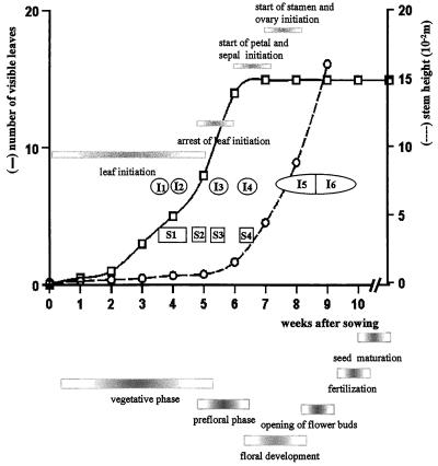
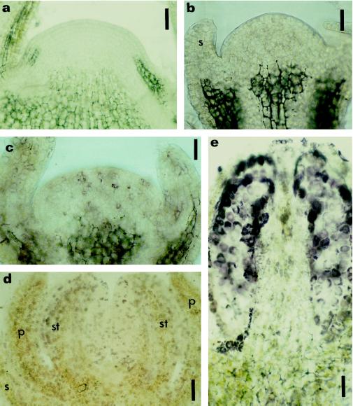
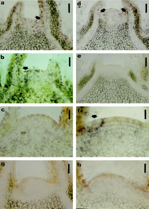
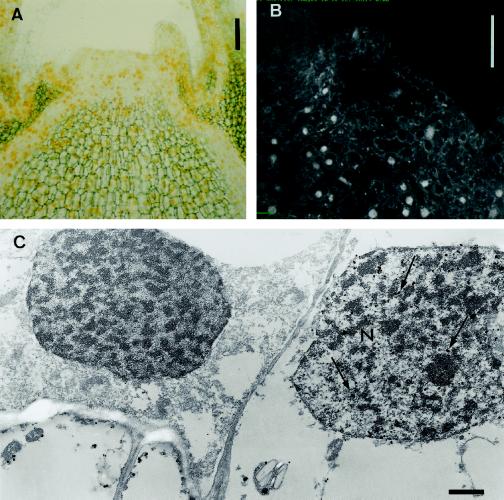
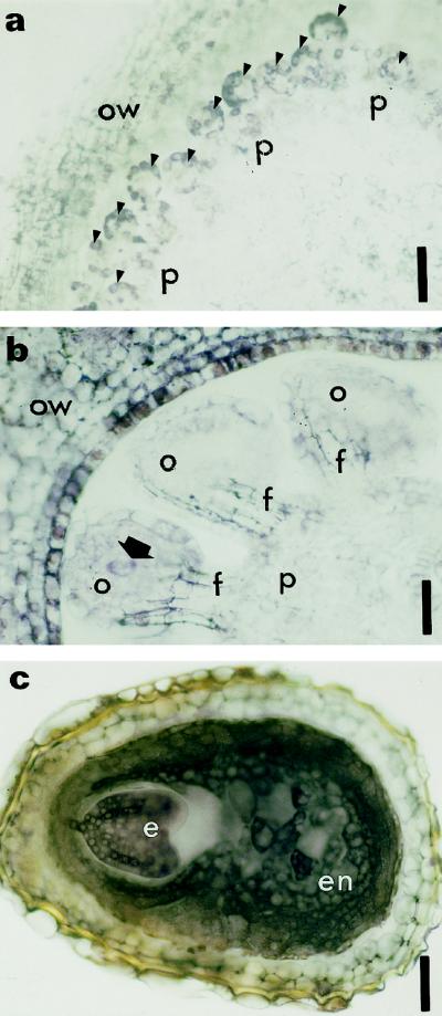
This study considered cytokinin distribution in tobacco (Nicotiana tabacum L.) shoot apices in distinct phases of development using immunocytochemistry and quantitative tandem mass spectrometry. In contrast to vegetative apices and flower buds, we detected no free cytokinin bases (zeatin, dihydrozeatin, or isopentenyladenine) in prefloral transition apices. We also observed a 3-fold decrease in the content of cytokinin ribosides (zeatin riboside, dihydrozeatin riboside, and isopentenyladenosine) during this transition phase. The group concluded that organ formation (e.g. leaves and flowers) is characterized by enhanced cytokinin content, in contrast to the very low endogenous cytokinin levels found in prefloral transition apices, which showed no organogenesis. The immunocytochemical analyses revealed a differing intracellular localization of the cytokinin bases. Dihydrozeatin and isopentenyladenine were mainly cytoplasmic and perinuclear, whereas zeatin showed a clear-cut nuclear labeling. To our knowledge, this is the first time that this phenomenon has been reported. Cytokinins do not seem to act as positive effectors in the prefloral transition phase in tobacco shoot apices. Furthermore, the differences in distribution at the cellular level may be indicative of a specific physiological role of zeatin in nuclear processes.


Figures

Figure 1
Figure 1
Summary of the main parameters of vegetative and reproductive development of the tobacco var Petit Havana cv SR1. The number of visible leaves and the stem height were recorded starting with an interval of 1 week, and were plotted against the number of weeks after sowing. Sampling points for both cytokinin quantification (S1–S4) and for immunolocalization of cytokinin bases (I1–I6) are located in the graph. The horizontal bars in the graph indicate the different morphogenetic events at the apical shoot meristem. The horizontal bars under the graph indicate the different phases of plant development.
Figure 5
Figure 5
Immunolocalization of zeatin and IP in prefloral and floral shoot apices. Longitudinal sections of a prefloral transition apex (a), early floral apices (b and c), and floral apices (d and e) subjected to immunohistochemistry for zeatin (a, c, d, and e) and IP (b). Localization was with AP-conjugated secondary antibodies and nitroblue tetrazolium/5-bromo-4-chloro-3-indolyl phosphate as the chromogenic substrate. a, Prefloral transition apex immunostained for zeatin, disclosing no reaction of the AP in the meristem. b and c, Early floral phase with sepal formation showing the absence of label for IP (b), but the presence of reaction with the anti-zeatin antibody was reflected in weak purple staining (c). d and e, Floral phase with sepal, petal, and stamen already initiated; a strong purple staining occurred in different cells throughout the developing flower after immunostaining of zeatin. e, Longitudinal section of a developing stamen immunolabeled with anti-zeatin, showing a strong signal in the sporogenic tissue. p, Petal; s, sepal; st, stamen. Bars = 90 μm (a) and 45 μm (b, c, d, and e).
Figure 2
Figure 2
DNA content distribution in apices during the vegetative and prefloral transition phase. Histogram of the nDNA content during the vegetative phase (a) and the prefloral transition (b). The percentage of cells in G0,G1 and S,G2 is also presented.
Figure 3
Figure 3
Immunolocalization of cytokinin bases within vegetative shoot apices. The location of cytokinin bases in longitudinal sections was determined using immunohistochemistry. Nitroblue tetrazolium/5-bromo-4-chloro-3-indolyl phosphate was used as chromogenic substrate for the AP, resulting in a purple reaction product. Immunolocalization of zeatin (a and d), DHZ (b and e), and IP (c and f) in vegetative shoot apices at developmental stages I1 (a, b, and c) and I2 (d, e, and f) (Fig. 1). Purple staining is present for zeatin (a and d) in the nucleus and cytoplasm, whereas for DHZ (b and e) and IP (c and f) a more perinuclear purple staining is visible. Arrows in a, b, d, and f point to staining in the lateral zones of the meristems. g and h, Sections (from stage I1, see Fig. 1) incubated with anti-zeatin (g) and anti-DHZ (h) antibodies saturated with zeatin and DHZ as a control, respectively. Bars = 90 μm (a, b, d, e, g, and h); 45 μm (c); and 22 μm (f).
Figure 4
Figure 4
Immunolocalization of zeatin in vegetative shoot apices. Zeatin was localized in the nucleus of vegetative meristem cells upon silver-enhanced gold-labeling by light and electron microscopy (A and C). A, Yellow color, originating from light diffraction by the silver particles, was distributed throughout the apex. C, A large number of silver particles (arrows) were detected in the nucleus (N) of some cells by electron microscopy. Confocal laser microscopy after immunolocalization with FITC as a probe revealed a strong fluorescence signal in the nucleus of some cells (B). Bars = 90 μm (A), 100 μm (B), and 1 μm (C).
Figure 6
Figure 6
Immunolocalization of zeatin at two stages of ovule formation and at the end of seed formation. Cross-sections of ovule primordia arising from the placenta (a, arrowheads) and developing ovule (b, arrow indicates the archesporial cell in a developing ovule). Purple staining resulting from labeling of zeatin was detected in ovule primordia (a, arrowheads), the ovary wall (a and b), and the developing ovules (b). Archesporial cells are also significantly labeled (b). c, Cross-section of a seed immunolabeled with anti-zeatin antibody. The cytoplasm of embryo and endosperm are heavily stained. e, Embryo; en, endosperm; f, funiculus; o, ovule; ow, ovary wall; p, placenta. Bars = 180 μm (a) and 90 μm (b and c).

References

    1. Bernier G. The control of floral evocation and morphogenesis. Annu Rev Plant Physiol Plant Mol Biol. 1988;39:175–219.
    1. Besnard-Wibaut C. Effectiveness of gibberellins and 6-benzyladenine of flowering of Arabidopsis thaliana. Physiol Plant. 1981;53:205–212.
    1. Beveridge CA, Murfet IC, Kerhoas L, Sotta B, Miginiac E, Rameau C. The shoot controls zeatin riboside export from pea roots: evidence from the branching mutant rms4. Plant J. 1997;11:339–345.
    1. Beveridge CA, Ross JJ, Murfet IC. Branching in pea. Action of genes Rms3 and Rms4. Plant Physiol. 1996;110:859–865. - PMC - PubMed
    1. Biermann BJ, Morehead AT, Tate ST, Price JF, Randhall SK, Crowell DN. Novel isoprenylated proteins identified by an expression library screen. J Biol Chem. 1994;269:25251–25254. - PubMed

LinkOut - more resources