. 1999 Jan 15;19(2):599-609.
doi: 10.1523/JNEUROSCI.19-02-00599.1999.

Subthalamic nucleus neurons switch from single-spike activity to burst-firing mode

Affiliations

Subthalamic nucleus neurons switch from single-spike activity to burst-firing mode

C Beurrier et al. J Neurosci. .

Abstract

The modification of the discharge pattern of subthalamic nucleus (STN) neurons from single-spike activity to mixed burst-firing mode is one of the characteristics of parkinsonism in rat and primates. However, the mechanism of this process is not yet understood. Intrinsic firing patterns of STN neurons were examined in rat brain slices with intracellular and patch-clamp techniques. Almost half of the STN neurons that spontaneously discharged in the single-spike mode had the intrinsic property of switching to pure or mixed burst-firing mode when the membrane was hyperpolarized from -41.3 +/- 1.0 mV (range, -35 to -50 mV; n = 15) to -51.0 +/- 1.0 mV (range, -42 to -60 mV; n = 20). This switch was greatly facilitated by activation of metabotropic glutamate receptors with 1S,3R-ACPD. Recurrent membrane oscillations underlying burst-firing mode were endogenous and Ca2+-dependent because they were largely reduced by nifedipine (3 microM), Ni2+ (40 microM), and BAPTA-AM (10-50 microM) at any potential tested, whereas TTX (1 microM) had no effect. In contrast, simultaneous application of TEA (1 mM) and apamin (0.2 microM) prolonged burst duration. Moreover, in response to intracellular stimulation at hyperpolarized potentials, a plateau potential with a voltage and ionic basis similar to those of spontaneous bursts was recorded in 82% of the tested STN neurons, all of which displayed a low-threshold Ni2+-sensitive spike. We propose that recurrent membrane oscillations during bursts result from the sequential activation of T/R- and L-type Ca2+ currents, a Ca2+-activated inward current, and Ca2+-activated K+ currents.


Figures
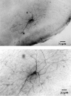
Fig. 1.
Fig. 1.
Microphotographs of a biocytin-filled STN neuron at two magnifications. The labeled neuron is located within the boundaries of the STN (top) and presents a dense dendritic arborization (top) and numerous spines on dendrites (bottom).
Fig. 2.
Fig. 2.
Single-spike mode. A, Tonic and regular activity (single-spike mode) of an STN neuron, recorded with intracellular techniques at resting membrane potential and (B) corresponding interspike interval histogram (mean interval, 66.1 ± 15.6 msec; bin width, 12.5 msec).C, Discharge frequency histogram of single-spike mode recorded in 41 STN neurons (mean frequency, 22.3 ± 1.5 Hz; bin width, 10 Hz). Spikes in A are truncated.
Fig. 3.
Fig. 3.
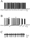
Burst-firing mode. Two types of burst mode recorded with patch-clamp techniques in two different STN neurons: pure burst mode (A) and mixed burst mode (B), consisting of long bursts (⋆) separated by sequences of short bursts (○). C, Spontaneous bursts recorded in the cell-attached configuration in voltage-clamp mode.
Fig. 4.
Fig. 4.
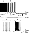
Switch of firing mode according to membrane potential. Pure burst mode (⋆) was triggered at resting membrane potential (I = 0, dotted line, middle) in a whole-cell-recorded neuron that displayed the single-spike mode (⋄) at a more depolarized membrane potential (I = +0.2 nA,left). At a more hyperpolarized potential (I = −0.6 nA, right), the cell became silent. The two bottom traces are taken from the above records and displayed at an expanded time scale. Spikes of the single-spike mode in the left part are truncated.
Fig. 5.
Fig. 5.
Ionic basis of the burst-firing mode I.A, TTX (1 μm) totally suppressed action potentials evoked during bursts while sparing the rhythmic oscillations of the membrane potential that underlie bursts. Note the increase in the duration of membrane oscillations in the presence of TTX (from 5.0 ± 0.0 sec to 16.4 ± 3.1 sec). B, The duration of bursts was irreversibly decreased by an application of nifedipine (3 μm, bottom trace) at all tested potentials, whereas the single-spike mode was unaffected.Traces in A and B were obtained from two different STN neurons recorded with patch-clamp techniques in whole-cell configuration. Spikes from the single-spike mode in B are truncated. Calibration is the same fortraces of each section.
Fig. 6.
Fig. 6.
Ionic basis of the burst-firing mode II.A, Bath application of BAPTA-AM (50 μm) decreased burst duration at any potential tested, although it spared the single-spike activity (left column).B, Simultaneous application of TEA (1 mm) and apamin (0.2 μm) prevented burst repolarization and locked membrane potential at −30 mV. Repolarizations were obtained by injecting brief hyperpolarizing current pulses (−80 pA, 100 msec). After each repolarizing command, the membrane spontaneously depolarized again (middle trace). As drugs washed out, bursts reappeared, but with a longer duration (2.9 ± 0.1 sec vs 1.7 ± 0.2 sec, bottom trace). Traces inA and B were obtained from two different STN neurons with patch-clamp recordings in whole-cell configuration. Spikes from the single-spike mode in A (on theleft) are truncated.
Fig. 7.
Fig. 7.
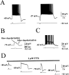
Plateau potentials. A, A short depolarizing (100 pA, 100 msec, left trace) or hyperpolarizing (−100 pA, 100 msec, right trace) current pulse triggered both a plateau potential (1.8 and 2.6 msec duration, respectively) that considerably outlasted the duration of the stimulus and was followed by a prominent AHP (28 and 17 mV amplitude, respectively). B, TTX (1 μm) revealed the presence of two different phases in the plateau potential: a slow depolarization triggered by the depolarizing current pulse (50 pA, 200 msec) and an afterdepolarization (267 msec) triggered at the break of the current pulse. C, Long duration plateau potential terminated by a short hyperpolarizing current pulse (−20 pA, 100 msec). D, Amplitude and duration of the plateau potential according to membrane potential. In the presence of TTX (1 μm), the same depolarizing current pulse (100 pA, 100 msec) evoked a plateau potential in the membrane potential range of −60 to −70 mV. At more depolarized (−40 mV, extreme left) or hyperpolarized (−80 mV, extreme right) potentials, the amplitude and duration of the plateau potential was considerably reduced. Traces in A,C, and D were obtained with patch-clamp recordings (whole-cell configuration), and traces inB were obtained with intracellular recordings. All spikes are truncated.
Fig. 8.
Fig. 8.
Ionic basis of the plateau response.A, TTX (1 μm) suppressed action potentials but not the plateau potential (middle trace) evoked by a 100 pA, 100 msec current pulse (left trace). In the presence of TTX, bath application of nifedipine (3 μm) suppressed the plateau potential (right trace).B, The duration of the plateau potential was also decreased by bath application of BAPTA-AM (50 μm). Note that the cell fired some action potentials during the current pulse (100 pA, 100 msec). C, In contrast, the plateau potential was significantly increased by simultaneous application of TEA (1 mm) and apamin (0.2 μm) from 0.5 sec (left trace) to 5.5 sec (right trace). All traces were obtained with patch-clamp recordings (whole-cell configuration).
Fig. 9.
Fig. 9.
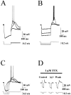
Voltage dependency and pharmacological properties of LTS. A, The three superimposed voltage traces show that LTS was recorded at the break of a hyperpolarizing current pulse (−300 pA, 200 msec) applied at a membrane potential of −63 mV. Its amplitude and rise time depended on the value of membrane potential at the end of the negative current pulse: at −78 mV, LTS was not evoked (bottom trace), whereas at a slightly more depolarized potential (approximately −72 mV, middle trace) it was present and gave rise to a spike. With increased depolarization, LTS amplitude increased, and spike delay decreased (top trace). B, Three superimposed responses to hyperpolarizing current pulses of increasing amplitude (−100, −250, and −350 pA) and fixed duration (300 msec) from Vm = −53 mV. LTS was only evoked when the membrane was held for 300 msec at a potential more hyperpolarized than −78 mV for 300 msec. C, Three superimposed voltage traces in response to hyperpolarizing current pulses of fixed amplitude (−150 pA) and increasing duration (40, 80, and 120 msec). LTS was evoked in a neuron maintained at −64 mV when the membrane was held at −85 mV for at least 80 msec during the application of a hyperpolarizing current pulse. D, In the presence of TTX (1 μm), LTS evoked in response to a hyperpolarizing current pulse (−130 pA, 200 msec) from Vm = −70 mV was not affected (Control and Wash), whereas it was reversibly suppressed by the concomitant application of Ni2+(40 μm). Traces in A,C, and D were obtained with intracellular recordings, and traces in B were obtained with patch-clamp recordings (whole-cell configuration). All spikes are truncated.
Fig. 10.
Fig. 10.
The hypothetical cascade of currents underlying the different phases of burst-firing mode. See Discussion for explanation.

References

    1. Albin RL, Young AB, Penney JB. The functional anatomy of the basal ganglia disorders. Trends Neurosci. 1989;12:366–375. - PubMed
    1. Aziz TZ, Peggs D, Sambrook MA, Crossman AR. Lesion of the subthalamic nucleus for the alleviation of 1-methyl-4-phenyl-1,2,3–6-tetrahydropyridine (MPTP)-induced parkinsonism in the primate. Mov Disord. 1991;6:288–292. - PubMed
    1. Bathia KP, Marsden CD. The behavioral and motor consequences of focal lesions of the basal ganglia in man. Brain. 1994;117:859–876. - PubMed
    1. Benazzouz A, Gross ChFéger J, Boraud Th, Bioulac B. Reversal of rigidity and improvement in motor performance by subthalamic high-frequency stimulation in MPTP-treated monkeys. Eur J Neurosci. 1993;5:382–389. - PubMed
    1. Benazzouz A, Piallat B, Pollak P, Limousin P, Gao DM, Krack P, Benabid AL. Single unit recordings of subthalamic nucleus and pars reticulata of substantia nigra in akineto-rigid parkinsonian patients. Soc Neurosci Abstr. 1996;22:91.18.

Publication types

MeSH terms

LinkOut - more resources