. 1999 Jan 15;19(2):653-63.
doi: 10.1523/JNEUROSCI.19-02-00653.1999.

Kainate receptor-mediated responses in the CA1 field of wild-type and GluR6-deficient mice

Affiliations

Kainate receptor-mediated responses in the CA1 field of wild-type and GluR6-deficient mice

I Bureau et al. J Neurosci. .

Abstract

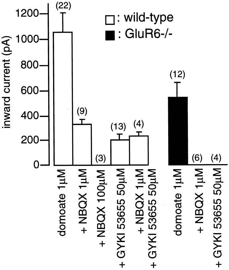
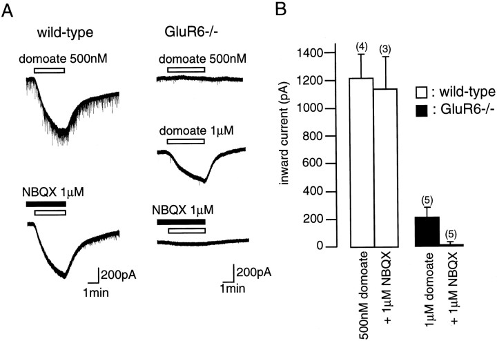
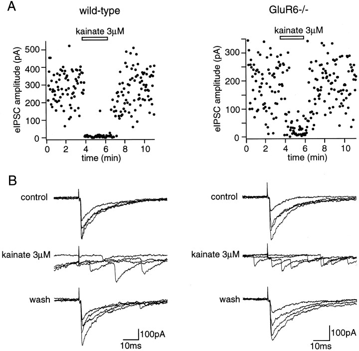
Kainate receptors are abundantly expressed in the hippocampus. Mice with disruption of kainate receptor subunits allow the genetic dissection of the role of each kainate receptor subunits in the synaptic physiology of the hippocampus, as well as in excitotoxic processes. We have compared the action of domoate and kainate on CA1 pyramidal neurons in slices from wild-type and GluR6-/- mice. The difference in the amplitude of inward currents evoked by domoate and kainate between wild-type and GluR6-/- mice demonstrates the presence of functional kainate receptors in CA1 pyramidal neurons. Block of domoate-activated inward currents by the AMPA receptor antagonists 2,3-dihydroxy-6-nitro-7-sulfonyl-benzo(F)quinoxaline (1 microM) and 1-(4-aminophenyl)-3-methylcarbamyl-4-methyl7, 8-methylenedioxy-3,4-dihydro-5H-2,3-benzodiazepine) (GYKI 53655) (50 microM) is complete in GluR6-/- mice but only partial in wild-type mice. In the presence of GYKI 53655, kainate receptor activation dramatically increases the frequency of spontaneous IPSCs in CA1 pyramidal cells from wild-type, as well as GluR6-/-, mice. This results from the kainate receptor-mediated activation of a sustained inward current and an increased action potential firing in afferent GABAergic interneurons of the CA1 field. These effects are observed in wild-type, as well as GluR6-/-, mice. Kainate receptors also decrease the amplitude of evoked IPSCs in CA1 pyramidal cells by increasing synaptic failures in wild-type and GluR6-/- mice. These results indicate that in CA1 pyramidal cells, distinct subtypes of kainate receptors mediate several functionally antagonistic effects.


Figures

Fig. 1.
Fig. 1.
Mapping of the mRNA encoding the five KAR subunits in the anterior dorsal hippocampus of 129SvJ mouse brain as assessed with 35S-labeled riboprobes. Photomicrographs were taken by the stereomicroscope (10×) under bright-field (A) or dark-field (B–F) illumination. A is a section processed for cresyl violet counterstaining. The dark-field representations in B–Freveal considerable differences in the expression pattern of each KAR subunit.
Fig. 2.
Fig. 2.
Cellular expression of GluR5 and GluR6 subunits at high magnification (200×). Expression of GluR5 and GluR6 subunits in the pyramidal cell layers and interneurons of the stratum oriens (s.o.) and stratum radiatum (s.r.) of CA1 and CA3 fields. GluR5 is highly expressed in the interneurons of the nonpyramidal layer, but some strongly labeled cells appear also among the pyramidal neurons. GluR6 is primarily expressed in the pyramidal cells, but scattered dimly labeled cells are also seen in the stratum oriens and radiatum of both CA1 and CA3 fields.
Fig. 3.
Fig. 3.
Comparison of the response of CA1 pyramidal cells to domoate and kainate in wild-type and GluR6−/− mice.A, Whole-cell currents activated by domoate in wild-type (left) and GluR6−/− (right) mice by domoate (open horizontal bar). At a concentration of 200 nm, domoate activated a small inward current in wild-type but not in GluR6−/− mice. At a concentration of 500 nm, domoate activated an inward current in GluR6−/− mice, which was much smaller than in wild-type mice. B, Pooled data of the average amplitudes of inward currents activated by domoate and kainate in wild-type (open bars) and GluR6−/− (filled bars) mice. The bar graphs show mean ± SEM values. Numbers in parentheses represent the number of cells tested. Membrane holding potential was −70 mV. All solutions were applied using a multibarrel perfusion system and contained TTX (500 nm), d,l-AP-5 (50 μm), and picrotoxin (100 μm).
Fig. 4.
Fig. 4.
Antagonism of domoate-activated inward currents in CA1 pyramidal cells by NBQX and GYKI 53655 in wild-type and GluR6−/− mice. Pooled data of the mean amplitude of currents activated by domoate (1 μm) in the presence and absence of NBQX or GYKI 53655. The bar graphs show mean ± SEM values.Numbers in parentheses represent the number of cells tested. All solutions were applied using a multibarrel perfusion system and contained TTX (500 nm), d,l-AP-5 (50 μm), and picrotoxin (100 μm).
Fig. 5.
Fig. 5.
Antagonism of domoate-activated inward currents in CA3 pyramidal cells by NBQX in wild-type and GluR6−/− mice.A, Whole-cell currents activated by domoate in the presence and absence of NBQX in wild-type (left traces) and GluR6−/− (right traces) mice. Domoate (500 nm) activated an inward current in wild-type but not in GluR6−/− mice. In wild-type mice, this current was only slightly inhibited by NBQX (1 μm). In GluR6−/− mice, domoate (1 μm) activated a small current, which was almost completely blocked by NBQX (1 μm). B, Pooled data of the mean amplitude of currents activated by domoate (500 nm or 1 μm) in the presence and absence of NBQX. The bar graphs show mean ± SEM values.Numbers in parentheses represent the number of cells tested. All solutions were bath-applied and contained TTX (500 nm) and picrotoxin (100 μm).
Fig. 6.
Fig. 6.
Antagonism of evoked EPSCs in CA1 pyramidal cells by NBQX and GYKI 53655 in wild-type and GluR6−/− mice. EPSCs were evoked by focal stimulations of the Schaeffer collaterals. Eachtrace represents the average of 10 consecutive recordings. A, Evoked EPSCs were primarily blocked by 1 μm NBQX and completely blocked by 3 μm NBQX in both wild-type and GluR6−/− mice. B, GYKI 53655 (50 μm) totally blocked evoked EPSCs in wild-type mice. All solutions were bath-applied and contained picrotoxin (100 μm) and d,l-AP-5 (50 μm).
Fig. 7.
Fig. 7.
Activation of KARs increases IPSC frequency in CA1 pyramidal cells from wild-type mice. A, Samples of continuous recording of spontaneous IPSCs in a CA1 pyramidal cell from a wild-type mouse. Domoate (500 nm) in the presence of GYKI 53655 (50 μm) lead to a reversible increase in IPSC frequency. B, Plot for the same cell of the amplitude of spontaneous IPSCs. C, Plot for the same cell of the mean frequency of IPSCs detected during 10 sec sample intervals against time. The slice was bathed with physiological saline supplemented with 4 mm CaCl2, 4 mmMgCl2, and 100 μmd,l-AP-5.
Fig. 8.
Fig. 8.
KAR-mediated increase in spontaneous IPSC frequency in CA1 pyramidal cells of GluR6−/− mice. A,Top trace, Sample of continuous recording of spontaneous IPSCs in a pyramidal cell of a GluR6−/− mouse. Domoate (500 nm) in the presence of GYKI 53655 (50 μm), applied during the time indicated by the horizontal bars, increased spontaneous IPSC frequency. Bottom trace, In the same cell, addition of 50 μm NBQX completely blocked the effects of domoate. B, Plot for the same cell of the mean frequency of spontaneous IPSCs detected during 10 sec sample intervals against time. Open horizontal bars, The time during which domoate was applied; gray horizontal bar, the time during which GYKI 53655 (50 μm) was applied; closed horizontal bar, the time during which NBQX (50 μm) was applied. The slice was bathed with physiological saline supplemented with 4 mmCaCl2, 4 mm MgCl2, and 100 μmd,l-AP-5.
Fig. 9.
Fig. 9.
Functional KARs in CA1 stratum oriens interneurons from wild-type and GluR6−/− mice. Left panels, Wild-type mice. Right panels, GluR6−/− mice.A, Domoate (1 μm) activated an inward current in stratum oriens interneurons of both wild-type and GluR6−/− mice in the presence of GYKI 53655 (50 μm).B, Samples of continuous recording of action potential firing activity in stratum oriens interneurons. In both wild-type and GluR6−/− mice, domoate (500 nm) induced a reversible increase in spike frequency in interneurons in the presence of GYKI 53655 (50 μm). C, Plots for the same cells of the mean frequency of spikes detected during 20 sec sample intervals against time. Open horizontal bars indicate the time during which domoate (500 nm) was applied.
Fig. 10.
Fig. 10.
KAR-mediated effect on evoked IPSCs in CA1 pyramidal cells from wild-type and GluR6−/− mice. Left panels, Wild-type mouse. Right panels, GluR6−/− mouse. A, Plots of the amplitude of IPSCs evoked in a CA1 pyramidal cell by focal stimulations in CA1 stratum oriens. Kainate (3 μm) in the presence of GYKI 53655 (50 μm), applied during the time indicated by thehorizontal bars, induced a reversible increase in the failure rate of evoked IPSCs in both wild-type and GluR6−/− mice.B, For the same cell, superposition of four consecutive recordings. Kainate (3 μm) lead to a reversible increase in spontaneous IPSC frequency and a failure rate of evoked IPSCs in the presence of GYKI 53655 (50 μm) in wild-type and GluR6−/− mice.

References

    1. Ankri N, Legendre P, Faber DS, Korn H. Automatic detection of spontaneous synaptic responses in central neurones. J Neurosci Methods. 1994;52:87–100. - PubMed
    1. Bettler B, Mulle C. AMPA and kainate receptors. Neuropharmacology. 1995;34:123–139. - PubMed
    1. Bischoff S, Barhanin J, Bettler B, Mulle C, Heinemann S. Spatial distribution of kainate receptor subunit mRNA in the mouse basal ganglia and ventral mesencephalon. J Comp Neurol. 1997;379:541–562. - PubMed
    1. Bleakman D, Ballyk BA, Schoepp DD, Palmer AJ, Bath CP, Sharpe EF, Woolley ML, Bufton HR, Kamboj RK, Tarnawa I, Lodge D. Activity of 2,3-benzodiazepines at native rat and recombinant human glutamate receptors in vitro: stereospecificity and selectivity profiles. Neuropharmacology. 1996;35:1689–1702. - PubMed
    1. Castillo PE, Malenka RC, Nicoll RA. Kainate receptors mediate a slow postsynaptic current in hippocampal CA3 neurons. Nature. 1997;388:182–186. - PubMed

Publication types

MeSH terms

LinkOut - more resources