. 1999 Jan 15;19(2):664-73.
doi: 10.1523/JNEUROSCI.19-02-00664.1999.

A role for MAPK/ERK in sympathetic neuron survival: protection against a p53-dependent, JNK-independent induction of apoptosis by cytosine arabinoside

Affiliations

A role for MAPK/ERK in sympathetic neuron survival: protection against a p53-dependent, JNK-independent induction of apoptosis by cytosine arabinoside

C N Anderson et al. J Neurosci. .

Abstract

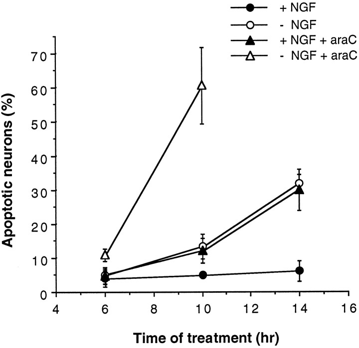
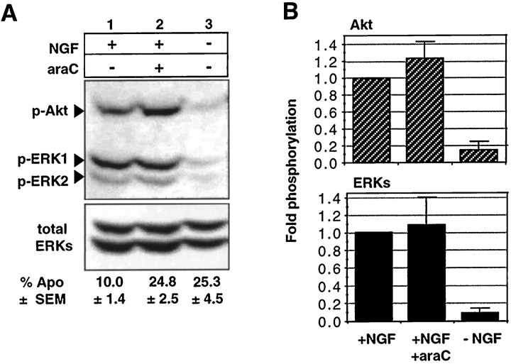
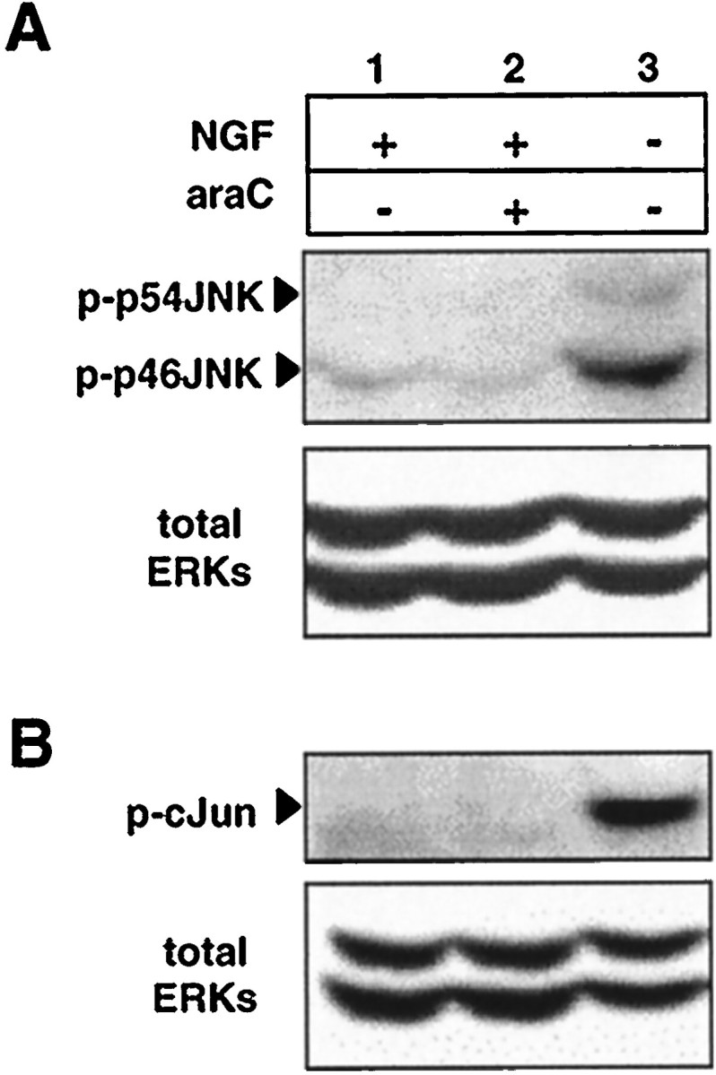
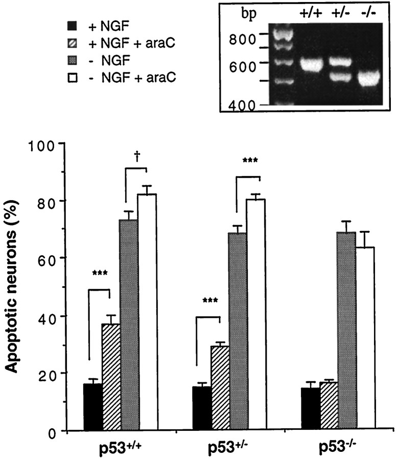
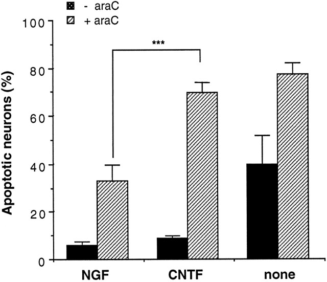
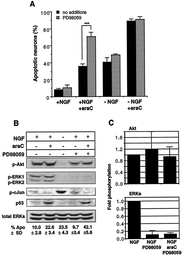
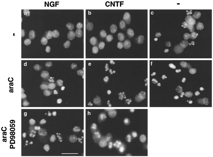
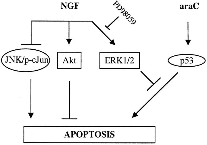
The antimitotic nucleoside cytosine arabinoside (araC) causes apoptosis in postmitotic neurons for which two mechanisms have been suggested: (1) araC directly inhibits a trophic factor-maintained signaling pathway required for survival, effectively mimicking trophic factor withdrawal; and (2) araC induces apoptosis by a p53-dependent mechanism distinct from trophic factor withdrawal. In rat sympathetic neurons, we found that araC treatment for 12 hr induced approximately 25% apoptosis without affecting NGF-maintained signaling; there was neither reduction in the activity of mitogen activated protein kinase/extracellular signal-regulated kinase (MAPK/ERK) or protein kinase B/Akt, a kinase implicated in NGF-mediated survival, nor was there c-Jun N-terminal kinase (JNK) activation or c-Jun N-terminal phosphorylation, events implicated in apoptosis induced by NGF withdrawal. However, araC treatment, but not NGF-withdrawal, elevated expression of p53 protein before and during apoptosis. Additionally, araC-induced apoptosis was suppressed in sympathetic neurons from p53 null mice. Although MAPK/ERK activity is not necessary for NGF-induced survival, it protected against toxicity by araC, because inhibition of the MAPK pathway by PD98059 resulted in a significant increase in the rate of apoptosis induced by araC in the presence of NGF. Consistent with this finding, ciliary neurotrophic factor, which does not cause sustained activation of MAPK/ERK, did not protect against araC toxicity. Our data show that, in contrast to NGF deprivation, araC induces apoptosis via a p53-dependent, JNK-independent mechanism, against which MAPK/ERK plays a substantial protective role. Thus, NGF can suppress apoptotic mechanisms in addition to those caused by its own deprivation.


Figures

Fig. 1.
Fig. 1.
Matching rates of apoptosis caused by NGF deprivation or araC treatment in the presence of NGF. Newly isolated neurons were plated into medium without NGF or with NGF (100 ng/ml) or araC (100 μm) for the indicated times before being fixed, stained, and counted. The results shown are the mean ± SD of three independent experiments. See Figure 8 for photomicrographs of stained nuclei under these conditions.
Fig. 2.
Fig. 2.
NGF-induced phosphorylation of p44-ERK1, p42-ERK2, and Akt is not inhibited by araC. SCG neurons were treated for 12 hr with NGF alone (20 ng/ml) (lane 1), NGF and 100 μm araC (lane 2), and no NGF or araC (lane 3), and the protein extracts were prepared and probed as described in Materials and Methods. A,Top panel shows phospho-Akt probed for phosphorylation at serine 473 and phospho-ERK1/2 probed with anti-active MAPK antibody;bottom panel shows total ERKs as an indication of protein loading. B, Fold-phosphorylation as determined by optical densitometry (see Materials and Methods). Top panel, Phospho-Akt; bottom panel, phospho-ERKs; both normalized to total ERKs. Data for Akt and ERKs are the mean ± range and mean ± SD for two and three independent experiments, respectively. Numbers below the lanesgive the mean ± SEM of percentage of apoptosis for six independent experiments.
Fig. 3.
Fig. 3.
JNK activation and c-Jun phosphorylation at serine 63 are not required for araC-induced apoptosis. SCG neurons were treated for 12 hr with NGF alone (lane 1), NGF and 100 μm araC (lane 2), and no NGF or araC (lane 3), and the protein extracts were prepared and processed as in Materials and Methods. A, The blot was probed first for JNK phosphorylated at residues threonine 183 and tyrosine 185 (top panel) and then for total ERKs as an indication of protein loading (bottom panel). For these experiments, NGF was used at 100 ng/ml. The result shown is representative of three independent experiments. B, In two other independent experiments, the amount of N-terminal phosphorylation of c-Jun was determined by probing with an antibody specific for c-Jun phosphorylated at residue serine 63 (top panel). Immunoblotting for total ERKs is shown (bottom panel) as an indication of protein loading. NGF was used at a concentration of 20 ng/ml. See Figure 2 for percentage of apoptosis for each condition.
Fig. 4.
Fig. 4.
SCG neurons from p53−/− mice are resistant to apoptosis induced by araC, but not NGF, withdrawal. SCG neurons were prepared from individual mice from litters born to p53+/− parents and cultured for 16 hr with or without NGF (100 ng/ml) and with or without araC (100 μm) before being fixed, stained, and counted as described in Materials and Methods. The p53 status of each animal tested was determined after counting had been completed. The results shown are the mean ± SEM for 6 (p53+/+), 13 (p53+/−), and 6 (p53−/−) animals, respectively. Student’st tests gave the significance values as indicated (***p < 0.001; †0.1 > p> 0.05). Inset, PCR results showing the three genotypes. Top band (642 bp) represents the presence of wild-type p53 gene; bottom band (510 bp) represents the absence of the p53 gene.
Fig. 5.
Fig. 5.
AraC causes an early and sustained elevation of p53 protein levels. Rat SCG neurons were treated for the indicated times before being collected, resolved on SDS-PAGE, and electroblotted as described in Materials and Methods. NGF was used at 100 ng/ml, and araC was used at 100 μm. A, Lanes 1–4, araC; lanes 1–5, NGF. Top panel shows the results of probing for p53; bottom panel shows results for total ERKs as an indication of protein loading. The results shown are representative of two independent experiments. B, Cultures were treated for 10 or 14 hr before collection, processing, and probing as in A.Lanes 1, 2, 5,6, NGF; lanes 2, 4,6, araC. The results shown are representative of three independent experiments.
Fig. 6.
Fig. 6.
AraC-induced apoptosis is delayed by NGF but not by CNTF. Neurons were cultured with 100 ng/ml CNTF, 20 ng/ml NGF, or with no growth factor additions (none) in the absence (black bars) or presence (hatched bars) of 100 μm araC as indicated. The cells were fixed at 14 hr before staining and counting. Results are the mean ± SD of three independent experiments. A Student–Newman–Keuls multiple comparisons test gave significant differences (***p< 0.001) for all comparisons, except between NGF versus CNTF (bars 1, 3), NGF with araC versus no growth factor additions (bars 2, 5), and CNTF with araC versus no growth factors with araC (bars 4, 6).
Fig. 7.
Fig. 7.
Suppression of ERK activity by PD98059 renders neurons more susceptible to araC: a comparison of signaling pathways.A, Neurons were cultured in the presence or absence of NGF (20 ng/ml), araC (100 μm), or PD98059 (50 μm) for 14 hr in the indicated conditions before being fixed, stained, and counted. All media contained 0.2% DMSO. Results shown are the mean ± SEM of three to five independent experiments (**p < 0.002; Student’s t test). See Figure 8 for photomicrographs of apoptotic nuclei.B, Neurons were treated with 20 ng/ml NGF (lanes 1, 2, 4, 5), 100 μm araC (lanes 2, 5), and 50 μm PD98059 (lanes 4, 5) for 12 hr and then processed for immunoblotting as in Materials and Methods. The panels show the results of probing with the following antibodies (top to bottom): anti-phospho-Akt (serine 473), anti-phospho-ERK1/2, anti-phospho-c-Jun (serine 63), anti-p53, and anti-ERK1/2. Results shown are representative of three independent experiments, except for anti-phospho-Akt and anti-phospho-c-Jun, which are representative of two independent experiments. The numbers below the lanes indicate the percentage of apoptosis and are the mean ± SD of three independent experiments.C, Fold-phosphorylation as determined by optical densitometry (see Materials and Methods): top panel, phospho-Akt (n = 2); bottom panel, phospho-ERKs (n = 3); both normalized to total ERK. Error bars represent the SD and range for ERK and Akt, respectively.
Fig. 8.
Fig. 8.
Nuclear morphologies of SCG neurons under various conditions as visualized with Hoechst 33342. Neurons were treated with 100 ng/ml NGF (a, d, g), 100 ng/ml CNTF (b, e, h), or without growth factors (-) (c,f). AraC was used at 100 μm(d–f), and PD98059 was used at 50 μm (g, h). Hoechst 33342 was added to the cultures at 14 hr, and photographs were taken before fixing and counting. Scale bar, 10 μm.
Fig. 9.
Fig. 9.
Scheme summarizing proposed interactions between NGF signaling and apoptotic pathways. NGF induces at least three signals: the sustained activation of MAPK/ERK and Akt, and suppression of JNK and c-Jun phosphorylation. In the absence of NGF, JNK is activated, whereas ERK1/2 and Akt are inactivated. In the presence of NGF, araC treatment causes elevation of p53 and apoptosis without affecting NGF signaling. ERK1/2, however, suppresses signaling downstream of p53, thereby delaying apoptosis. Inhibition of the ERK pathway with PD98059 primarily eliminates this suppression, suggesting that the lack of ERK activity caused by NGF withdrawal may therefore account for the acceleration in the rate of apoptosis induced by araC.

References

    1. Alessi DR, Andjelkovic M, Caudwell B, Cron P, Morrice N, Cohen P, Hemmings BA. Mechanism of activation of protein kinase B by insulin and IGF-1. EMBO J. 1996;15:6541–6551. - PMC - PubMed
    1. Andjelkovic M, Jakubowicz T, Cron P, Ming XF, Han JW, Hemmings BA. Activation and phosphorylation of a pleckstrin homology domain-containing protein-kinase (RAC-PK/PKB) promoted by serum and protein phospatase inhibitors. Proc Natl Acad Sci USA. 1996;93:5699–5704. - PMC - PubMed
    1. Baker WJ, Royer GLJ, Weiss RB. Cytarabine and neurologic toxicity. J Clin Oncol. 1991;9:679–693. - PubMed
    1. Buckmaster A, Nobes CD, Edwards SN, Tolkovsky AM. NGF is required for induction of c-fos immunoreactivity by serum, depolarisation, cyclic AMP or trauma in cultured rat sympathetic neurons. Eur J Neurosci. 1991;3:698–707. - PubMed
    1. Clarke AR, Purdie CA, Harrison DJ, Morris RG, Bird CC, Hooper ML, Wyllie AH. Thymocyte apoptosis induced by p53-dependent and independent pathways. Nature. 1993;362:849–852. - PubMed

Publication types

MeSH terms