. 1999 Jan 15;19(2):737-46.
doi: 10.1523/JNEUROSCI.19-02-00737.1999.

ATP stimulates sympathetic transmitter release via presynaptic P2X purinoceptors

Affiliations

ATP stimulates sympathetic transmitter release via presynaptic P2X purinoceptors

S Boehm. J Neurosci. .

Abstract

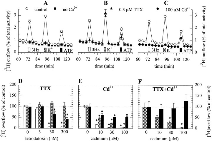
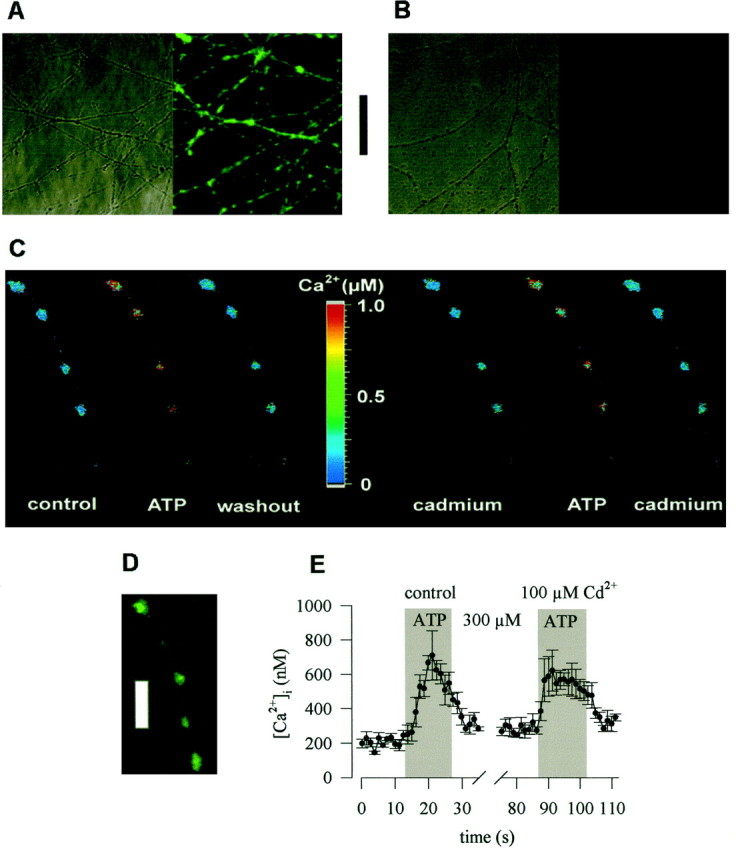
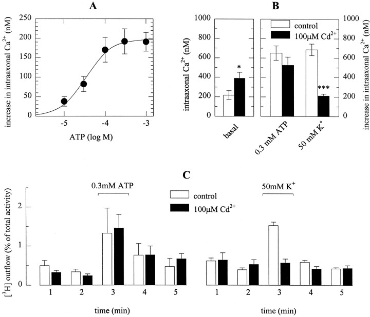
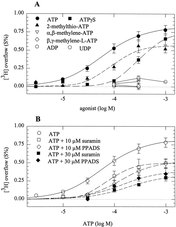
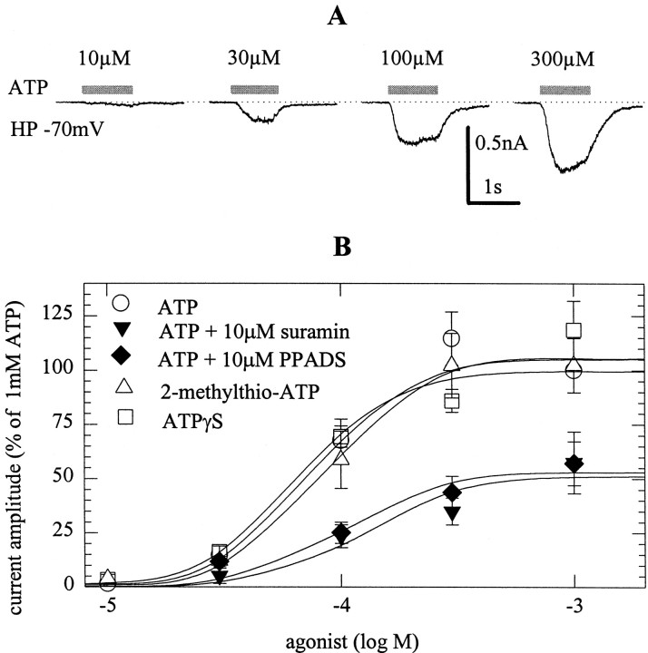
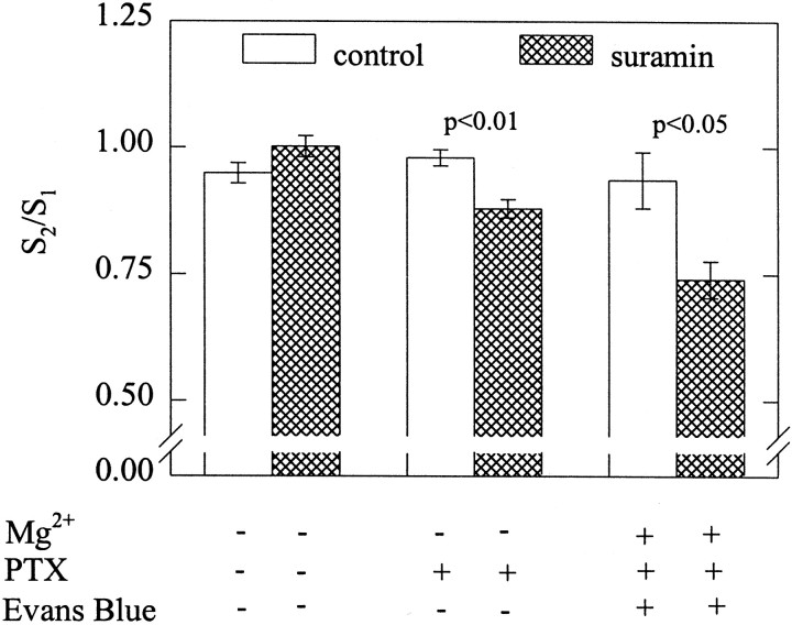
ATP is a fast transmitter in sympathetic ganglia and at the sympathoeffector junction. In primary cultures of dissociated rat superior cervical ganglion neurons, ATP elicits noradrenaline release in an entirely Ca2+-dependent manner. Nevertheless, ATP-evoked noradrenaline release was only partially reduced (by approximately 50%) when either Na+ or Ca2+ channels were blocked, which indicates that ATP receptors themselves mediated transmembrane Ca2+ entry. An "axonal" preparation was obtained by removing ganglia from explant cultures, which left a network of neurites behind; immunostaining for axonal and dendritic markers revealed that all of these neurites were axons. In this preparation, ATP raised intraaxonal Ca2+ and triggered noradrenaline release, and these actions were not altered when Ca2+ channels were blocked by Cd2+. Hence, Ca2+-permeable ATP-gated ion channels, i.e., P2X purinoceptors, are located at presynaptic sites and directly mediate Ca2+-dependent transmitter release. These presynaptic P2X receptors displayed a rank order of agonist potency of ATP >/= 2-methylthio-ATP > ATPgammaS >> alpha,beta-methylene-ATP approximately beta,gamma-methylene-L-ATP and were blocked by suramin or PPADS. ATP, 2-methylthio-ATP, and ATPgammaS also evoked inward currents measured at neuronal somata, but there these agonists were equipotent. Hence, presynaptic P2X receptors resemble the cloned P2X2 subtype, but they appear to differ from somatodendritic P2X receptors in terms of agonist sensitivity. Suramin reduced depolarization-evoked noradrenaline release by up to 20%, when autoinhibitory mechanisms were inactivated by pertussis toxin. These results indicate that presynaptic P2X purinoceptors mediate a positive, whereas G-protein-coupled P2Y purinoceptors mediate a negative, feedback modulation of sympathetic transmitter release.


Figures

Fig. 1.
Fig. 1.
ATP-evoked noradrenaline release from SCG neurons is entirely Ca2+-dependent, but partially TTX- and Cd2+-resistant. After loading with [3H]noradrenaline, neurons were superfused, and subsequent to a 1 hr washout period, 4 min fractions of superfusate were collected. After 72 min of superfusion, 90 electrical pulses were delivered, after 92 min, 50 mmK+ (Na+ was reduced accordingly) was applied for 30 sec, and after 112 min, 0.3 mm ATP was present for 30 sec. In cultures depicted by filled symbols, Ca2+-free buffer was supplied (A) and either 0.3 μm TTX (B) or 100 μmCd2+ (C) were present in Ca2+-containing buffer, all from 50 min of superfusion onward. The outflow of tritium is shown as percentage of the total radioactivity in the cultures; n = 6.D, Concentration dependence of the inhibition of electrically (open bars), K+(hatched bars)-, and ATP (filled bars)-evoked tritium overflow by TTX; n = 6. E, Concentration dependence of the inhibition of electrically (open bars), K+(hatched bars)-, and ATP (filled bars)-evoked overflow by Cd2+;n = 6. F, Concentration dependence of the inhibition of K+ (hatched bars)- and ATP (filled bars)-evoked overflow by Cd2+ in the continuous presence of TTX, which was added to the buffer after 50 min of superfusion;n = 6. Asterisks indicate significant differences versus corresponding controls atp < 0.05.
Fig. 2.
Fig. 2.
Characterization of “axonal” preparations and ATP-induced Ca2+ entry into axonal varicosities. Explant cultures of SCG were prepared as described in Materials and Methods. A shows a transmission and an indirect immunofluorescence image of the neurite network in such cultures probed with the monoclonal antibody PC1C6 directed against the tau protein.B displays a similar picture after staining with antibody SMI32 directed against the nonphosphorylated form of neurofilament H. The bar between A andB represents 50 μm. In C, an explant culture was loaded with fura-2 AM, and the ganglion was removed, leaving only the axonal network behind. ATP was applied to the axon shown in either the absence (left) or presence (right) of 100 μm Cd2+. The picture displays the pseudocolour representation of the ratio of fluorescence intensity obtained at excitation wavelengths of 340 and 380 nm and the according values of Ca2+concentrations. D shows indirect immunofluorescence of a neurite in an explant culture probed with anitbody SY38 directed against the vesicle protein synaptophysin. The barrepresents 10 μm. E shows the time course of Ca2+ concentrations averaged for the four axonal varicosities shown in C.
Fig. 3.
Fig. 3.
The ATP-triggered rise of Ca2+in, and noradrenaline release from, sympathetic axons is Cd2+-resistant. Explant cultures were labeled with fura-2 AM, and experiments were performed as described in the legend to Figure 2. A shows the averaged concentration–response relation for the ATP-induced rise in intraaxonal Ca2+ concentrations as determined in 22–25 neurites. B depicts basal intraaxonal Ca2+ concentrations (left) and the rises in Ca2+ concentrations (right) evoked by either 0.3 mm ATP or 50 mmK+ (Na+ was reduced accordingly) in 13 neurites. Open bars represent control conditions, whereas filled bars represent experiments performed in the presence of 100 μm Cd2+; *p < 0.05 and ***p < 0.001 versus corresponding controls. The results in A andB have been obtained in different preparations.C shows tritium outflow from axonal preparations labeled with [3H]noradrenaline. After a 1 hr washout period, cultures were incubated for 1 min periods in buffer containing either no (open bars) or 100 μm(filled bars) Cd2+. During the third incubation period, 0.3 mm ATP (left) or 50 mm K+ (Na+ was reduced accordingly; right) were present;n = 5–8.
Fig. 4.
Fig. 4.
Agonist and antagonist sensitivity of presynaptic P2X receptors. After loading with [3H]noradrenaline, neurons were superfused, and subsequent to a 1 hr washout period, 4 min fractions of superfusate were collected. I added 100 μm Cd2+ to the buffer after 50 min of superfusion. At 72, 92, or 112 min of superfusion the indicated concentrations of purinoceptor agonists and/or antagonists were present for 30 sec. A depicts the concentration–response curves for tritium overflow induced by ATP, 2-methylthio-ATP, ATPγS, α,β-methylene-ATP, β,γ-methylene-l-ATP, ADP, and UDP.n = 6–12, with the exception of ATP, wheren = 29–51. B depicts the concentration–response curves for tritium overflow induced by ATP in either the absence or the presence of 10 or 30 μm suramin or PPADS. n = 7–15, again with the exception of ATP applied alone, where the results of A are shown again.
Fig. 5.
Fig. 5.
Agonist and antagonist sensitivity of somatic P2X receptors. At a holding potential of −70 mV, whole-cell currents were evoked by the application of ATP, 2-methylthio-ATP, or ATPγS, applied either alone or in the continuous presence of suramin or PPADS.A shows original traces of ATP-evoked currents.B depicts concentration–response curves for agonist-induced peak currents, and amplitudes are expressed as percentage of the amplitude obtained with 1 mm ATP in the very same cell; n = 6–7.
Fig. 6.
Fig. 6.
P2X receptors mediate positive feedback modulation of noradrenaline release. After loading with [3H]noradrenaline, neurons were superfused with a Mg2+-free buffer, and subsequent to a 1 hr washout period, 4 min fractions of superfusate were collected. At 72 (S1) and 92 (S2) min, superfusion was stopped, 180 electrical pulses were delivered at 3 Hz, and superfusion was continued thereafter. I included 30 μmsuramin in the buffer 8 min before the second stimulation in the cultures represented by hatched bars. Where indicated, cultures had been treated with pertussis toxin (100 ng/ml for 24 hr), and the buffer contained 2 mmMg2+ and 100 μm Evans blue, respectively; n = 6. Levels of significances of the differences between results obtained in either the presence or the absence of suramin are indicated.

References

    1. Augustine GJ, Neher E. Neuronal Ca2+ signalling takes the local route. Curr Opin Neurobiol. 1992;2:302–307. - PubMed
    1. Boehm S. Noradrenaline release from rat sympathetic neurons evoked by P2-purinoceptor activation. Naunyn Schmiedebergs Arch Pharmacol. 1994;350:454–458. - PubMed
    1. Boehm S. Selective inhibition of M-type K+ channels in rat sympathetic neurons by uridine nucleotide preferring receptors. Br J Pharmacol. 1998;124:1261–1269. - PMC - PubMed
    1. Boehm S, Betz H. Somatostatin inhibits excitatory transmission at rat hippocampal synapses via presynaptic receptors. J Neurosci. 1997;17:4066–4075. - PMC - PubMed
    1. Boehm S, Huck S. α2-Adrenoceptor mediated inhibition of acetylcholine-induced noradrenaline release from rat sympathetic neurons: an action at voltage-gated Ca2+ channels. Neuroscience. 1995;69:221–231. - PubMed

Publication types

MeSH terms

LinkOut - more resources