

Peripheral inflammation facilitates Abeta fiber-mediated synaptic input to the substantia gelatinosa of the adult rat spinal cord
- PMID: 9880605
- PMCID: PMC6782212
- DOI: 10.1523/JNEUROSCI.19-02-00859.1999
Peripheral inflammation facilitates Abeta fiber-mediated synaptic input to the substantia gelatinosa of the adult rat spinal cord
Abstract
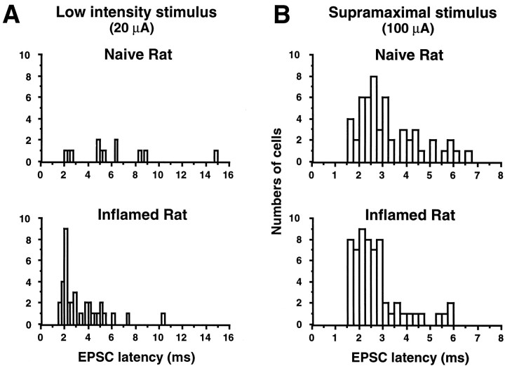
Whole-cell patch-clamp recordings were made from substantia gelatinosa (SG) neurons in thick adult rat transverse spinal cord slices with attached dorsal roots to study changes in fast synaptic transmission induced by peripheral inflammation. In slices from naive rats, primary afferent stimulation at Abeta fiber intensity elicited polysynaptic EPSCs in only 14 of 57 (25%) SG neurons. In contrast, Abeta fiber stimulation evoked polysynaptic EPSCs in 39 of 62 (63%) SG neurons recorded in slices from rats inflamed by an intraplantar injection of complete Freund's adjuvant (CFA) 48 hr earlier (p < 0.001). Although the peripheral inflammation had no significant effect on the threshold and conduction velocities of Abeta, Adelta, and C fibers recorded in dorsal roots, the mean threshold intensity for eliciting EPSCs was significantly lower in cells recorded from rats with inflammation (naive: 33.2 +/- 15.1 microA, n = 57; inflamed: 22.8 +/- 11.3 microA, n = 62, p < 0.001), and the mean latency of EPSCs elicited by Abeta fiber stimulation in CFA-treated rats was significantly shorter than that recorded from naive rats (3.3 +/- 1.8 msec, n = 36 vs 6.0 +/- 3.5 msec, n = 12; p = 0.010). Abeta fiber stimulation evoked polysynaptic IPSCs in 4 of 25 (16%) cells recorded from naive rat preparations and 14 of 26 (54%) SG neurons from CFA-treated rats (p < 0.001). The mean threshold intensity for IPSCs was also significantly lower in CFA-treated rats (naive: 32.5 +/- 15.7 microA, n = 25; inflamed: 21. 9 +/- 9.9 microA, n = 26, p = 0.013). The facilitation of Abeta fiber-mediated input into the substantia gelatinosa after peripheral inflammation may contribute to altered sensory processing.
Figures


References
-
- Beal JA, Bicknell HR. Development and maturation of neurons in the substantia gelatinosa(SG) of the rat spinal cord. In: Rowe M, Willis WD Jr, editors. Development, organization and processing in somatosensory pathway. Wiley-Liss; New York: 1985. pp. 23–30.
-
- Bennett GJ, Abdelmoumene M, Hayashi H, Dubner R. Physiology and morphology of substantia gelatinosa neurons intracellularly stained with horseradish peroxidase. J Comp Neurol. 1980;194:809–827. - PubMed
-
- Bleazard L, Hill RG, Morris R. The correlation between the distribution of the NK1 receptor and the action of tachykinin agonists in the dorsal horn of the rat indicates that substance P does not have a functional role on substantia gelatinosa (lamina II) neurons. J Neurosci. 1994;14:7655–7664. - PMC - PubMed
-
- Brown AG. Organization in the spinal cord: the anatomy and physiology of identified neurones. Springer-Verlag; Berlin: 1981.
Publication types
MeSH terms
Grants and funding
LinkOut - more resources
Full Text Sources
Other Literature Sources