. 1999 Feb;73(2):897-906.
doi: 10.1128/JVI.73.2.897-906.1999.

Apoptosis induced by infection of primary brain cultures with diverse human immunodeficiency virus type 1 isolates: evidence for a role of the envelope

Affiliations

Apoptosis induced by infection of primary brain cultures with diverse human immunodeficiency virus type 1 isolates: evidence for a role of the envelope

A Ohagen et al. J Virol. 1999 Feb.

Abstract

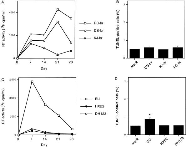
Apoptosis of neurons and astrocytes is induced by human immunodeficiency type 1 (HIV-1) infection in vitro and has been demonstrated in brain tissue from patients with AIDS. We analyzed a panel of diverse HIV-1 primary isolates for the ability to replicate and induce neuronal and astrocyte apoptosis in primary human brain cultures. Apoptosis was induced three- to eightfold by infection with the blood-derived HIV-1 isolates 89.6, SG3, and ADA. In contrast, the brain-derived HIV-1 isolates YU2, JRFL, DS-br, RC-br, and KJ-br did not induce significant levels of apoptosis. The ability of HIV-1 isolates to induce apoptosis was independent of their replication capacity. Studies of recombinant chimeras between the SG3 and YU2 viruses showed that replacement of the YU2 Env with the SG3 Env was sufficient to confer the ability to induce apoptosis to the YU2 virus. Replacement of the Env V3 regions alone largely conferred the phenotypes of the parental clones. The SG3 Env used CXCR4 and CCR3 as coreceptors for virus entry, whereas YU2 used CCR5 and CCR3. The V3 regions of SG3 and YU2 conferred the ability to use CXCR4 and CCR5, respectively. In contrast, the 3' region of Env, particularly the C3V4 region, was required in conjunction with the V3 region for efficient use of CCR3. These results provide evidence that Env is a major determinant of neurodegenerative mechanisms associated with HIV-1 infection in vitro and raise the possibility that blood-derived viruses which emerge during the late stages of disease may affect disease progression in the central nervous system.


Figures

FIG. 1
FIG. 1
Apoptosis induced by infection of primary brain cultures with different HIV-1 isolates. Cultures were infected by incubation with 89.6, SG3, ADA, YU2, or JRFL virus stock (20,000 3H cpm RT units) or mock infected with control supernatants. (A) Virus replication measured by RT assay of culture supernatants (means of triplicate samples). The background level for RT assays in mock infections was ≤200 3H cpm RT units/ml. (B to D) Detection of apoptosis on day 30 after infection by TUNEL staining (B), propidium iodide staining (C), and detection of cytosolic histone-associated DNA fragments by ELISA (D) (mean ± SD, n = 2). ∗, P < 0.05 by ANOVA with Bonferroni posttest correction compared with mock-infected control cultures. Results are representative of three independent experiments. O.D., optical density.
FIG. 2
FIG. 2
Detection of apoptosis in primary brain cultures infected with primary HIV-1 isolates derived from brain tissue (A and B) or blood (C and D) of AIDS patients. (A and B) Cultures were infected with RC-br, DS-br, or KJ-br (20,000 3H cpm RT units) or mock infected with control supernatants. (A) Virus replication measured by RT assay of culture supernatants (means of quadruplicate samples). The background level for RT assays in mock infections was ≤200 3H cpm RT units/ml. (B) Apoptosis on day 30 after infection, detected by TUNEL staining (mean ± SD, n = 2). (C and D) Cultures were infected with ELI, DH123, or HXB2 or mock infected with control supernatants, and virus replication (C) and apoptosis (D) were detected as for panels A and B. ∗, P < 0.05 by ANOVA with Bonferroni posttest correction compared with mock-infected control cultures. Similar results were obtained in two independent experiments.
FIG. 3
FIG. 3
Structure of SG3/YU2 envelope chimeras. Chimeric env genes were constructed by using the indicated restriction enzyme sites in the HIV-1 genome (top). Scores for syncytium formation in CEMx174 or MT2 cells infected with the indicated viruses as described in Materials and Methods are shown at the right as follows: 1+, 1 to 10%; 2+, 10 to 25%; 3+, 25 to 50%; 4+, >50%. Similar results were obtained in three independent experiments.
FIG. 4
FIG. 4
Replication of the SG3/YU2 chimeric viruses in PBMC and CEMx174 cells. PBMC (A) and CEMx174 cells (B) were infected with equal amounts (250,000 35S cpm RT units) of YU2, SG3, SG26, SG29, SG52, SG57, or SG84 virus stock. Virus replication was monitored by measuring RT activity in culture supernatants every third day. The background level for RT assays in mock infections was ≤1,500 35S cpm RT units/25 μl. Results are representative of three independent experiments.
FIG. 5
FIG. 5
Induction of apoptosis in primary brain cultures infected with SG3/YU2 chimeric viruses. Cultures were infected with YU2, SG3, SG26, SG29, SG52, or SG57 virus stock normalized for equivalent amounts of RT activity. (A) Virus replication measured by RT activity of culture supernatants (means of triplicate samples). The background level for RT assays in mock-infected cultures was ≤200 3H cpm RT units. (B) Detection of apoptosis on day 30 after infection by TUNEL staining. ∗, P < 0.05 by ANOVA with Bonferroni posttest correction compared with mock-infected control cultures. Similar results were obtained in three independent experiments.
FIG. 6
FIG. 6
Expression of HIV-1 envelope chimeras. 293T cells were transfected with expression plasmids containing the SG3, YU2, SG52, SG57, SG68, or SG84 chimeric env gene as shown in Fig. 3. Cell lysates were analyzed by Western blotting using rabbit anti-gp120. Positions of gp160 and gp120 are indicated on the right; positions of molecular weight markers are indicated on the left.
FIG. 7
FIG. 7
CAT activities in transfected U87 cells infected with HIV-1 reporter viruses. U87 cells cotransfected with a plasmid expressing CD4 and a plasmid expressing the chemokine receptor CCR5, CCR3, or CXCR4 were infected with CAT viruses with either no Env protein or Env proteins of HXB2, SG3, YU2, SG52, SG57, SG68, or SG84. Results of the CAT assay performed with the U87 cell lysates are shown. Similar results were obtained in three independent experiments.

References

    1. Adie-Biassette H, Levy Y, Colombel M, Poron F, Natcher S, Keohane C, Gray F. Neuronal apoptosis in HIV infection in adults. Neuropathol Appl Neurobiol. 1995;21:218–227. - PubMed
    1. Bagasra O, Lavi E, Bobroski L, Khalili K, Pestaner J P, Tawadros R, Pomerantz R. Cellular reservoirs of HIV-1 in the central nervous system of infected individuals: identification by the combination of in situ polymerase chain reaction and immunohistochemistry. AIDS. 1996;10:573–585. - PubMed
    1. Bieniasz P D, Fridell R A, Aramori I, Ferson S S G, Caron M G, Cullen B R. HIV-1-induced cell fusion is mediated by multiple regions within both the viral envelope and the CCR-5 co-receptor. EMBO J. 1997;16:2599–2609. - PMC - PubMed
    1. Björndal Å, Deng H, Jansson M, Fiore J R, Colognesi C, Karlsson A, Albert J, Scarlatti G, Littman D R, Fenyö E M. Coreceptor usage of primary human immunodeficiency virus type 1 isolates varies according to biological phenotype. J Virol. 1997;71:7478–7487. - PMC - PubMed
    1. Brew B J, Evans L, Byrne C, Pemberton L, Hurren L. The relationship between AIDS dementia complex and the presence of macrophage tropic and non-syncytium inducing isolates of human immunodeficiency virus type 1 in the cerebrospinal fluid. J Neurovirol. 1996;2:152–157. - PubMed

Publication types

MeSH terms