. 1999 Feb;73(2):1023-35.
doi: 10.1128/JVI.73.2.1023-1035.1999.

Epstein-Barr virus-encoded latent membrane protein 1 activates the JNK pathway through its extreme C terminus via a mechanism involving TRADD and TRAF2

Affiliations

Epstein-Barr virus-encoded latent membrane protein 1 activates the JNK pathway through its extreme C terminus via a mechanism involving TRADD and TRAF2

A G Eliopoulos et al. J Virol. 1999 Feb.

Abstract

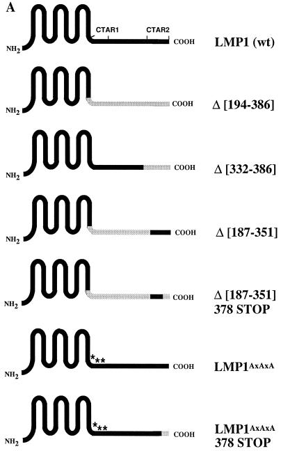
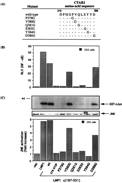
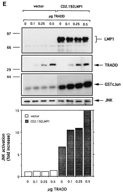
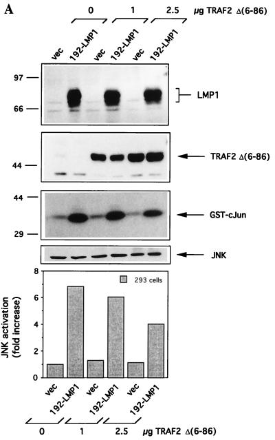
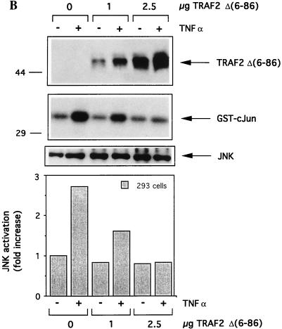
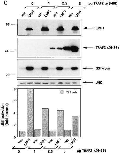
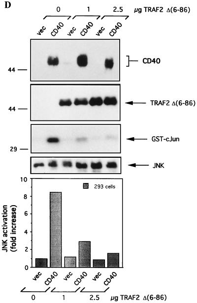
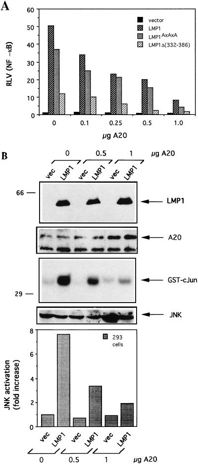
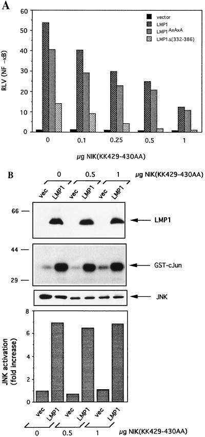
The transforming Epstein-Barr virus-encoded latent membrane protein 1 (LMP1) activates signalling on the NF-kappaB axis through two distinct domains in its cytoplasmic C terminus, namely, CTAR1 (amino acids [aa] 187 to 231) and CTAR2 (aa 351 to 386). The ability of CTAR1 to activate NF-kappaB appears to be attributable to the direct interaction of tumor necrosis factor (TNF) receptor-associated factor 2 (TRAF2), while recent work indicates that CTAR2-induced NF-kappaB is mediated through its association with TNF receptor-associated death domain (TRADD). LMP1 expression also results in activation of the c-Jun N-terminal kinase (JNK) (also known as stress-activated protein kinase) cascade, an effect which is mediated exclusively through CTAR2 and can be dissociated from NF-kappaB induction. The organization and signalling components involved in LMP1-induced JNK activation are not known. In this study we have dissected the extreme C terminus of LMP1 and have identified the last 8 aa of the protein (aa 378 to 386) as being important for JNK signalling. Using a series of fine mutants in which single amino acids between codons 379 and 386 were changed to glycine, we have found that mutations of Pro379, Glu381, Ser383, or Tyr384 diminish the ability of LMP1 CTAR2 to engage JNK signalling. Interestingly, this region was also found to be essential for CTAR2-mediated NF-kappaB induction and coincides with the LMP1 amino acid sequences shown to bind TRADD. Furthermore, we have found that LMP1-mediated JNK activation is synergistically augmented by low levels of TRADD expression, suggesting that this adapter protein is critical for LMP1 signalling. TRAF2 is known to associate with TRADD, and expression of a dominant-negative N-terminal deletion TRAF2 mutant was found to partially inhibit LMP1-induced JNK activation in 293 cells. In addition, the TRAF2-interacting protein A20 blocked both LMP1-induced JNK and NF-kappaB activation, further implicating TRAF2 in these phenomena. While expression of a kinase-inactive mutated NF-kappaB-inducing kinase (NIK), a mitogen-activated protein kinase kinase kinase which also associates with TRAF2, impaired LMP1 signalling on the NF-kappaB axis, it did not inhibit LMP1-induced JNK activation, suggesting that these two pathways may bifurcate at the level of TRAF2. These data further define a role for TRADD and TRAF2 in JNK activation and confirm that LMP1 utilizes signalling mechanisms used by the TNF receptor/CD40 family to elicit its pleiotropic activities.


Figures

FIG. 1
FIG. 1
The last 8 aa of LMP1 are critical for JNK and NF-κB signalling. (A) Schematic representation of the LMP1 protein and the deleted LMP1 gene sequences used in this study. Solid black lines represent wild-type (wt) LMP1 sequences, and dotted lines denote deleted LMP1 sequences. CTAR1 is located at residues 194 to 232, and CTAR2 is located at residues 351 to 386. The asterisks represent a triple P204xQ206xT208→AxAxA mutation. (B) Induction of NF-κB-dependent transcriptional activity by LMP1 and LMP1 deletion mutants. HEK 293 cells were transfected with 1 μg of pSG5-based constructs in the presence of 50 ng of NF-κB-regulated luciferase reporter plasmid 3Enh.κBconA-Luc and 50 ng of β-galactosidase expression vector. Relative luciferase values (RLV), which represent the luciferase values normalized on the basis of β-galactosidase expression, were determined at 36 h posttransfection. The data shown represent fold increases in RLV relative to the vector control (vec), which was given the arbitrary value of 1, and are representative of at least five independent experiments. (C) Effects of wild-type and mutated LMP1 expression on JNK activity. HEK 293 cells were transfected with 1 μg of pSG5 or pSG5-based LMP1 expression vectors in the presence of 0.5 μg of the HA-tagged JNK1 expression vector HA-p46SAPKγ-pCDNA3. At 36 h posttransfection, HA-JNK was immunoprecipitated from 250 μg of cell lysates by using anti-HA antibody, and kinase assays were performed as described in Materials and Methods. JNK activity was assessed by the ability of the immunoprecipitate to phosphorylate GST–c-Jun substrate. Results of a representative assay are shown (second panel). The same lysates were analyzed for wild-type or mutated LMP1 (upper panel) and JNK (third panel) expression. Numbers on the left are molecular weights in thousands. Relative levels of JNK activation were quantitated on a phosphorimager and are presented in histogram form (lower panel). At least four independent experiments were performed and gave similar results.
FIG. 1
FIG. 1
The last 8 aa of LMP1 are critical for JNK and NF-κB signalling. (A) Schematic representation of the LMP1 protein and the deleted LMP1 gene sequences used in this study. Solid black lines represent wild-type (wt) LMP1 sequences, and dotted lines denote deleted LMP1 sequences. CTAR1 is located at residues 194 to 232, and CTAR2 is located at residues 351 to 386. The asterisks represent a triple P204xQ206xT208→AxAxA mutation. (B) Induction of NF-κB-dependent transcriptional activity by LMP1 and LMP1 deletion mutants. HEK 293 cells were transfected with 1 μg of pSG5-based constructs in the presence of 50 ng of NF-κB-regulated luciferase reporter plasmid 3Enh.κBconA-Luc and 50 ng of β-galactosidase expression vector. Relative luciferase values (RLV), which represent the luciferase values normalized on the basis of β-galactosidase expression, were determined at 36 h posttransfection. The data shown represent fold increases in RLV relative to the vector control (vec), which was given the arbitrary value of 1, and are representative of at least five independent experiments. (C) Effects of wild-type and mutated LMP1 expression on JNK activity. HEK 293 cells were transfected with 1 μg of pSG5 or pSG5-based LMP1 expression vectors in the presence of 0.5 μg of the HA-tagged JNK1 expression vector HA-p46SAPKγ-pCDNA3. At 36 h posttransfection, HA-JNK was immunoprecipitated from 250 μg of cell lysates by using anti-HA antibody, and kinase assays were performed as described in Materials and Methods. JNK activity was assessed by the ability of the immunoprecipitate to phosphorylate GST–c-Jun substrate. Results of a representative assay are shown (second panel). The same lysates were analyzed for wild-type or mutated LMP1 (upper panel) and JNK (third panel) expression. Numbers on the left are molecular weights in thousands. Relative levels of JNK activation were quantitated on a phosphorimager and are presented in histogram form (lower panel). At least four independent experiments were performed and gave similar results.
FIG. 2
FIG. 2
Single point mutations within aa 379 to 385 severely impair CTAR2-mediated NF-κB and JNK activation. (A) Schematic representation of LMP1 CTAR2 mutants with single amino acid substitutions. (B) Activation of NF-κB in HEK 293 cells by the CTAR2 fine mutants described in panel A. Relative luciferase values (RLV) (fold increase) of full-length LMP1 (bar 2), LMP1Δ(187-351) (bar 3), and LMP1Δ(187-351)/378 STOP (bar 4) were also determined and are shown for comparison. The results shown are representative of those from three independent experiments. Bars correspond to those in panel C. (C) Activation of JNK signalling by fine CTAR2 mutants. HEK 293 cells were transfected with the pSG5-based constructs described in panel A in the presence of 0.5 μg of p46SAPKγ-pcDNA3, and JNK activity was determined by immune complex kinase assays with GST–c-Jun as the substrate (upper panel). Immunoblot analysis of anti-HA immunoprecipitates with a JNK-specific antibody was also performed to demonstrate that comparable amounts of HA-p46SAPKγ-pcDNA3 were analyzed in cotransfection experiments (middle panel). Levels of GST–c-Jun phosphorylation were quantitated on a phosphorimager. The data shown represent fold increases in JNK activation relative to the vector control (vec), which was given the arbitrary value of 1. Three independent experiments were performed and gave similar results. Consistent levels of LMP1 expression were verified by immunofluorescence staining (data not shown). wt, wild type.
FIG. 3
FIG. 3
LMP1-mediated JNK activation requires oligomerization of its cytoplasmic C terminus and is synergistically augmented by TRADD expression. (A) Schematic representation of LMP1 (left panel) and a chimera (CD2.192-LMP1) comprising the extracellular and transmembrane domains of CD2 fused to the cytoplasmic terminus of LMP1 (right panel). (B) Schematic representation of the plasma membrane localization of LMP1 and the CD2.192-LMP1 chimera. LMP1 spontaneously forms functional homo-oligomers, while CD2.192-LMP1 chimeric molecules are distributed on the cell membrane essentially as inactive monomers. (C) Following CD2 cross-linking with OX34 anti-CD2 MAb and IgG, the CD2.192-LMP1 chimera aggregates on the cell membrane and forms oligomers, thereby mimicking the constitutive aggregation of LMP1. (D) The CD2.192-LMP1 chimera activates the JNK pathway following receptor aggregation. HEK 293 cells were transiently transfected with 1 μg of pSG5CD2.192-LMP1 or control vector in the presence of 0.5 μg of p46SAPKγ-pcDNA3 and 36 h later were treated with OX34 anti-CD2 MAb and cross-linking mouse IgG for 0, 0.5, 1, 2, or 6 h. Cell lysates were then analyzed for LMP1 expression by immunoblotting (upper panel) and for JNK activity with immune complex kinase assays and GST–c-Jun as the substrate (middle and lower panels). Numbers on the left are molecular weights in thousands. vec, vector. (E) LMP1-mediated JNK activation is synergistically augmented by TRADD expression. HEK 293 cells were transfected with CD2.192-LMP1 or control vector and p46SAPKγ-pcDNA3 as described above, in the presence of 1 μg of crmA expression vector and increasing concentrations of pRK-TRADD (0, 0.1, 0.25, or 0.5 μg). Twenty-four hours later, cells were treated with OX34 and IgG for 2 h before being analyzed for LMP1 and TRADD expression (upper two panels) and JNK activity (third panel). JNK protein levels from HA immunoprecipitates were also determined (fourth panel). JNK activities were quantitated on a phosphorimager, and results are depicted in histogram form (lower panel). The data shown represent fold increases in JNK activation relative to the untreated control (bar 1), which was given the arbitrary value of 1. At least two independent experiments were performed and gave similar results.
FIG. 3
FIG. 3
LMP1-mediated JNK activation requires oligomerization of its cytoplasmic C terminus and is synergistically augmented by TRADD expression. (A) Schematic representation of LMP1 (left panel) and a chimera (CD2.192-LMP1) comprising the extracellular and transmembrane domains of CD2 fused to the cytoplasmic terminus of LMP1 (right panel). (B) Schematic representation of the plasma membrane localization of LMP1 and the CD2.192-LMP1 chimera. LMP1 spontaneously forms functional homo-oligomers, while CD2.192-LMP1 chimeric molecules are distributed on the cell membrane essentially as inactive monomers. (C) Following CD2 cross-linking with OX34 anti-CD2 MAb and IgG, the CD2.192-LMP1 chimera aggregates on the cell membrane and forms oligomers, thereby mimicking the constitutive aggregation of LMP1. (D) The CD2.192-LMP1 chimera activates the JNK pathway following receptor aggregation. HEK 293 cells were transiently transfected with 1 μg of pSG5CD2.192-LMP1 or control vector in the presence of 0.5 μg of p46SAPKγ-pcDNA3 and 36 h later were treated with OX34 anti-CD2 MAb and cross-linking mouse IgG for 0, 0.5, 1, 2, or 6 h. Cell lysates were then analyzed for LMP1 expression by immunoblotting (upper panel) and for JNK activity with immune complex kinase assays and GST–c-Jun as the substrate (middle and lower panels). Numbers on the left are molecular weights in thousands. vec, vector. (E) LMP1-mediated JNK activation is synergistically augmented by TRADD expression. HEK 293 cells were transfected with CD2.192-LMP1 or control vector and p46SAPKγ-pcDNA3 as described above, in the presence of 1 μg of crmA expression vector and increasing concentrations of pRK-TRADD (0, 0.1, 0.25, or 0.5 μg). Twenty-four hours later, cells were treated with OX34 and IgG for 2 h before being analyzed for LMP1 and TRADD expression (upper two panels) and JNK activity (third panel). JNK protein levels from HA immunoprecipitates were also determined (fourth panel). JNK activities were quantitated on a phosphorimager, and results are depicted in histogram form (lower panel). The data shown represent fold increases in JNK activation relative to the untreated control (bar 1), which was given the arbitrary value of 1. At least two independent experiments were performed and gave similar results.
FIG. 4
FIG. 4
Involvement of TRAF2 in LMP1-mediated JNK activation. (A) The effects of dominant-negative N-terminally deleted TRAF2 [TRAF2Δ(6-86)] on LMP1-mediated JNK activation were determined by using the CD2.192-LMP1 chimera. HEK 293 cells were transiently transfected with 1 μg of CD2.192-LMP1 or control vector (vec) in the presence of increasing amounts of TRAF2Δ(6-86) (0, 1, or 2.5 μg) and 0.5 μg of p46SAPKγ-pcDNA3 and 36 h later were treated for 2 h with OX34 and IgG before being analyzed for LMP1 and mutant TRAF2 expression (upper two panels), JNK activity with GST–c-Jun as the substrate (third panel) and HA-JNK levels (fourth panel). Numbers on the left are molecular weights in thousands. GST–c-Jun phosphorylation levels were quantitated on a phosphorimager, and results are depicted in histogram form (lower panel). Data shown represent fold increases in JNK activation relative to the untreated control (bar 1), which was given the arbitrary value of 1. Three independent experiments were performed and gave similar results. TRAF2Δ(6-86) conferred only a partial inhibition of LMP1-induced JNK activation. (B) Expression of dominant-negative TRAF2 mutant abrogates TNF-mediated JNK activation. HEK 293 cells transiently transfected with p46SAPKγ-pcDNA3 and TRAF2Δ(6-86) as described above were treated for 30 min with 15 ng of TNF-α per ml before being analyzed for mutant TRAF2 expression (upper panel), JNK activity (second panel), and JNK levels (third panel). JNK activities were quantitated on a phosphorimager, and results are depicted in histogram form (lower panel), with the untreated control (bar 1) given the arbitrary value of 1. Three independent experiments were performed and gave similar results. (C) Effect of TRAF2Δ(6-86) on JNK activation mediated by expression of full-length LMP1. HEK 293 cells were transiently transfected with 1 μg of pSG5-LMP1 or control vector in the presence of increasing amounts of TRAF2Δ(6-86) (0, 1, 2.5, or 5 μg) and 0.5 μg of p46SAPKγ-pcDNA3 and 36 h later were analyzed for LMP1 and mutant TRAF2 expression (upper two panels), JNK activity with GST–c-Jun (aa 1 to 79) as the substrate (third panel), and HA-JNK levels (fourth panel). GST–c-Jun phosphorylation levels were quantitated on a phosphorimager, and results are depicted in histogram form (lower panel). Mutant TRAF2 expression was detected at 1 μg following a longer exposure of the film. At least five independent experiments were performed and gave similar results. TRAF2Δ(6-86) conferred only a partial (40 to 60%) inhibition of full-length LMP1-induced JNK activation. (D) Unlike LMP1, TRAF2Δ(6-86) induces a dramatic decrease in CD40-mediated JNK activation. HEK 293 cells were transiently transfected with 1 μg of pcDNA3-CD40 or control vector in the presence of increasing amounts of TRAF2Δ(6-86) (0, 1, or 2.5 μg) and 0.5 μg of p46SAPKγ-pcDNA3, and 36 h later cell lysates were analyzed for CD40 and mutant TRAF2 expression (upper two panels), JNK activity (third panel), and JNK expression in HA immunoprecipitates (fourth panel). Kinase activities were quantitated on a phosphorimager, and results are depicted in histogram form (lower panel). Data are representative of those from two independent experiments.
FIG. 4
FIG. 4
Involvement of TRAF2 in LMP1-mediated JNK activation. (A) The effects of dominant-negative N-terminally deleted TRAF2 [TRAF2Δ(6-86)] on LMP1-mediated JNK activation were determined by using the CD2.192-LMP1 chimera. HEK 293 cells were transiently transfected with 1 μg of CD2.192-LMP1 or control vector (vec) in the presence of increasing amounts of TRAF2Δ(6-86) (0, 1, or 2.5 μg) and 0.5 μg of p46SAPKγ-pcDNA3 and 36 h later were treated for 2 h with OX34 and IgG before being analyzed for LMP1 and mutant TRAF2 expression (upper two panels), JNK activity with GST–c-Jun as the substrate (third panel) and HA-JNK levels (fourth panel). Numbers on the left are molecular weights in thousands. GST–c-Jun phosphorylation levels were quantitated on a phosphorimager, and results are depicted in histogram form (lower panel). Data shown represent fold increases in JNK activation relative to the untreated control (bar 1), which was given the arbitrary value of 1. Three independent experiments were performed and gave similar results. TRAF2Δ(6-86) conferred only a partial inhibition of LMP1-induced JNK activation. (B) Expression of dominant-negative TRAF2 mutant abrogates TNF-mediated JNK activation. HEK 293 cells transiently transfected with p46SAPKγ-pcDNA3 and TRAF2Δ(6-86) as described above were treated for 30 min with 15 ng of TNF-α per ml before being analyzed for mutant TRAF2 expression (upper panel), JNK activity (second panel), and JNK levels (third panel). JNK activities were quantitated on a phosphorimager, and results are depicted in histogram form (lower panel), with the untreated control (bar 1) given the arbitrary value of 1. Three independent experiments were performed and gave similar results. (C) Effect of TRAF2Δ(6-86) on JNK activation mediated by expression of full-length LMP1. HEK 293 cells were transiently transfected with 1 μg of pSG5-LMP1 or control vector in the presence of increasing amounts of TRAF2Δ(6-86) (0, 1, 2.5, or 5 μg) and 0.5 μg of p46SAPKγ-pcDNA3 and 36 h later were analyzed for LMP1 and mutant TRAF2 expression (upper two panels), JNK activity with GST–c-Jun (aa 1 to 79) as the substrate (third panel), and HA-JNK levels (fourth panel). GST–c-Jun phosphorylation levels were quantitated on a phosphorimager, and results are depicted in histogram form (lower panel). Mutant TRAF2 expression was detected at 1 μg following a longer exposure of the film. At least five independent experiments were performed and gave similar results. TRAF2Δ(6-86) conferred only a partial (40 to 60%) inhibition of full-length LMP1-induced JNK activation. (D) Unlike LMP1, TRAF2Δ(6-86) induces a dramatic decrease in CD40-mediated JNK activation. HEK 293 cells were transiently transfected with 1 μg of pcDNA3-CD40 or control vector in the presence of increasing amounts of TRAF2Δ(6-86) (0, 1, or 2.5 μg) and 0.5 μg of p46SAPKγ-pcDNA3, and 36 h later cell lysates were analyzed for CD40 and mutant TRAF2 expression (upper two panels), JNK activity (third panel), and JNK expression in HA immunoprecipitates (fourth panel). Kinase activities were quantitated on a phosphorimager, and results are depicted in histogram form (lower panel). Data are representative of those from two independent experiments.
FIG. 5
FIG. 5
The zinc finger, TRAF2-interacting protein A20 potently inhibits both LMP1-induced NF-κB and JNK activation. (A) Effects of A20 expression on NF-κB activity induced by wild-type LMP1, CTAR1, [LMP1Δ(332-386)], and CTAR2 effector (LMP1AxAxA). HEK 293 cells were transiently transfected with NF-κB-driven luciferase reporter and β-galactosidase expression plasmids and with 1 μg of pSG5 or pSG5-based LMP1-expressing constructs in the presence of increasing amounts of A20 (0, 0.1, 0.25, 0.5, or 1 μg). Relative luciferase values (RLV) are depicted in histogram form; the RLV of vector control-transfected cells was given the arbitrary value of 1. Data are representative of those from at least three independent experiments. (B) Effects of A20 expression on LMP1-mediated JNK activation. HEK 293 cells were transiently transfected with 1 μg of pSG5 (vec) (lanes 1, 3, and 5) or pSG5-LMP1 (lanes 2, 4, and 6) in the presence of increasing concentrations of A20 (0, 0.5, or 1 μg) and 0.5 μg of p46SAPKγ-pcDNA3. Thirty-six hours later cell lysates were analyzed for LMP1 (upper panel) or A20 (second panel) expression. The lower band in the A20 immunoblot represents nonspecific protein. The same lysates (250 μg) were subjected to immune complex kinase assays with GST–c-Jun (aa 1 to 79) as the substrate (third panel). Numbers on the left are molecular weights in thousands. Data were analyzed on a phosphorimager and are depicted in histogram form as fold increases compared to the vector control (bar 1), which was given the arbitrary value of 1 (lower panel). Immunoprecipitates were also analyzed for JNK levels (fourth panel).
FIG. 6
FIG. 6
NIK is a component of LMP1-mediated NF-κB but not JNK signalling. (A) Transfection of kinase-inactive NIK [NIK(KK429-430AA)] blocks NF-κB activation induced by 1 μg of wild-type LMP1, CTAR1 [LMP1Δ(332-386)], and CTAR2 effector (LMP1AxAxA). Relative luciferase values are depicted in histogram form; the RLV of vector control-transfected cells was given the arbitrary value of 1. Results are representative of those from three independent experiments. (B) Kinase-inactive NIK does not inhibit LMP1-induced JNK activation. HEK 293 cells were transiently transfected with 1 μg of pSG5 (vec) (lanes 1, 3, and 5) or pSG5-LMP1 (lanes 2, 4, and 6) in the presence of increasing concentrations of NIK(KK429-430AA) (0, 0.5, or 1 μg) and 0.5 μg of p46SAPKγ-pcDNA3. Thirty-six hours later cell lysates were analyzed for LMP1 expression (upper panel) by immunoblotting or subjected to immune complex kinase assays with GST–c-Jun (aa 1 to 79) as the substrate (second panel). Numbers on the left are molecular weights in thousands. Data were quantitated on a phosphorimager and are depicted in histogram form as fold increases compared to the vector control (bar 1), which was given the arbitrary value of 1 (lower panel). Immunoprecipitates were also analyzed for JNK levels (fourth panel).

Similar articles

Cited by

References

    1. Baichwal V R, Sudgen B. Transformation of Balb 3T3 cells by the BNLF1 gene of Epstein-Barr virus. Oncogene. 1988;2:461–467. - PubMed
    1. Brodeur S R, Cheng G, Baltimore D, Thorley-Lawson D A. Localization of the major NF-κB -activating site and the sole TRAF3 binding site of LMP1 defines two distinct signaling motifs. J Biol Chem. 1997;272:19777–19784. - PubMed
    1. Cao Z, Xiong J, Takeuchi M, Kurama T, Goeddel D V. TRAF6 is a signal transducer for interleukin-1. Nature (London) 1996;383:443–446. - PubMed
    1. Cheng G, Baltimore D. TANK, a co-inducer with TRAF2 of TNF- and CD40L-mediated NF-κB activation. Genes Dev. 1996;10:963–973. - PubMed
    1. Dawson C W, Rickinson A B, Young L S. Epstein-Barr virus latent membrane protein inhibits human epithelial cell differentiation. Nature (London) 1990;344:777–780. - PubMed

Publication types

MeSH terms

Substances