

Interaction of poliovirus with its receptor affords a high level of infectivity to the virion in poliovirus infections mediated by the Fc receptor
- PMID: 9882307
- PMCID: PMC103926
- DOI: 10.1128/JVI.73.2.1066-1074.1999
Interaction of poliovirus with its receptor affords a high level of infectivity to the virion in poliovirus infections mediated by the Fc receptor
Abstract
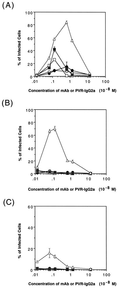
Poliovirus infects susceptible cells through the poliovirus receptor (PVR), which functions to bind virus and to change its conformation. These two activities are thought to be necessary for efficient poliovirus infection. How binding and conformation conversion activities contribute to the establishment of poliovirus infection was investigated. Mouse L cells expressing mouse high-affinity Fcgamma receptor molecules were established and used to study poliovirus infection mediated by mouse antipoliovirus monoclonal antibodies (MAbs) (immunoglobulin G2a [IgG2a] subtypes) or PVR-IgG2a, a chimeric molecule consisting of the extracellular moiety of PVR and the hinge and Fc portion of mouse IgG2a. The antibodies and PVR-IgG2a showed the same degree of affinity for poliovirus, but the infectivities mediated by these molecules were different. Among the molecules tested, PVR-IgG2a mediated the infection most efficiently, showing 50- to 100-fold-higher efficiency than that attained with the different MAbs. A conformational change of poliovirus was induced only by PVR-IgG2a. These results strongly suggested that some specific interaction(s) between poliovirus and the PVR is required for high-level infectivity of poliovirus in this system.
Figures
Similar articles
-
Multiple pathways for establishment of poliovirus infection.Virus Res. 1999 Aug;62(2):97-105. doi: 10.1016/s0168-1702(99)00040-4. Virus Res. 1999. PMID: 10507320
-
Characterization of the poliovirus 147S particle: new insights into poliovirus uncoating.Virology. 2003 Jan 5;305(1):55-65. doi: 10.1006/viro.2002.1736. Virology. 2003. PMID: 12504541
-
Homolog-scanning mutagenesis reveals poliovirus receptor residues important for virus binding and replication.J Virol. 1994 Apr;68(4):2578-88. doi: 10.1128/JVI.68.4.2578-2588.1994. J Virol. 1994. PMID: 8139037 Free PMC article.
-
Early events in poliovirus infection: virus-receptor interactions.Proc Natl Acad Sci U S A. 1996 Oct 15;93(21):11378-81. doi: 10.1073/pnas.93.21.11378. Proc Natl Acad Sci U S A. 1996. PMID: 8876143 Free PMC article. Review.
-
Poliovirus Receptor: More than a simple viral receptor.Virus Res. 2017 Oct 15;242:1-6. doi: 10.1016/j.virusres.2017.09.001. Epub 2017 Sep 8. Virus Res. 2017. PMID: 28870470 Free PMC article. Review.
Cited by
-
Foot-and-mouth disease virus exhibits an altered tropism in the presence of specific immunoglobulins, enabling productive infection and killing of dendritic cells.J Virol. 2011 Mar;85(5):2212-23. doi: 10.1128/JVI.02180-10. Epub 2010 Dec 22. J Virol. 2011. PMID: 21177807 Free PMC article.
-
An attenuated strain of enterovirus 71 belonging to genotype a showed a broad spectrum of antigenicity with attenuated neurovirulence in cynomolgus monkeys.J Virol. 2007 Sep;81(17):9386-95. doi: 10.1128/JVI.02856-06. Epub 2007 Jun 13. J Virol. 2007. PMID: 17567701 Free PMC article.
-
Antibody dependent enhancement infection of enterovirus 71 in vitro and in vivo.Virol J. 2011 Mar 8;8:106. doi: 10.1186/1743-422X-8-106. Virol J. 2011. PMID: 21385398 Free PMC article.
-
Development of a particle agglutination method with soluble virus receptor for identification of poliovirus.J Clin Microbiol. 2010 Aug;48(8):2698-702. doi: 10.1128/JCM.00207-10. Epub 2010 Jun 2. J Clin Microbiol. 2010. PMID: 20519462 Free PMC article.
-
Neutralization of hepatitis A virus (HAV) by an immunoadhesin containing the cysteine-rich region of HAV cellular receptor-1.J Virol. 2001 Jan;75(2):717-25. doi: 10.1128/JVI.75.2.717-725.2001. J Virol. 2001. PMID: 11134285 Free PMC article.
References
-
- Aoki J, Koike S, Ise I, Sato-Yoshida Y, Nomoto A. Amino acid residues on human poliovirus receptor involved in interaction with poliovirus. J Biol Chem. 1994;269:8431–8438. - PubMed
-
- Bernhardt G, Harber J, Zibert A, deCrombrugghe M, Wimmer E. The poliovirus receptor: identification of domains and amino acid residues critical for virus binding. Virology. 1994;203:344–356. - PubMed
Publication types
MeSH terms
Substances
LinkOut - more resources
Full Text Sources
Research Materials
