. 1999 Feb;73(2):1080-91.
doi: 10.1128/JVI.73.2.1080-1091.1999.

The inhibitory activity of the AU-rich RNA element in the human papillomavirus type 1 late 3' untranslated region correlates with its affinity for the elav-like HuR protein

Affiliations

The inhibitory activity of the AU-rich RNA element in the human papillomavirus type 1 late 3' untranslated region correlates with its affinity for the elav-like HuR protein

M Sokolowski et al. J Virol. 1999 Feb.

Abstract

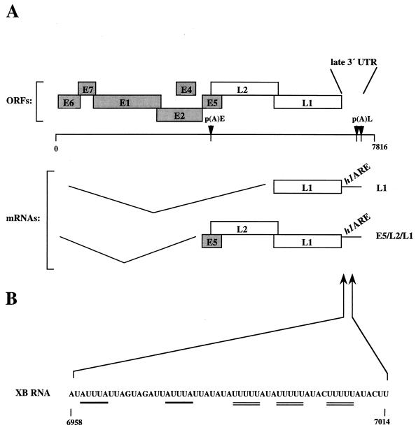
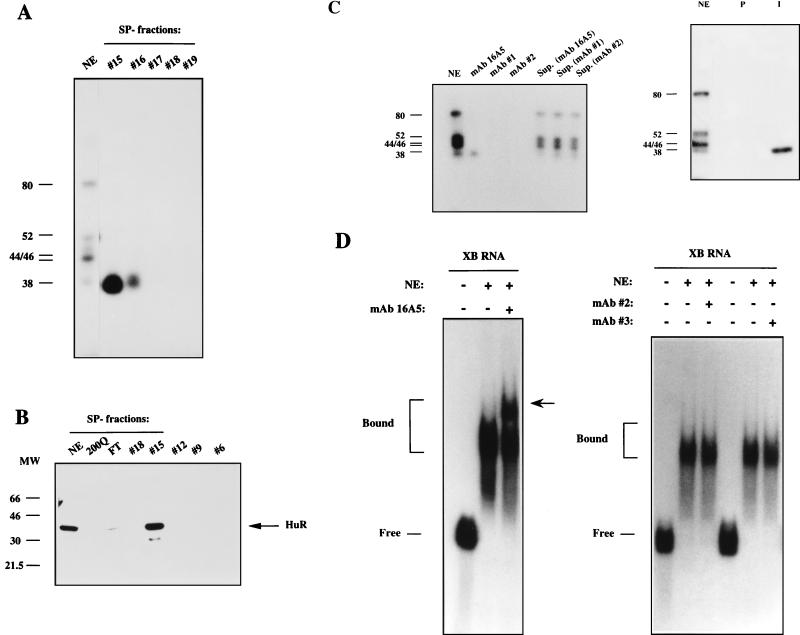
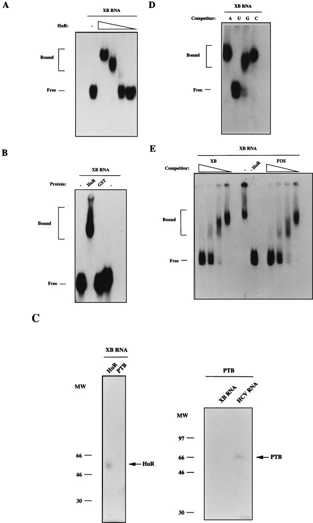
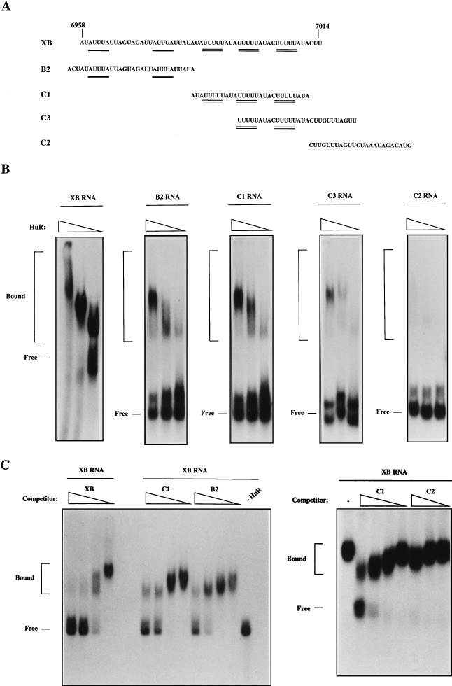
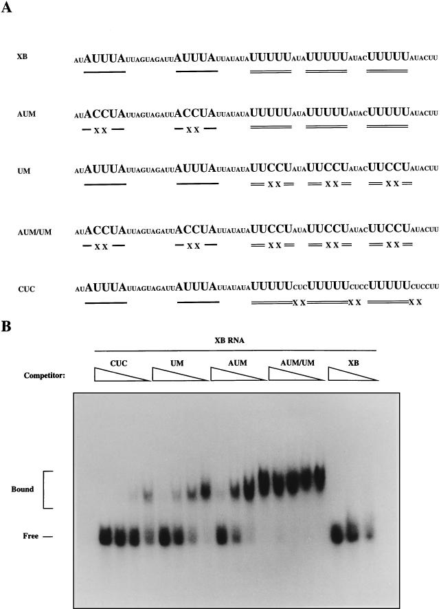
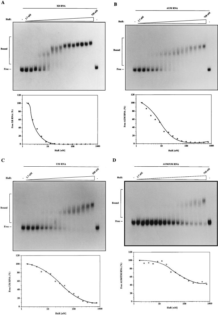
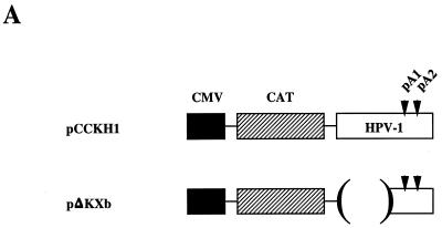
A 57-nucleotide adenosine- and uridine-rich RNA instability element in the human papillomavirus type 1 late 3' untranslated region termed h1ARE has previously been shown to interact specifically with three nuclear proteins that failed to bind to an inactive mutant RNA. Two of those were identified as the heterogeneous ribonucleoproteins C1 and C2, whereas the third, a 38-kDa, poly(U) binding protein (p38), remained unidentified. Here we show that partially purified p38 reacts with a monoclonal antibody raised against the recently identified elav-like HuR protein, indicating that p38 is the HuR protein. Indeed, recombinant glutathione S-transferase (GST)-HuR protein binds specifically to sites within the h1ARE. Determination of the apparent Kd value of GST-HuR for the h1ARE and the inactive mutant thereof revealed that GST-HuR bound with a more than 50-fold-higher affinity to the wild-type sequence. Therefore, the binding affinity of GST-HuR for the wild-type and mutant h1AREs correlates with their inhibitory activities in transfected cells, strongly suggesting that the HuR protein is involved in the posttranscriptional regulation of human papillomavirus type 1 late-gene expression.


Figures

FIG. 1
FIG. 1
(A) Schematic illustration of the HPV-1 genome and multiply spliced late mRNAs (2). Numbers refer to nucleotide positions in the HPV-1a genomic clone (11). The early (E1 to E7) and late (L1 and L2) ORFs are indicated as grey and white boxes respectively, and the early and late poly(A) signals [p(A)E and p(A)L] are shown as black triangles. The position of the HPV-1 AU-rich element (h1ARE) in the late 3′ UTR is shown. (B) The minimal h1ARE sequence used as the RNA probe named XB is shown. The functionally important sequence motifs are underlined.
FIG. 2
FIG. 2
The partially purified, 38-kDa h1ARE binding protein is detected with anti-HuR MAb. (A) UV cross-linking of fractions eluted from an SP column (fractions 15 to 19) to radiolabeled RNA XB (Fig. 1), as described in Materials and Methods. Numbers on the left indicate the sizes (in kilodaltons) of the previously characterized proteins that UV cross-link to XB RNA (43, 52). NE, nuclear extract. (B) Western immunoblot analysis of selected SP and Mono Q column fractions with anti-HuR MAb 16A5, as described in Materials and Methods. The migration of the HuR protein is indicated by an arrow. MW, molecular weight markers (in thousands). FT, flowthrough fraction from the Mono Q column; 200Q, fraction eluted from the Mono Q column with 200 mM KCl; NE, nuclear extract. (C) Left: HeLa nuclear extract (20 μg) was UV cross-linked to 4 pmol of XB RNA followed by immunoprecipitation with anti-HuR MAb 16A5 or with MAbs against two unrelated human RNA binding proteins (MAb1 and MAb2). The supernatants (Sup.) from the immunoprecipitations are shown. Lane NE shows UV cross-linking of nuclear extract to HPV-1 XB RNA. Right: HeLa nuclear extract (20 μg) was UV cross-linked to 4 pmol of XB RNA, and the products of four cross-linking reactions were pooled and immunoprecipitated with anti-HuR MAb 16A5 (I) or with sera from nonimmunized mice (P). In lane NE, the products of one reaction of UV cross-linked nuclear extract to HPV-1 XB RNA were loaded. (D) Left: an RNA gel shift assay with nuclear extract and XB RNA was performed in the absence or presence of the anti-HuR MAb 16A5. The arrow indicates the supershift induced by the MAb. Right: An RNA gel shift assay in the absence or presence of MAbs against two unrelated human RNA binding proteins (MAb2 and MAb3).
FIG. 3
FIG. 3
GST-HuR interacts specifically with the h1ARE. (A) RNA gel shift assay with radiolabeled XB RNA in the absence of GST-HuR protein (−) or in the presence of threefold serial dilutions of 1 μg of purified GST-HuR protein. The positions of free and bound RNAs are indicated. (B) RNA gel shift assay with radiolabeled XB RNA and 1 μg of GST-HuR protein (lane HuR) or 1 μg of GST (lane GST). The positions of free and bound RNAs are indicated. −, gel shift in the absence of GST-HuR protein. (C) Left panel: UV cross-linking of 1 μg of GST-HuR (lane HuR) or 1 μg of GST-polypyrimidine tract binding (lane PTB) fusion proteins to 1 pmol of radiolabeled XB RNA (molar ratio of RNA to GST-HuR, 1:15,000). Right panel: UV cross-linking of PTB to XB RNA or hepatitis C virus 5′ UTR (HCV RNA). MW, molecular weight marker (in thousands). (D) RNA gel shift assay with radiolabeled XB RNA and 1 μg of purified GST-HuR protein after preincubation of GST-HuR with poly(A), poly(U), poly(G), or poly(C) homoribopolymer competitors (A, U, G, and C, respectively). The migrations of free and bound RNAs are indicated. (E) RNA gel shift assay with radiolabeled XB RNA and 1 μg of purified GST-HuR protein after preincubation of GST-HuR with a 10-, 3.3-, 1.1-, and 0.38-fold excess of unlabeled h1ARE (XB) or c-fos ARE (FOS) RNA competitors. The positions of free and bound RNAs are indicated. −, no competitor; −HuR, gel shift in the absence of the GST-HuR protein.
FIG. 4
FIG. 4
The h1ARE contains multiple binding sites for GST-HuR. (A) Schematic illustration of the XB RNA and the overlapping B2, C1, C3, and C2 RNAs. The AUUUA and UUUUU motifs are underlined. (B) RNA gel shift assays with radiolabeled XB, B2, C1, C3, or C2 RNAs and threefold serial dilutions of 1 μg of purified GST-HuR protein. The positions of free and bound RNAs are indicated. (C) Left: RNA gel shift assays with radiolabeled XB RNA and 1 μg of purified GST-HuR protein preincubated with a 10-, 3.3-, 1.1-, and 0.38-fold excess of unlabeled XB, C1, or B2 RNAs as competitors. The positions of free and bound RNAs are indicated. −, no competitor; −HuR, gel shift in the absence of GST-HuR protein. Right: RNA gel shift assays with radiolabeled XB RNA and 1 μg of purified GST-HuR protein preincubated with a 10-, 3.3-, 1.1-, and 0.38-fold excess of unlabeled C1 RNA or a 10-, 3.3-, and 1.1-fold excess of unlabeled C2 RNA as competitors. The positions of free and bound RNAs are indicated. −, no competitor.
FIG. 5
FIG. 5
GST-HuR interacts specifically with the functionally important motifs within the h1ARE. (A) Nucleotide sequence of the XB RNA and the AUM, UM, AUM/UM, and CUC mutant RNAs. The AUUUA and UUUUU motifs are underlined. Substitution mutations of uracil (U) to cytidine (C) are marked with X. (B) RNA gel shift assay with radiolabeled XB RNA and 1 μg of purified GST-HuR protein preincubated with a 10-, 3.3-, 1.1-, and 0.38-fold excess of unlabeled CUC, UM, or AUM/UM mutant RNAs, a 10-, 3.3-, and 1.1-fold excess of XB RNA, or a 7.1-, 2.3-, 0.78-, and 0.27-fold excess of AUM. The positions of free and bound RNAs are indicated.
FIG. 6
FIG. 6
Affinity determination of GST-HuR for the wild-type h1ARE or h1ARE substitution mutants. The panels show representative RNA gel shift assays with 4 fmol of radiolabeled XB (A), AUM (B), UM (C), or AUM/UM (D) RNAs with 1.5-fold serial dilutions of the GST-HuR protein, starting with 750 nM. The positions of free and bound RNAs are indicated. −, gel shift in the absence of GST-HuR protein. The graphs show the mean values of percent free XB, AUM, UM, or AUM/UM RNA quantified in three independent RNA gel shift experiments plotted against the concentration of serially diluted GST-HuR protein. The 100% values correspond to the concentration of free RNA in the presence of 1.7 nM GST-HuR protein. Free RNA was quantified by using a phosphorimager.
FIG. 7
FIG. 7
Overexpression of HuR in HeLa cells. (A) Schematic illustration of the plasmids. Plasmid names are indicated on the left. The cytomegalovirus immediate-early promoter (CMV), the CAT ORF, the HPV-1 late 3′ UTR-containing sequences, and the HPV-1 late poly(A) signals (pA1 and pA2) are indicated. Brackets mark the limits of a deletion, and lines between the boxes represent vector sequences. (B) Histogram showing the quantified CAT levels produced from pCCKH1 or pΔKXb normalized to β-gal levels produced from pCMV-LacZ in HeLa cells infected with vTF7-3 in the absence (−HuR) or presence (+HuR) of the HuR-producing plasmid pT7HuR. Fold inhibition represents the CAT levels produced from pΔKXb divided by the CAT levels produced from pCCKH1. A representative experiment performed in triplicate is shown; results are given as mean ± standard deviation. (C) Western immunoblotting on the cell extracts from the transfections with pCCKH1 in the absence (−HuR) or presence (+HuR) of pT7HuR. The HuR protein was detected with MAb 16A5.
FIG. 7
FIG. 7
Overexpression of HuR in HeLa cells. (A) Schematic illustration of the plasmids. Plasmid names are indicated on the left. The cytomegalovirus immediate-early promoter (CMV), the CAT ORF, the HPV-1 late 3′ UTR-containing sequences, and the HPV-1 late poly(A) signals (pA1 and pA2) are indicated. Brackets mark the limits of a deletion, and lines between the boxes represent vector sequences. (B) Histogram showing the quantified CAT levels produced from pCCKH1 or pΔKXb normalized to β-gal levels produced from pCMV-LacZ in HeLa cells infected with vTF7-3 in the absence (−HuR) or presence (+HuR) of the HuR-producing plasmid pT7HuR. Fold inhibition represents the CAT levels produced from pΔKXb divided by the CAT levels produced from pCCKH1. A representative experiment performed in triplicate is shown; results are given as mean ± standard deviation. (C) Western immunoblotting on the cell extracts from the transfections with pCCKH1 in the absence (−HuR) or presence (+HuR) of pT7HuR. The HuR protein was detected with MAb 16A5.

Similar articles

Cited by

References

    1. Ali N, Siddiqui A. Interaction of polypyrimidine tract-binding protein with the 5′ noncoding region of the hepatitis C virus RNA genome and its functional requirement in internal initiation of translation. J Virol. 1995;69:6367–6375. - PMC - PubMed
    1. Baker C, Calef C. Maps of papillomavirus mRNA transcripts. In: Billakanti S R, Calef C E, Farmer A D, Halpern A L, Myers G L, editors. Human papillomaviruses: a compilation and analysis of nucleic acid and amino acid sequences. Los Alamos, N.M: Los Alamos National Laboratory; 1997. pp. 3–10.
    1. Baker C C. Post-transcriptional regulation of papillomavirus gene expression. In: Billakanti S R, Calef C E, Farmer A D, Halpern A L, Myers G L, editors. Human papillomaviruses: a compilation and analysis of nucleic acid and amino acid sequences. Los Alamos, N.M: Los Alamos National Laboratory; 1997. pp. 11–16.
    1. Baker C C, Howley P M. Differential promoter utilization by the bovine papillomavirus in transformed cells and productively infected wart tissues. EMBO J. 1987;6:1027–1035. - PMC - PubMed
    1. Beelman C A, Parker R. Degradation of mRNA in eucaryotes. Cell. 1995;81:179–183. - PubMed

Publication types