. 1999 Feb;73(2):1127-37.
doi: 10.1128/JVI.73.2.1127-1137.1999.

Bovine leukemia virus-induced persistent lymphocytosis in cattle does not correlate with increased ex vivo survival of B lymphocytes

Affiliations

Bovine leukemia virus-induced persistent lymphocytosis in cattle does not correlate with increased ex vivo survival of B lymphocytes

F Dequiedt et al. J Virol. 1999 Feb.

Abstract

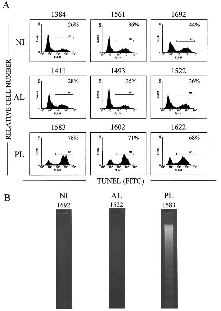
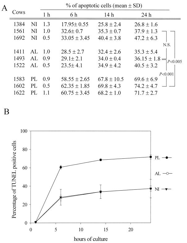
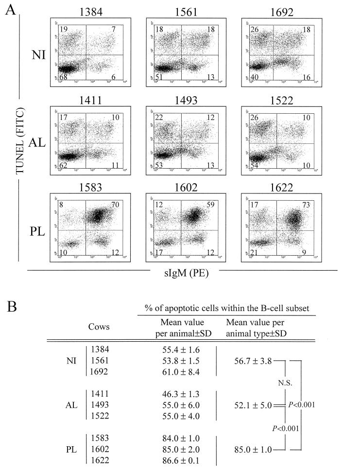
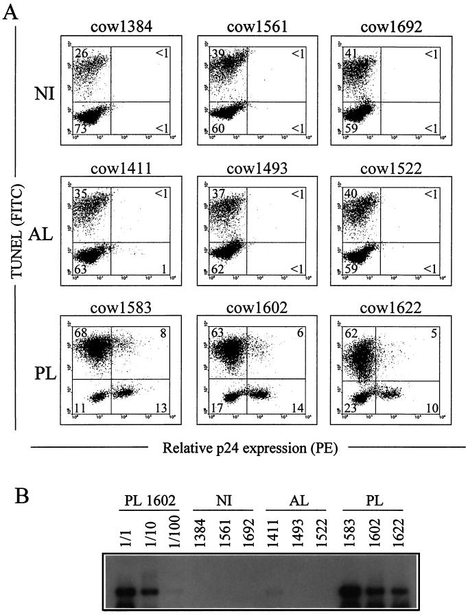
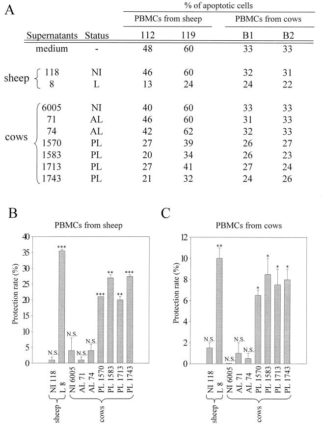
Bovine leukemia virus (BLV) is an oncogenic retrovirus associated with B-cell lymphocytosis, leukemia, and lymphosarcoma in the ovine and bovine species. We have recently reported that in sheep, BLV protects the total population of peripheral blood mononuclear cells (PBMCs) from ex vivo spontaneous apoptosis. This global decrease in the apoptosis rates resulted from both direct and indirect mechanisms which allow extension of cell survival. Although sheep are not natural hosts for BLV, these animals are prone to develop virus-induced leukemia at very high frequencies. Most infected cattle, however, remain clinically healthy. This difference in the susceptibilities to development of leukemia in these two species might be related to alterations of the apoptotic processes. Therefore, we designed this study to unravel the mechanisms of programmed cell death in cattle. We have observed that PBMCs from persistently lymphocytotic BLV-infected cows were more susceptible to spontaneous ex vivo apoptosis than cells from uninfected or aleukemic animals. These higher apoptosis rates were the consequence of an increased proportion of B cells exhibiting lower survival abilities. About one-third of the BLV-expressing cells did not survive the ex vivo culture conditions, demonstrating that viral expression is not strictly associated with cell survival in cattle. Surprisingly, culture supernatants from persistently lymphocytotic cows exhibited efficient antiapoptotic properties on both uninfected bovine and uninfected ovine cells. It thus appears that indirect inhibition of cell death can occur even in the presence of high apoptosis rates. Together, these results demonstrate that the protection against spontaneous apoptosis associated with BLV is different in cattle and in sheep. The higher levels of ex vivo apoptosis occurring in cattle might indicate a decreased susceptibility to development of leukemia in vivo.


Figures

FIG. 1
FIG. 1
Detection of apoptosis in PBMC cultures from BLV-infected PL, BLV-infected AL, and NI cows. (A) After 24 h of culture, PBMCs were fixed and the DNA strand breaks were labeled by the TUNEL procedure. Incorporation of FITC-dUTP was assessed by flow cytometry on the FL1 channel over 10,000 events. For each sample, distributions of the cells according to the relative fluorescence on the FL1 channel [TUNEL (FITC) on the x axis] are represented as a histogram. The percentage on each histogram corresponds to the proportion of apoptotic cells (M1). Results from one representative experiment are shown. (B) Visualization of internucleosomal DNA fragmentation by agarose gel electrophoresis. After 24 h of culture, the LMW fraction of the DNA was isolated, electrophoresed through a 1.5% agarose gel, and stained with ethidium bromide. The samples are from PL cow 1583, AL cow 1522, and NI cow 1692.
FIG. 2
FIG. 2
Kinetic analysis of the apoptosis levels in cultures of PBMCs from PL, AL, and NI cows. (A) PBMCs were cultured for 1, 6, 14, and 24 h and processed for detection of DNA strand breaks by the TUNEL procedure. Data are mean values from two independent experiments performed in duplicate. SD, standard deviation. The statistical evaluation of the differences between NI, AL, and PL animals was performed with a Student t test. N.S., not statistically significant. (B) Mean values were calculated from the data presented in panel A, according to the stage of the disease. These values are schematically represented for a kinetic analysis after 1, 6, 14, and 24 h of culture.
FIG. 3
FIG. 3
Detection of apoptosis in B lymphocytes from BLV-infected PL (no. 1583, 1602, and 1622), BLV-infected AL (no. 1411, 1493, and 1522) and NI (no. 1384, 1561, and 1692) cows. PBMCs were cultured for 24 h and labeled with the PIg45A2 MAb (directed against bovine IgM) and a phycoerythrin (PE)-conjugated secondary antibody. The cells were then fixed and processed by the TUNEL procedure to detect apoptosis, and samples were then analyzed by dual-immunofluorescence flow cytometric analysis. (A) Results from a representative experiment are presented as dot plots. On the basis of control staining, each distribution was divided into four quadrants. The numbers indicate the percentage of PBMCs in each quadrant. (B) Percentages of apoptotic cells within the B-lymphocyte population. The mean values and standard deviations (SD) were calculated with three animals from two independent experiments. The significance of the differences in mean values between groups of animals was established by a Student t test. N.S., not statistically significant.
FIG. 4
FIG. 4
In situ detection of apoptosis in BLV-expressing cells. PBMCs from PL cows 1583, 1602, and 1622, AL cows 1411, 1493, and 1522, and NI cows 1384, 1561, and 1692 were cultured for 24 h and fixed in paraformaldehyde-ethanol. Cells were then labeled with an anti-p24 MAb (4′G9) and a phycoerythrin (PE)-coupled secondary antibody. The DNA strand breaks were labeled by the TUNEL procedure. Samples were then analyzed by dual-flow cytometric immunofluorescence analysis. (A) Results from a representative experiment are presented as dot plots. Numbers represent the percentages of positively stained cells in each quadrant. (B) Semiquantitative PCR amplification of viral sequences. The DNAs were prepared from 500-μl aliquots of blood and resuspended in PCR buffer. Specific proviral sequences were amplified by 26 cycles of PCR. The amplification products were resolved on a 1% agarose gel and analyzed by Southern blotting with a BLV probe. Blood samples from NI cows 1384, 1561, and 1692 were used as controls for PCR contamination. The semiquantitative analysis was done by amplification of serial dilutions (1/1, 1/10, and 1/100) from PL cow 1602.
FIG. 5
FIG. 5
Protection of ovine and bovine uninfected PBMCs by culture supernatants from PBMCs from cows with PL. (A) PBMCs from a BLV-infected sheep (leukemic [L] sheep no. 8), an NI sheep (no. 118), an NI cow (no. 6005), two BLV-infected AL cows (no. 71 and 74), or PL cows (no. 1570, 1583, 1713, and 1743) were cultured for 48 h. The corresponding cell culture supernatants were added to PBMCs from NI sheep 112 and 119 or NI cows B1 and B2 at a 1/20 dilution. After 20 h of culture, the percentages of apoptotic cells were determined by the TUNEL procedure and flow cytometry analysis. As a control, apoptosis rates in culture medium alone were also measured. (B) The protection rate corresponds to the difference between the percentage measured in medium alone and the percentage measured with a conditioned supernatant. Mean values (ranging from 1 to 35.5%) and standard deviations obtained with uninfected PBMCs from sheep 112 and 119 are illustrated in a histogram. (C) The mean values (ranging from 0 to 10%) and standard deviations of protection rates obtained with uninfected PBMCs from cows B1 and B2 are illustrated in a histogram. The significance of the protection rates was analyzed by using a Student t test (N.S., not statistically significant; ∗, P < 0.05; ∗∗, P < 0.005; ∗∗∗, P < 0.0005).

References

    1. Arai M, Kannagi M, Matsuoka M, Sato T, Yamamoto N, Fuji M. Expression of FAP-1 (Fas-associated phosphatase) and resistance to Fas-mediated apoptosis in T cell lines derived from human T cell leukemia virus type 1-associated myelopathy/tropical spastic paraparesis patients. AIDS Res Hum Retroviruses. 1998;14:261–267. - PubMed
    1. Burny A, Cleuter Y, Kettmann R, Mammerickx M, Marbaix G, Portetelle D, Van den Broeke A, Willems L, Thomas R. Bovine leukemia: facts and hypotheses derived from the study of an infectious cancer. Adv Vet Sci Comp Med. 1988;32:149–170. - PubMed
    1. Chevallier N, Barthelemy M, Le Rhun D, Lainé V, Levy D, Schwartz-Cornil I. Bovine leukemia virus-induced lymphocytosis and increased cell survival involve the CD11b+ B-lymphocyte subset in sheep. J Virol. 1998;72:4413–4420. - PMC - PubMed
    1. Chlichlia K, Moldenhauer G, Daniel P T, Busslinger M, Gazzolo L, Schirrmacher V, Khazaie K. Immediate effects of reversible HTLV-I tax function: T-cell activation and apoptosis. Oncogene. 1995;10:269–277. - PubMed
    1. Chlichlia K, Busslinger M, Peter M E, Walczak H, Krammer P H, Schirrmacher V, Khazaie K. ICE-proteases mediate HTLV-I Tax induced apoptotic cell death. Oncogene. 1997;14:2265–2272. - PubMed

Publication types

Substances

LinkOut - more resources