. 1999 Feb;73(2):1219-26.
doi: 10.1128/JVI.73.2.1219-1226.1999.

Translation initiation at the CUU codon is mediated by the internal ribosome entry site of an insect picorna-like virus in vitro

Affiliations

Translation initiation at the CUU codon is mediated by the internal ribosome entry site of an insect picorna-like virus in vitro

J Sasaki et al. J Virol. 1999 Feb.

Abstract

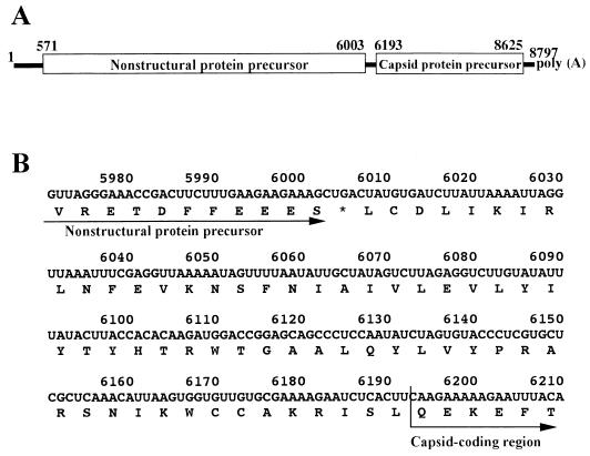
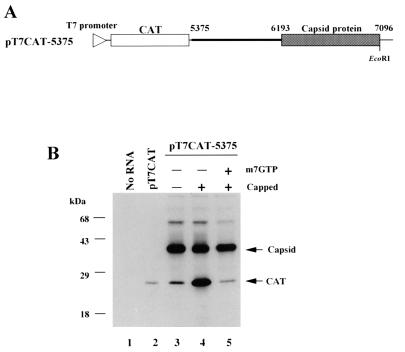
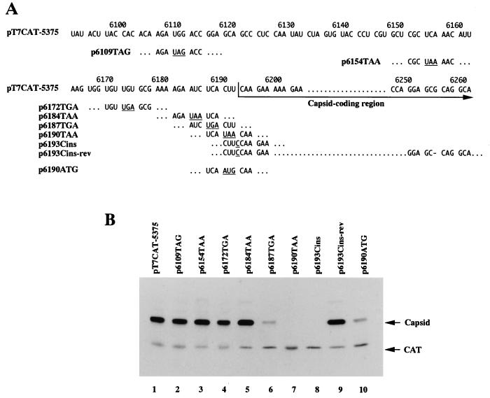
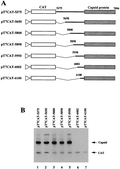
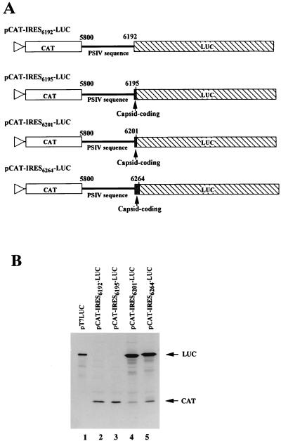
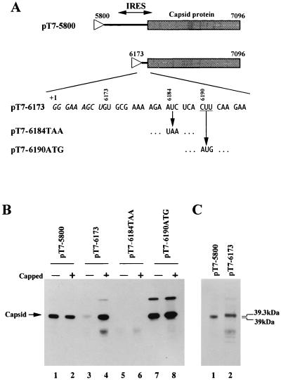
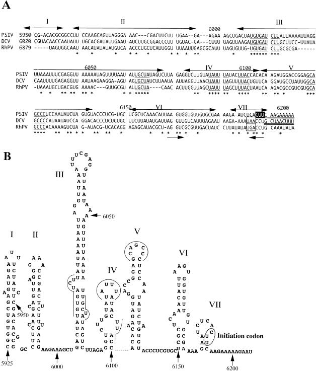
AUG-unrelated translation initiation was found in an insect picorna-like virus, Plautia stali intestine virus (PSIV). The positive-strand RNA genome of the virus contains two nonoverlapping open reading frames (ORFs). The capsid protein gene is located in the 3'-proximal ORF and lacks an AUG initiation codon. We examined the translation mechanism and the initiation codon of the capsid protein gene by using various dicistronic and monocistronic RNAs in vitro. The capsid protein gene was translated cap independently in the presence of the upstream cistron, indicating that the gene is translated by internal ribosome entry. Deletion analysis showed that the internal ribosome entry site (IRES) consisted of approximately 250 bases and that its 3' boundary extended slightly into the capsid-coding region. The initiation codon for the IRES-mediated translation was identified as the CUU codon, which is located just upstream of the 5' terminus of the capsid-coding region by site-directed mutagenesis. In vitro translation assays of monocistronic RNAs lacking the 5' part of the IRES showed that this CUU codon was not recognized by scanning ribosomes. This suggests that the PSIV IRES can effectively direct translation initiation without stable codon-anticodon pairing between the initiation codon and the initiator methionyl-tRNA.


Figures

FIG. 1
FIG. 1
Genome organization of PSIV. (A) Schematic diagram of the PSIV genome. ORFs are shown in open boxes. The numbers indicate nucleotide positions. The first nucleotide of the capsid protein gene represents the 5′-terminal nucleotide of the capsid-coding region. (B) Nucleotide and deduced amino acid sequences of the segment between the nonstructural protein gene and the capsid-coding region. The asterisk indicates the stop codon for the nonstructural protein gene.
FIG. 2
FIG. 2
(A) Schematic diagram of pT7CAT-5375. The thin line indicates the vector sequence, and the triangle on the line represents the location of the T7 RNA polymerase promoter. The thick line indicates the PSIV sequence. The open and shaded boxes show the CAT gene and PSIV capsid-coding region, respectively. The nucleotide positions in the PSIV genome are indicated above the line. The EcoRI site used to linearize the plasmid is also shown. (B) Cap influence on in vitro translation of the RNAs transcribed from pT7CAT-5375. Capped and uncapped RNAs were translated in a rabbit reticulocyte lysate with or without a cap analog, m7GTP. To examine the electrophoretic mobility of the CAT protein, uncapped RNA from pT7CAT, which contains only a CAT gene, was also translated. Each product was separated by sodium dodecyl sulfate-polyacrylamide gel electrophoresis (12% polyacrylamide), blotted onto a polyvinylidene difluoride membrane, and then detected by enhanced chemiluminescence. The positions of the translation products (CAT and capsid protein) and molecular mass markers are indicated on the right and left of the panel, respectively. The 55-kDa bands observed in lanes 3 to 5 were thought to be insufficiently denatured proteins translated from the second cistron (see Materials and Methods).
FIG. 3
FIG. 3
Identification of the initiation codon for capsid protein translation. (A) RNA sequences of mutants derived from pT7CAT-5375. The numbers above the sequence indicate nucleotide positions. Stop codons and inserted nucleotides introduced by site-directed mutagenesis are underlined, and a deleted nucleotide is shown by a dash (p6193Cins-rev). (B) In vitro translation products from uncapped RNAs synthesized from pT7CAT-5375 and the site-directed mutants shown in panel A. The positions of the translation products (CAT and capsid protein) are indicated on the right.
FIG. 4
FIG. 4
Mapping the 5′ boundary of IRES for PSIV capsid protein translation. (A) Schematic diagrams of pT7CAT-5375 and a series of deletion mutants. The thin lines indicate vector sequences, and the triangles represent the location of the T7 promoter. The thick lines indicate the PSIV sequence. The CAT genes and PSIV capsid-coding regions are shown by open and shaded boxes, respectively. The numbers above the lines indicate nucleotide positions. Deleted regions are shown by angled lines. (B) In vitro translation products of uncapped RNAs synthesized from pT7CAT-5375 and the deletion mutants shown in panel A. The positions of the translation products (CAT and capsid protein) are indicated on the right.
FIG. 5
FIG. 5
Mapping the 3′ boundary of IRES for PSIV capsid protein translation. (A) Schematic diagrams of the pCAT-IRES-LUC series of constructs. The triangles represent the location of the T7 promoter. The CAT and LUC genes are shown by open and hatched boxes, respectively. The thick lines indicate the PSIV sequences, and the shaded boxes show the PSIV capsid-coding regions. The numbers above the lines indicate the positions of the 5′- and 3′-terminal nucleotides of PSIV sequences. (B) In vitro translation products of uncapped RNAs synthesized from the pCAT-IRES-LUC series of constructs. To examine the electrophoretic mobility of the LUC protein, uncapped RNA synthesized from pT7LUC, which contains only a LUC gene, was also analyzed. The positions of the translation products (CAT and LUC) are indicated on the right of the panel.
FIG. 6
FIG. 6
(A) Schematic diagrams of plasmids used to synthesize monocistronic RNAs. The lines indicate PSIV sequences and the capsid-coding regions are shown as shaded boxes. Triangles represent the location of the T7 promoter. The 5′-terminal nucleotide sequence of the RNA transcribed from the plasmid pT7-6173 is shown. Italic and roman letters indicate the vector and PSIV sequences, respectively. +1 represents the transcription start site for T7 RNA polymerase. The CUU initiation codon is underlined. For pT7-6184TAA and pT7-6190ATG, only mutated codons are shown. (B) In vitro translation products of uncapped and capped RNAs synthesized from pT7-5800, pT7-6173, pT7-6184TAA, and pT7-6190ATG. The position of the translation products (capsid protein) is indicated on the left of the panel. (C) Comparison of the molecular masses of the translation products from pT7-5800 and pT7-6173. To show the difference in mobility, the same amounts of products were electrophoresed.
FIG. 7
FIG. 7
(A) Multiple alignment of nucleotide sequences upstream of PSIV (28), DCV (11), and RhPV (19) (accession no. AB006531, AF014388, and AF022937, respectively) capsid-coding regions. The numbers on the left indicate the starting nucleotide positions of the aligned sequences, and the numbers above the sequences represent the nucleotide positions in the PSIV sequence. The initiation codon for the PSIV capsid protein translation is shown in reverse type. In the PSIV and DCV sequences, the capsid-coding regions are doubly underlined. The DCV sequence has a stop codon (boxed) 2 codons upstream of the 5′ terminus of the capsid-coding region, and the RhPV sequence also has a stop codon in the same position. Asterisks indicate nucleotides conserved in all three viruses, and the conserved short RNA segments are underlined. Two arrows below the sequences show an inverted repeat. The double-headed arrows above the sequences represent stem-loop segments in the secondary structure predicted for the PSIV RNA, and the roman numerals correspond to those shown in panel. (B) Computer-predicted secondary structure of the PSIV RNA sequence containing the IRES for the capsid protein translation. The numbers indicate nucleotide positions. The initiation codon is circled. The lines and curves indicate the conserved short RNA segments. Stem-loop structures are numbered from I to VII.

References

    1. Agol V I. The 5′-untranslated region of picornaviral genomes. Adv Virus Res. 1991;40:103–180. - PMC - PubMed
    1. Belsham G J, Sonenberg N. RNA-protein interactions in regulation of picornavirus RNA translation. Microbiol Rev. 1996;60:499–511. - PMC - PubMed
    1. Boeck R, Curran J, Matsuoka Y, Compans R, Kolakofsky D. The parainfluenza virus type 1 P/C gene uses a very efficient GUG codon to start its C′ protein. J Virol. 1992;66:1765–1768. - PMC - PubMed
    1. Curran J, Kolakofsky D. Ribosomal initiation from an ACG codon in the Sendai virus P/C mRNA. EMBO J. 1988;7:245–251. - PMC - PubMed
    1. Graff J, Ehrenfeld E. Coding sequences enhance internal initiation of translation by hepatitis A virus RNA in vitro. J Virol. 1998;72:3571–3577. - PMC - PubMed

Publication types

LinkOut - more resources