. 1999 Feb;73(2):1245-53.
doi: 10.1128/JVI.73.2.1245-1253.1999.

Analysis of synthesis, stability, phosphorylation, and interacting polypeptides of the 34-kilodalton product of open reading frame 6 of the early region 4 protein of human adenovirus type 5

Affiliations

Analysis of synthesis, stability, phosphorylation, and interacting polypeptides of the 34-kilodalton product of open reading frame 6 of the early region 4 protein of human adenovirus type 5

D Boivin et al. J Virol. 1999 Feb.

Abstract

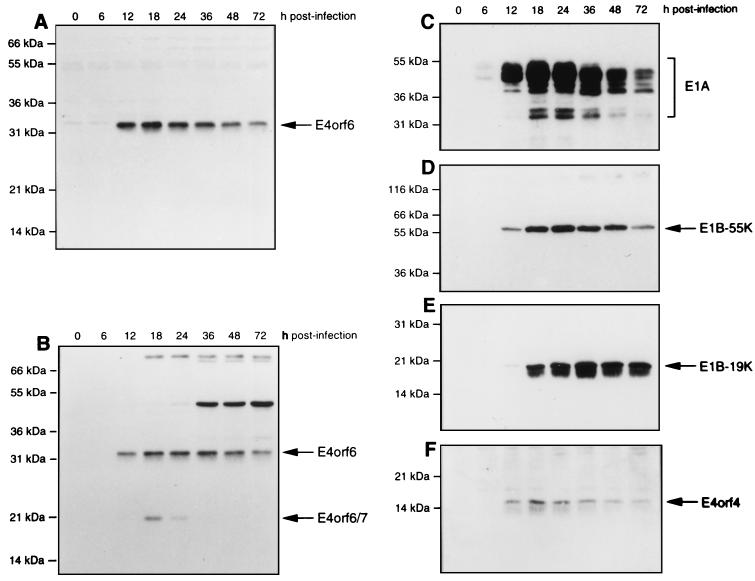
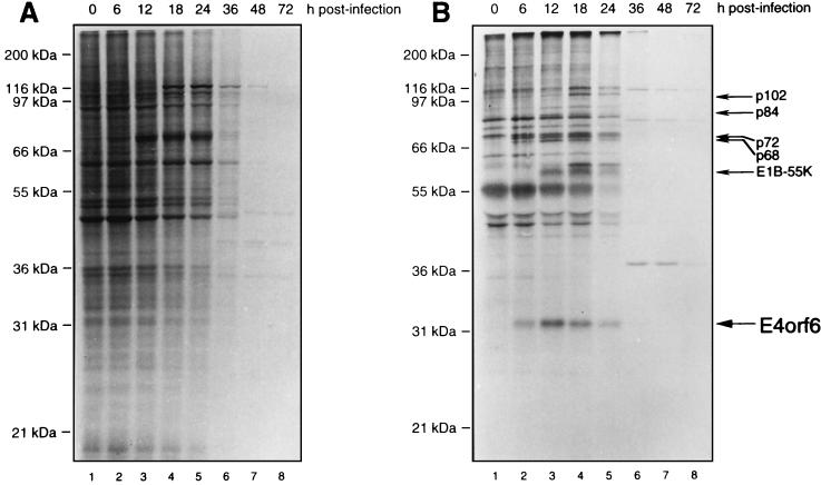
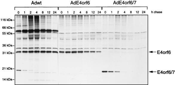
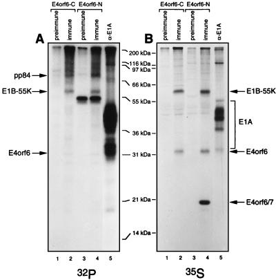
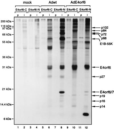
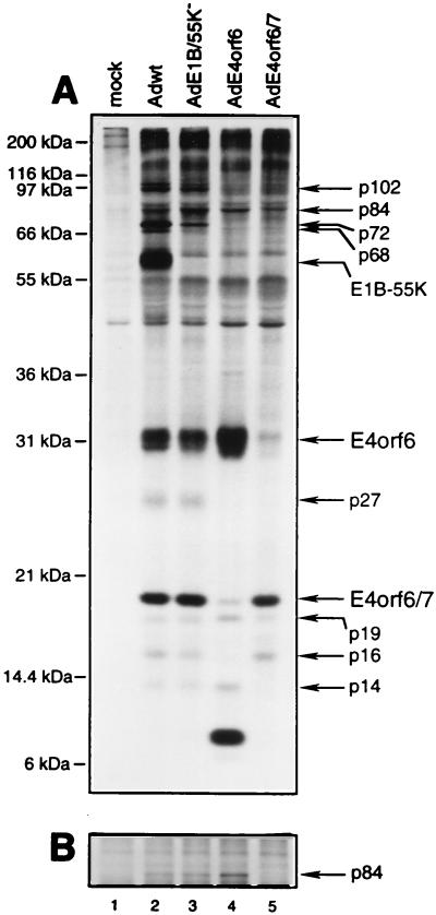
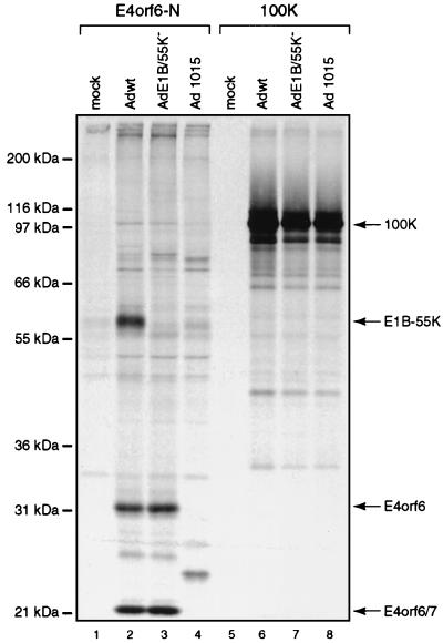
The 34-kDa early-region 4 open reading frame 6 (E4orf6) product of human adenovirus type 5 forms complexes with both the cellular tumor suppressor p53 and the viral E1B 55-kDa protein (E1B-55kDa). E4orf6 can inhibit p53 transactivation activity, as can E1B-55kDa, and in combination these viral proteins cause the rapid turnover of p53. In addition, E4orf6-55kDa complexes play a critical role at later times in the regulation of viral mRNA transport and shutoff of host cell protein synthesis. In the present study, we have further characterized some of the biological properties of E4orf6. Analysis of extracts from infected cells by Western blotting indicated that E4orf6, like E1A and E1B products, is present at high levels until very late times, suggesting that it is available to act throughout the infectious cycle. This pattern is similar to that of E4orf4 but differs markedly from that of another E4 product, E4orf6/7, which is present only transiently. Synthesis of E4orf6 is maximal at early stages but ceases completely with the onset of shutoff of host protein synthesis; however, it was found that unlike E4orf6/7, E4orf6 is very stable, thus allowing high levels to be maintained even at late times. E4orf6 was shown to be phosphorylated at low levels. Coimmunoprecipitation studies in cells lacking p53 indicated that E4orf6 interacts with a number of other proteins. Five of these were shown to be viral or virally induced proteins ranging in size from 102 to 27 kDa, including E1B-55kDa. One such species, of 72 kDa, was shown not to represent the E2 DNA-binding protein and thus remains to be identified. Another appeared to be the L4 100-kDa nonstructural adenovirus late product, but it appeared to be present nonspecifically and not as part of an E4orf6 complex. Apart from p53, three additional cellular proteins, of 84, 19, and 14 kDa were detected by using an adenovirus vector that expresses only E4orf6. The 19-kDa species and a 16-kDa cellular protein were also shown to interact with E4orf6/7. It is possible that complex formation with these viral and cellular proteins plays a role in one or more of the biological activities associated with E4orf6 and E4orf6/7.


Figures

FIG. 1
FIG. 1
Expression of E4orf6, E4orf6/7, and other Ad proteins in HeLa cells infected with wt Ad5. HeLa cells were infected with wt Ad5, and cell extracts were prepared at various times after infection. Equal amounts of whole-cell protein were separated by SDS-PAGE, and following transfer to nitrocellulose, the levels of E4orf6 and other viral proteins were determined by Western blotting. (A) E4orf6 with E4orf6-C serum. (B) E4orf6 and E4orf6/7 with E4orf6-N serum. (C) E1A proteins with M73 monoclonal antibody. (D) E1B-55kDa with 2A6 monoclonal antibody. (E) E1B-19kDa with 19-C1 serum. (F) E4orf4 levels with E4orf4-C serum. The positions of migration of molecular mass markers are shown on the left, and those of the viral proteins are shown on the right.
FIG. 2
FIG. 2
Time course of host cell shutoff and E4orf6 synthesis in Ad5-infected HeLa cells. HeLa cells were infected with wt Ad5, and at various times after infection they were labeled for 1 h with [35S]methionine-[35S]cysteine and cell extracts were prepared. (A) Analysis of whole-cell protein synthesis. A 5-μg portion of total-cell protein from each sample was analyzed by SDS-PAGE followed by fluorography. (B) Analysis of E4orf6 synthesis. Equal aliquots of the cell extracts shown in panel A were immunoprecipitated with E4orf6-C serum, and immunoprecipitates were analyzed by SDS-PAGE followed by fluorography. The positions of molecular mass markers are shown on the left, and that of E4orf6 is shown on the right.
FIG. 3
FIG. 3
Pulse-chase analysis of E4orf6 and E4orf6/7 proteins in HeLa cells. HeLa cells were infected with wt Ad5, AdE4orf6, or AdE4orf6/7. At 12 h p.i., the cells were labeled for 1 h with [35S]methionine-[35S]cysteine and then incubated further for various times with medium containing an excess of cold methionine. Cell extracts were subjected to immunoprecipitation with E4orf6-N serum, which recognizes both E4orf6 and E4orf6/7, and immunoprecipitates were analyzed by SDS-PAGE followed by autoradiography.
FIG. 4
FIG. 4
Phosphorylation of E4orf6. HeLa cells were infected with wt Ad5 or mock infected, and at 15 h p.i. they were labeled either with [32P]orthophosphate for 4 h (A) or with [35S]methionine-[35S]cysteine for 2 h (B). Cell extracts were immunoprecipitated with E4orf6-C (lanes 1 and 2) or E4orf6-N (lanes 3 and 4) serum or the corresponding preimmune serum or with M73 monoclonal antibody (lanes 5), and the precipitates were analyzed by SDS-PAGE followed by autoradiography. The positions of molecular mass markers are shown in the middle, and those of relevant proteins are shown at the sides.
FIG. 5
FIG. 5
Detection of E4orf6-binding proteins. p53-null H1299 cells were mock infected (lanes 1 to 4) or infected with either wt Ad5 (lanes 5 to 8) or AdE4orf6 (lanes 9 to 12). Cells were labeled at 18 h p.i. with [35S]methionine-[35S]cysteine for 2 h, and cell extracts were prepared under mild conditions and immunoprecipitated with preimmune (lanes p) or immune (lanes i) E4orf6-C or E4orf6-N serum. The proteins were separated on an SDS–12% polyacrylamide gel, and the labeling pattern was visualized by fluorography. The positions of migration of molecular mass markers are shown on the left, and those of relevant proteins are shown on the right.
FIG. 6
FIG. 6
Complex formation between E4orf6 and viral or cellular proteins. An experiment similar to that described in the legend to Fig. 5, using H1299 cells and E4orf6-N serum, was performed with mock-infected cells (lane 1) or cells infected with wt Ad5 (lane 2), mutant E1B/55K (lane 3), AdE4orf6 (lane 4), or AdE4orf6/7 (lane 5). (A) Precipitates were analyzed by SDS-PAGE with gels containing 12% polyacrylamide. (B) Portion of a gel containing similar samples that were analyzed with gels containing 8% polyacrylamide. The positions of migration of molecular mass markers are shown on the left, and those of relevant proteins are shown on the right.
FIG. 7
FIG. 7
Complex formation between E4orf6 and the L4 100-kDa protein. An experiment similar to that described in the legend to Fig. 5, using H1299 cells and E4orf6-N serum or 2100K-1 anti-L4 100-kDa protein antibody, was performed with mock-infected cells (lanes 1 and 5) or those infected with wt Ad5 (lanes 2 and 6), mutant E1B/55K (lanes 3 and 7), or dl1015 (lanes 4 and 8). Precipitates were analyzed by SDS-PAGE with gels containing 12% polyacrylamide. The positions of migration of molecular mass markers are shown on the left, and those of relevant proteins are shown on the right.

References

    1. Babiss L E, Ginsberg H S. Adenovirus type 5 early region 1b gene product is required for efficient shutoff of host protein synthesis. J Virol. 1984;50:202–212. - PMC - PubMed
    1. Babiss L E, Ginsberg H S, Darnell J J. Adenovirus E1B proteins are required for accumulation of late viral mRNA and for effects on cellular mRNA translation and transport. Mol Cell Biol. 1985;5:2552–2558. - PMC - PubMed
    1. Bacchetti S, Graham F L. Inhibition of cell proliferation by an adenovirus vector expressing the human wild type p53 protein. Int J Oncol. 1993;3:781–788. - PubMed
    1. Bayley S T, Mymryk J S. Adenovirus E1A proteins and transformation. Int J Oncol. 1994;5:425–444. - PubMed
    1. Boivin, D. Unpublished data.

Publication types