. 1999 Feb;73(2):1309-19.
doi: 10.1128/JVI.73.2.1309-1319.1999.

Cloning and characterization of adeno-associated virus type 5

Affiliations

Cloning and characterization of adeno-associated virus type 5

J A Chiorini et al. J Virol. 1999 Feb.

Abstract

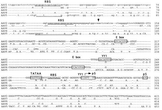
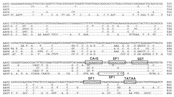
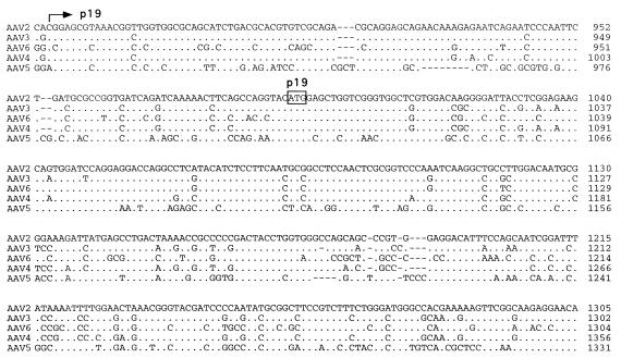
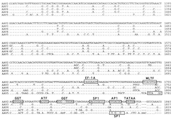
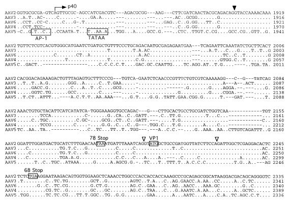
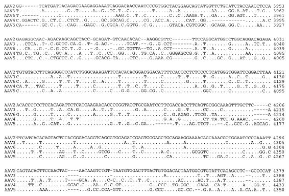
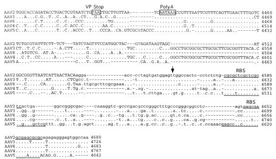
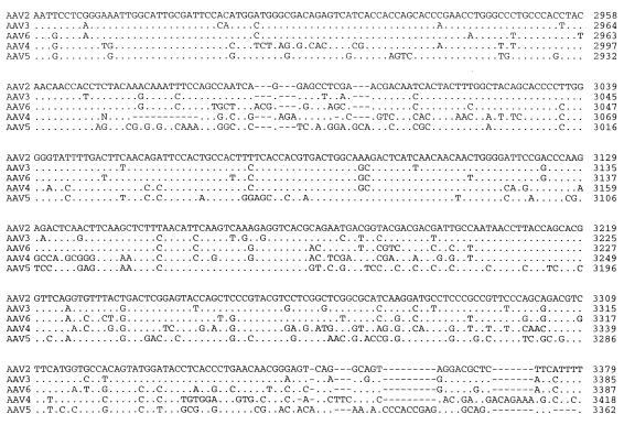
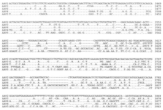
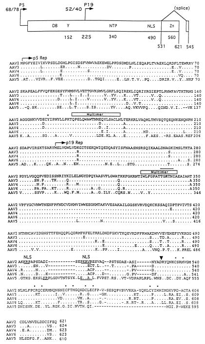
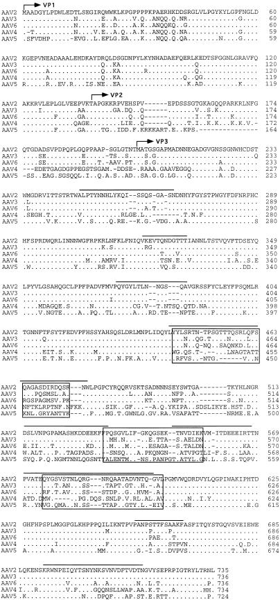
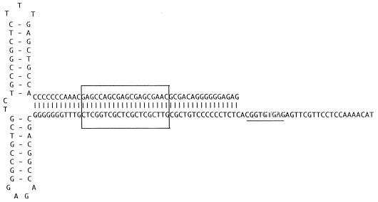
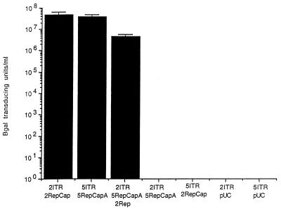
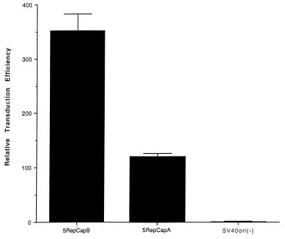
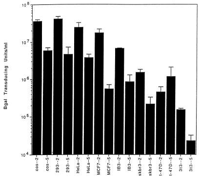
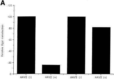
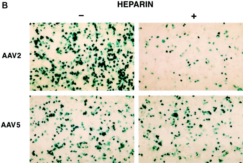
Adeno-associated virus type 5 (AAV5) is distinct from other dependovirus serotypes based on DNA hybridization and serological data. To better understand the biology of AAV5, we have cloned and sequenced its genome and generated recombinant AAV5 particles. The single-stranded DNA genome is similar in length and genetic organization to that of AAV2. The rep gene of AAV5 is 67% homologous to AAV2, with the majority of the changes occurring in the carboxyl and amino termini. This homology is much less than that observed with other reported AAV serotypes. The inverted terminal repeats (ITRs) are also unique compared to those of the other AAV serotypes. While the characteristic AAV hairpin structure and the Rep DNA binding site are retained, the consensus terminal resolution site is absent. These differences in the Rep proteins and the ITRs result in a lack of cross-complementation between AAV2 and AAV5 as measured by the production of recombinant AAV particles. Alignment of the cap open reading frame with that of the other AAV serotypes identifies both conserved and variable regions which could affect tissue tropism and particle stability. Comparison of transduction efficiencies in a variety of cells lines and a lack of inhibition by soluble heparin indicate that AAV5 may utilize a distinct mechanism of uptake compared to AAV2.


Figures

FIG. 1
FIG. 1
Sequence of the AAV5 genome. The genomes of AAV2, AAV3, AAV4, AAV5, and AAV6 were aligned by using the Align program (Scientific and Educational Software). The sequence of the ITR region is presented in lowercase type, and the rest of the genome is shown in capitals. trs are indicated by vertical arrows. Proposed transcription factor binding sites are boxed, with the name of the site above, as is the single polyadenylation site. Identified promoters are indicated by horizontal arrows, with their corresponding AAV2 map position indicated. Initiation (ATG) and termination (STOP) codons are marked. Splice donor and acceptor sites are indicated by solid and open triangles, respectively.
FIG. 1
FIG. 1
Sequence of the AAV5 genome. The genomes of AAV2, AAV3, AAV4, AAV5, and AAV6 were aligned by using the Align program (Scientific and Educational Software). The sequence of the ITR region is presented in lowercase type, and the rest of the genome is shown in capitals. trs are indicated by vertical arrows. Proposed transcription factor binding sites are boxed, with the name of the site above, as is the single polyadenylation site. Identified promoters are indicated by horizontal arrows, with their corresponding AAV2 map position indicated. Initiation (ATG) and termination (STOP) codons are marked. Splice donor and acceptor sites are indicated by solid and open triangles, respectively.
FIG. 2
FIG. 2
(Top) Rep functional domains. Within the rep gene, two promoters, located at map units 5 and 19, direct the expression of four proteins, Rep78 and Rep68 and Rep52 and Rep40, respectively. Two proteins are produced from each promoter, one spliced, Rep68 and Rep40, and one unspliced, Rep78 and Rep52. Within the rep ORF, several functional domains have been identified and are indicated at the top. These include DNA binding (DB), trs (Y), NTP binding pocket (NTP), nuclear localization domain (NLS), zinc finger (Zn), and splice sites, with their relative amino acid positions indicated below. (Bottom) Comparison of rep ORF. The rep ORFs of AAV2, AAV3, AAV4, AAV6, and AAV5 were compared by using the Palign program (Pcgene). Identical amino acids are indicated by a dot. Dashes indicate gaps in the sequence added by the alignment program. The initiator codon of the p5 Rep proteins Rep78 and Rep68 and the p19 Rep proteins Rep52 and Rep40 are indicated by a horizontal arrow. Tyrosine 152 which has been shown to be important in trs activity is indicated by an asterisk. The NTP binding site is overlined, and lysine 340 is in bold. Regions necessary for multimerization are indicated above the sequence. Basic amino acids of the nuclear localization signal (NLS) are underlined and indicated above. A vertical arrow indicates the point of divergence for the Rep78 and Rep52 proteins from Rep68 and Rep40 as a result of the splicing event. The coordinating cystine and histidine amino acids which form the zinc finger structure in the carboxy terminus are indicated by dots.
FIG. 3
FIG. 3
Comparison of cap ORF. The capsid ORFs of AAV2, AAV3, AAV4, AAV5, and AAV6 were compared, using the Palign program (Pcgene). Identical amino acids are indicated by a single dot. Dashes indicate gaps in the sequence added by the alignment program. The theoretical initiator codons of VP2 and VP3 are indicated by horizontal arrows and identified. Regions which have been proposed to be on the surface of AAV are overlined (3a). Divergent regions are boxed.
FIG. 4
FIG. 4
AAV5 ITR. The sequence of the ITR is shown in the hairpin conformation. The putative Rep binding site is boxed, and the region homologous to the AAV2 trs is underlined, with the sequence differences in the trs outlined.
FIG. 5
FIG. 5
Complementation assay. Recombinant AAV particles were produced in cos cells by using plasmids which contain the rep and cap genes of AAV2 (2RepCap), AAV5 (5RepCapA), or AAV2 Rep alone (2Rep) and a nucleus-localized beta-galactosidase gene with an RSV promoter and either AAV2 ITRs (2ITR) or AAV5 ITRs (5ITR). After electroporation, the cos cells were infected with adenovirus and cleared viral lysate was prepared and used to transduce cos cells.
FIG. 6
FIG. 6
Effect of SV40ori on particle production. cos cells were electroporated with a vector plasmid containing AAV5 ITRs and a nucleus-localized beta-galactosidase gene with an RSV promoter (AAV5RnlacZ) and one of three helper plasmids: AAV5RepCapA, AAV5RepCapB, and SV40ori(−). Cells were infected with adenovirus, and at 48 h postinfection, clear viral lysate was prepared and serial dilutions of the virus were used to transduce cos cells. The relative transduction efficiencies are reported compared to the transduction efficiency of the SV40ori(−) helper plasmid.
FIG. 7
FIG. 7
Titers for rAAV2LacZ and rAAV5LacZ. Different cell lines were transduced with an equal number of particles in serial dilutions of either rAAV2 or rAAV5 expressing LacZ (see Materials and Methods) and are expressed as transducing units per milliliter.
FIG. 8
FIG. 8
Heparin competition. (A) Effect of soluble heparin sulfate on AAV5 and AAV2 transduction as determined by preincubation of rAAV5 virus (MOI, 100) containing the beta-galactosidase gene with 20 μg of heparin sulfate/ml prior to transduction of cos cells. Free virus was washed from the cells after a 1-h incubation and stained for beta-galactosidase activity. Transduction efficiency was then normalized to the heparin-minus condition. (B) Effect of heparin sulfate on AAV5 and AAV2 transduction at an MOI of 1,000.
FIG. 8
FIG. 8
Heparin competition. (A) Effect of soluble heparin sulfate on AAV5 and AAV2 transduction as determined by preincubation of rAAV5 virus (MOI, 100) containing the beta-galactosidase gene with 20 μg of heparin sulfate/ml prior to transduction of cos cells. Free virus was washed from the cells after a 1-h incubation and stained for beta-galactosidase activity. Transduction efficiency was then normalized to the heparin-minus condition. (B) Effect of heparin sulfate on AAV5 and AAV2 transduction at an MOI of 1,000.

References

    1. Bantel-Schall U, zur Hausen H. Characterization of the DNA of a defective human parvovirus isolated from a genital site. Virology. 1984;134:52–63. - PubMed
    1. Blacklow N R, Hoggan M D, Rowe W P. Serologic evidence for human infection with adeno-associated viruses. J Natl Cancer Inst. 1968;40:319–327. - PubMed
    1. Buller R M L, Rose J A. Characterization of adenovirus-associated virus-induced polypeptides in KB cells. J Virol. 1978;25:3331–3338. - PMC - PubMed
    1. Chapman M S, Rossmann M G. Structure, sequence, and function correlations among parvoviruses. Virology. 1993;194:491–508. - PubMed
    1. Chejanovsky N, Carter B J. Mutagenesis of an AUG codon in the adeno-associated virus rep gene: effects on viral DNA replication. Virology. 1989;173:120–128. - PubMed

Associated data

LinkOut - more resources