. 1999 Feb;73(2):1350-61.
doi: 10.1128/JVI.73.2.1350-1361.1999.

Interactions of the cytoplasmic domains of human and simian retroviral transmembrane proteins with components of the clathrin adaptor complexes modulate intracellular and cell surface expression of envelope glycoproteins

Affiliations

Interactions of the cytoplasmic domains of human and simian retroviral transmembrane proteins with components of the clathrin adaptor complexes modulate intracellular and cell surface expression of envelope glycoproteins

C Berlioz-Torrent et al. J Virol. 1999 Feb.

Abstract

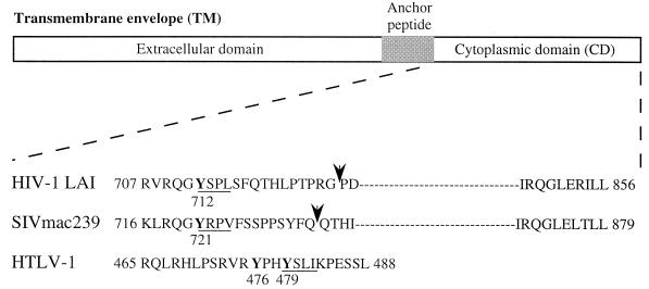
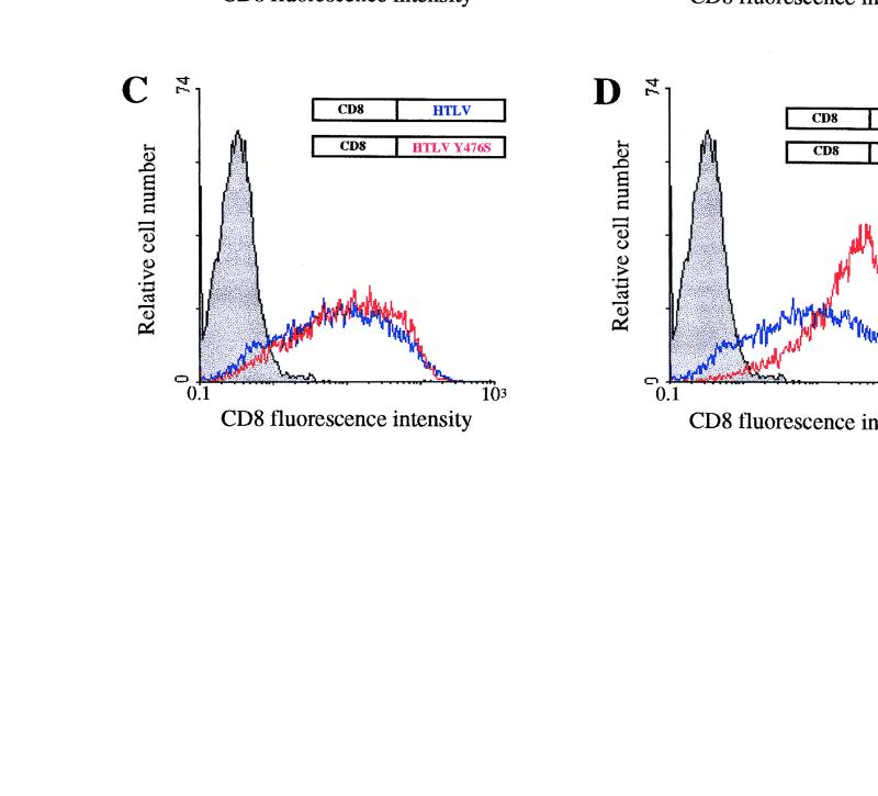
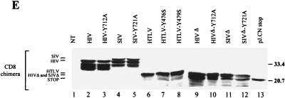
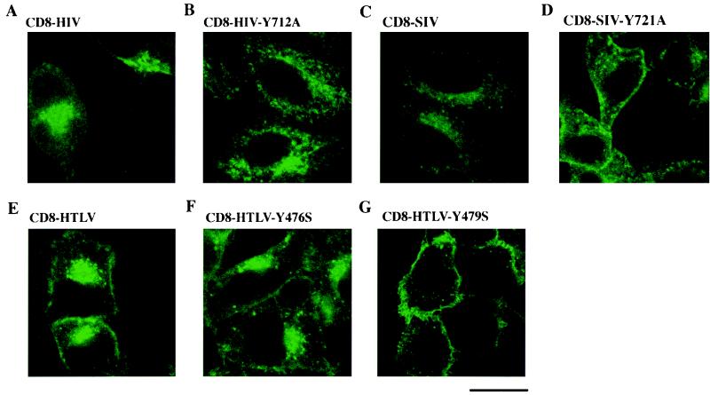
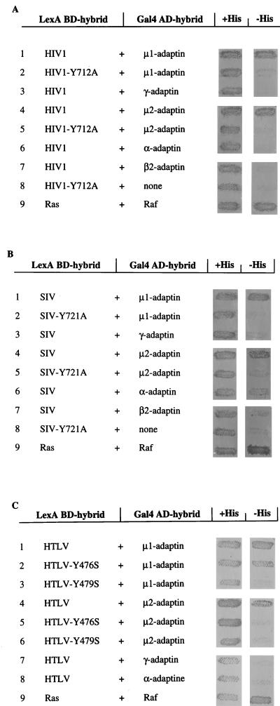
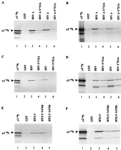
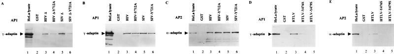
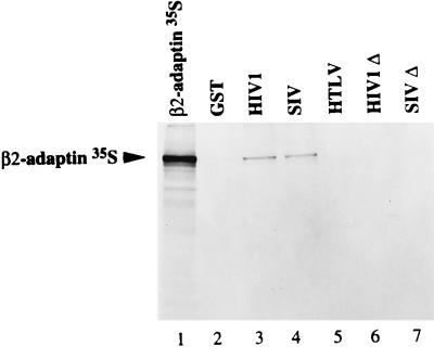
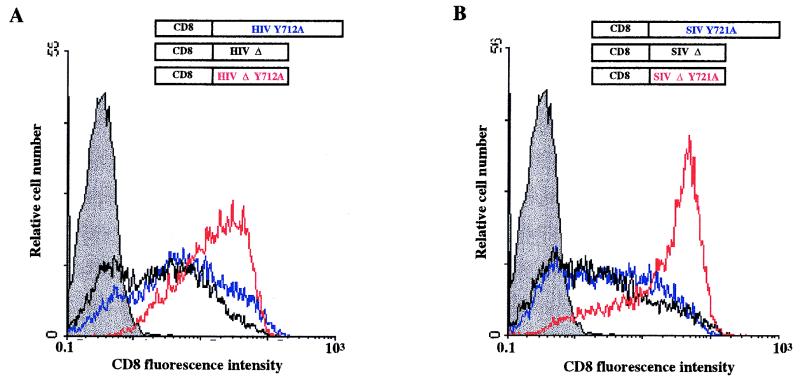
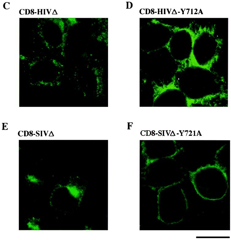
The cytoplasmic domains of the transmembrane (TM) envelope proteins (TM-CDs) of most retroviruses have a Tyr-based motif, YXXO, in their membrane-proximal regions. This signal is involved in the trafficking and endocytosis of membrane receptors via clathrin-associated AP-1 and AP-2 adaptor complexes. We have used CD8-TM-CD chimeras to investigate the role of the Tyr-based motif of human immunodeficiency virus type 1 (HIV-1), simian immunodeficiency virus (SIV), and human T-leukemia virus type 1 (HTLV-1) TM-CDs in the cell surface expression of the envelope glycoprotein. Flow cytometry and confocal microscopy studies showed that this motif is a major determinant of the cell surface expression of the CD8-HTLV chimera. The YXXO motif also plays a key role in subcellular distribution of the envelope of lentiviruses HIV-1 and SIV. However, these viruses, which encode TM proteins with a long cytoplasmic domain, have additional determinants distal to the YXXO motif that participate in regulating cell surface expression. We have also used the yeast two-hybrid system and in vitro binding assays to demonstrate that all three retroviral YXXO motifs interact with the micro1 and micro2 subunits of AP complexes and that the C-terminal regions of HIV-1 and SIV TM proteins interact with the beta2 adaptin subunit. The TM-CDs of HTLV-1, HIV-1, and SIV also interact with the whole AP complexes. These results clearly demonstrate that the cell surface expression of retroviral envelope glycoproteins is governed by interactions with adaptor complexes. The YXXO-based signal is the major determinant of this interaction for the HTLV-1 TM, which contains a short cytoplasmic domain, whereas the lentiviruses HIV-1 and SIV have additional determinants distal to this signal that are also involved.


Figures

FIG. 1
FIG. 1
Structures of retroviral TM proteins. The external domain, anchor peptide, and cytoplasmic domain are indicated. Numbering corresponds to the position of the beginning and the end of the cytoplasmic domains. Arrows indicate the position of the truncation corresponding to the membrane-proximal region of HIV-1 LAI and SIVmac239 TM-CD used in two-hybrid and in vitro binding assays (HIV-1 707-726 and SIV 716-733). Canonical YXXØ motifs are underlined and mutated tyrosine residues are in boldface.
FIG. 2
FIG. 2
Cell surface expression of CD8-TM-CD chimeras in HeLa cells. (A to D) HeLa cells were cotransfected by electroporation with 10 μg of the pCD8-HIV (A), pCD8-SIV (B), or pCD8-HTLV (C and D) (blue curves) or 10 μg of the pCD8-HIV-Y712A (A), pCD8-SIV-Y721A (B), pCD8-HTLV-Y476S (C), or pCD8-HTLV-Y479S (D) vector (red curves) along with 4 μg of pRSV-GFP vector. Surface expression of CD8 hybrids in GFP+ cells was analyzed by flow cytometry 48 h later. Grey curves, cells transfected with the pRSV-GFP vector alone, as the negative control; black curve, cells transfected with the pJ.CNstop vector, as the positive control (A). Data are representative of three independent experiments. (E) CD8-TM-CD chimera expression in HeLa cells. Cells were transfected with 10 μg of pCD8-HIV (lane 2), pCD8-HIV-Y712A (lane 3), pCD8-SIV (lane 4), pCD8-SIV-Y721A (lane 5), pCD8-HTLV (lane 6), pCD8-HTLV-Y476S (lane 7), pCD8-HTLV-Y479S (lane 8), pCD8-HIVΔ (lane 9), pCD8-HIVΔ-Y712A (lane 10), pCD8-SIVΔ (lane 11), pCD8-SIVΔ-Y721A (lane 12), and pJ.CN stop (lane 13) vectors. Identical quantities of lysates from transfected HeLa cells were analyzed by Western blotting with anti-CD8 antibody H-160 (Santa Cruz Biotechnology). NT, nontransfected control cells. Sizes are indicated in kilodaltons on the right.
FIG. 2
FIG. 2
Cell surface expression of CD8-TM-CD chimeras in HeLa cells. (A to D) HeLa cells were cotransfected by electroporation with 10 μg of the pCD8-HIV (A), pCD8-SIV (B), or pCD8-HTLV (C and D) (blue curves) or 10 μg of the pCD8-HIV-Y712A (A), pCD8-SIV-Y721A (B), pCD8-HTLV-Y476S (C), or pCD8-HTLV-Y479S (D) vector (red curves) along with 4 μg of pRSV-GFP vector. Surface expression of CD8 hybrids in GFP+ cells was analyzed by flow cytometry 48 h later. Grey curves, cells transfected with the pRSV-GFP vector alone, as the negative control; black curve, cells transfected with the pJ.CNstop vector, as the positive control (A). Data are representative of three independent experiments. (E) CD8-TM-CD chimera expression in HeLa cells. Cells were transfected with 10 μg of pCD8-HIV (lane 2), pCD8-HIV-Y712A (lane 3), pCD8-SIV (lane 4), pCD8-SIV-Y721A (lane 5), pCD8-HTLV (lane 6), pCD8-HTLV-Y476S (lane 7), pCD8-HTLV-Y479S (lane 8), pCD8-HIVΔ (lane 9), pCD8-HIVΔ-Y712A (lane 10), pCD8-SIVΔ (lane 11), pCD8-SIVΔ-Y721A (lane 12), and pJ.CN stop (lane 13) vectors. Identical quantities of lysates from transfected HeLa cells were analyzed by Western blotting with anti-CD8 antibody H-160 (Santa Cruz Biotechnology). NT, nontransfected control cells. Sizes are indicated in kilodaltons on the right.
FIG. 3
FIG. 3
Localization of CD8-TM-CD hybrids in HeLa cells. HeLa cells were transfected with 10 μg of the pCD8-HIV (A), pCD8-HIV-Y712A (B), pCD8-SIV (C), pCD8-SIV-Y721A (D), pCD8-HTLV (E), pCD8-HTLV-Y476S (F), and pCD8-HTLV-Y479S (G) vectors; 48 h later, the cells were fixed, permeabilized, and stained with an anti-CD8-FITC antibody. The distribution of CD8 hybrids was examined by immunofluorescence staining and confocal microscopy analysis. A representative medial section is shown. Scale bar, 20 μm. Data are representative of three independent experiments.
FIG. 4
FIG. 4
Interaction of the HIV-1, SIV, and HTLV-1 TM-CD Tyr-based motifs with μ1 and μ2 subunits in the yeast two-hybrid system. The yeast reporter strain L40 was cotransformed with plasmids encoding various Gal4 AD-adaptin hybrids and plasmids encoding the LexA BD fused to TM-CD707-726 of HIV-1 LAI (A), TM-CD716-733 of SIVmac239 (B), or complete TM-CD of HTLV-1 (C). Cotransformants were analyzed for histidine auxotrophy. They were patched on medium with histidine (+His) and then replica plated on medium without histidine (−His). Growth in the absence of histidine indicates interaction between hybrid proteins. The positive control was the interaction between Ras and Raf proteins, which bind to each other efficiently (lanes 9). Binding specificity was verified by the absence of interaction between the retroviral tyrosine-based motifs TM-CD and the Gal4 AD alone (A and B, lanes 8).
FIG. 5
FIG. 5
Interaction of the Tyr-based motifs or full-length TM-CDs of HIV-1, SIV, and HTLV-1 with in vitro-translated μ1 and μ2. μ1 (A, C, and E) and μ2 (B, D, and F) were translated in vitro in rabbit reticulocyte lysate and incubated with identical quantities of GST (lanes 2), GST-HIVΔ (A and B, lanes 3), GST-HIVΔ-Y712A (A and B, lanes 4), GST-SIVΔ (A and B, lanes 5), GST-SIVΔ-Y721A (A and B, lanes 6), GST-HIV (C and D, lanes 3), GST-HIV-Y712A (C and D, lanes 4), GST-SIV (C and D, lanes 5), GST-SIV-Y721A (C and D, lanes 6), GST-HTLV (E and F, lanes 3), GST-HTLV-Y476S (E and F, lanes 4), and GST-HTLV-Y479S (E and F, lanes 5). Bound labeled material was analyzed by SDS-PAGE and autoradiography. One-fifth of the input of μ1 and μ2 in vitro-translated products used for the binding assay was run on lane 1 of each panel.
FIG. 6
FIG. 6
Interaction of HIV-1, SIV, and HTLV-1 TM-CDs with the AP-1 and AP-2 complexes. Identical quantities of GST (lanes 2), GST-HIVΔ (A, lane 3), GST-HIVΔ-Y712A (A, lane 4), GST-SIVΔ (A, lane 5), GST-SIVΔ-Y721A (A, lane 6), GST-HIV (B and C, lanes 3), GST-HIV-Y712A (B and C, lanes 4), GST-SIV (B and C, lanes 5), GST-SIV-Y721A (B and C, lanes 6), GST-HTLV (D and E, lanes 3), GST-HTLV-Y476S (D and E, lanes 4), and GST-HTLV-Y479S (D and E, lanes 5), were incubated with HeLa cell lysates (25 × 106 cells). The binding of AP-1 and AP-2 complexes to GST fusion proteins was revealed by Western blotting with anti-γ adaptin MAb (A, B, and D) and anti-α adaptin MAb (C and E). Positions of the α-adaptin (Mr, ∼100,000) and γ-adaptin MAb (C and E). Positions of the α-adaptin (Mr, ∼100,000) and γ-adaptin (Mr, ∼ 104,000) are indicated in the crude cell lysate from 106 cells (lanes 1).
FIG. 7
FIG. 7
Interaction of full-length HIV-1, SIV, and HTLV-1 TM-CDs with in vitro-translated β2-adaptin. β2 subunit was translated in vitro in rabbit reticulocyte lysate and incubated with identical quantities of GST (lane 2), GST-HIV TM-CD (lane 3), GST-SIV TM-CD (lane 4), GST-HTLV TM-CD (lane 5), GST-HIVΔ TM-CD (lane 6), and GST-SIVΔ TM-CD (lane 7). Bound labeled material was analyzed by SDS-PAGE and autoradiography. One-fifth of the input of β2 in vitro-translated product used for the binding assay was run on lane 1.
FIG. 8
FIG. 8
Cell surface and intracellular distribution of deleted and mutated CD8-HIVΔ-Y712A and CD8-SIVΔ-Y721A chimeras in HeLa cells. (A and B) HeLa cells were cotransfected by electroporation with 10 μg of the pCD8-HIV-Y712A (blue curve), pCD8-HIVΔ-Y712A (red curve), or pCD8-HIVΔ (black curve) vector (A) or the pCD8-SIV (blue curve), pCD8-SIVΔ-Y721A (red curve), or pCD8-SIVΔ (black curve) (B) vector along with 4 μg of pRSV-GFP vector; 48 h later, the surface expression of CD8 hybrid in GFP+ cells was analyzed by flow cytometry. Grey curves represent cells transfected with the pRSV-GFP vector alone, as the negative control. Data are representative of three independent experiments. (C to F) HeLa cells were transfected with 10 μg of the pCD8-HIVΔ (C), pCD8-HIVΔ-Y712A (D), pCD8-SIVΔ (E), or pCD8-SIVΔ-Y721A (F) vector; 48 h later, cells were fixed, permeabilized, and stained with an anti-CD8-FITC antibody. The distribution of CD8 hybrids was examined by immunofluorescence staining and confocal microscopy analysis. A representative medial section is shown. Scale bar, 20 μm. Data are representative of three independent experiments.
FIG. 8
FIG. 8
Cell surface and intracellular distribution of deleted and mutated CD8-HIVΔ-Y712A and CD8-SIVΔ-Y721A chimeras in HeLa cells. (A and B) HeLa cells were cotransfected by electroporation with 10 μg of the pCD8-HIV-Y712A (blue curve), pCD8-HIVΔ-Y712A (red curve), or pCD8-HIVΔ (black curve) vector (A) or the pCD8-SIV (blue curve), pCD8-SIVΔ-Y721A (red curve), or pCD8-SIVΔ (black curve) (B) vector along with 4 μg of pRSV-GFP vector; 48 h later, the surface expression of CD8 hybrid in GFP+ cells was analyzed by flow cytometry. Grey curves represent cells transfected with the pRSV-GFP vector alone, as the negative control. Data are representative of three independent experiments. (C to F) HeLa cells were transfected with 10 μg of the pCD8-HIVΔ (C), pCD8-HIVΔ-Y712A (D), pCD8-SIVΔ (E), or pCD8-SIVΔ-Y721A (F) vector; 48 h later, cells were fixed, permeabilized, and stained with an anti-CD8-FITC antibody. The distribution of CD8 hybrids was examined by immunofluorescence staining and confocal microscopy analysis. A representative medial section is shown. Scale bar, 20 μm. Data are representative of three independent experiments.

References

    1. Aiken C, Konner J, Landau N R, Lenburg M E, Trono D. Nef induces CD4 endocytosis: requirement for a critical dileucine motif in the membrane-proximal CD4 cytoplasmic domain. Cell. 1988;76:853–864. - PubMed
    1. Alconada A, Bauer U, Holflack B. A tyrosine-based motif and a casein kinase II phosphorylation site regulate the intracellular trafficking of the varicella-zoster virus glycoprotein I, a protein localized in the trans-golgi network. EMBO J. 1996;15:6096–6110. - PMC - PubMed
    1. Beltzer J P, Spiess M. In vitro binding of the asialoglycoprotein receptor to the β adaptin of plasma membrane coated vesicles. EMBO J. 1991;10:3735–3742. - PMC - PubMed
    1. Benichou S, Bomsel M, Bodeus M, Durand H, Doute M, Letourneur F, Camonis J, Benarous R. Physical interaction between the HIV-1 Nef protein and β-COP, an essential component for membrane traffic. J Biol Chem. 1994;269:30073–30076. - PubMed
    1. Benmerah A, Begue B, Dautry-Varsat A, Cerf-Bensussan N. The ear of α-adaptin interacts with the COOH-terminal domain of the Eps15 protein. J Biol Chem. 1996;271:12111–12116. - PubMed

Publication types

MeSH terms

Substances