. 1999 Feb;73(2):1382-91.
doi: 10.1128/JVI.73.2.1382-1391.1999.

RNA-binding and capping activities of proteins in rotavirus open cores

Affiliations

RNA-binding and capping activities of proteins in rotavirus open cores

J T Patton et al. J Virol. 1999 Feb.

Abstract

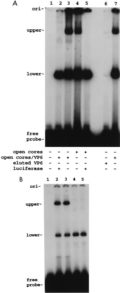
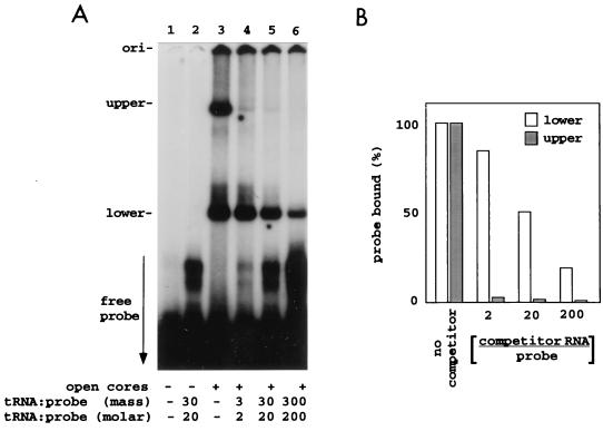
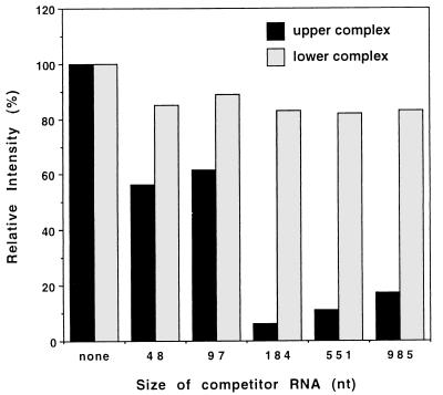
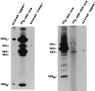
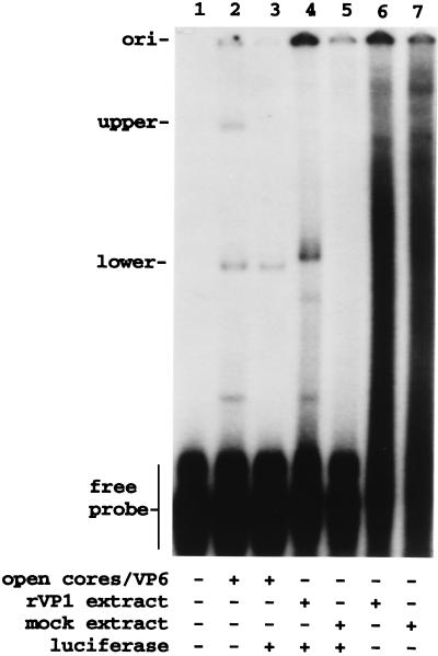
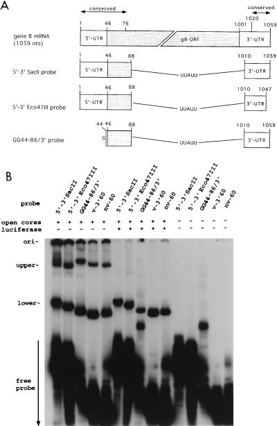
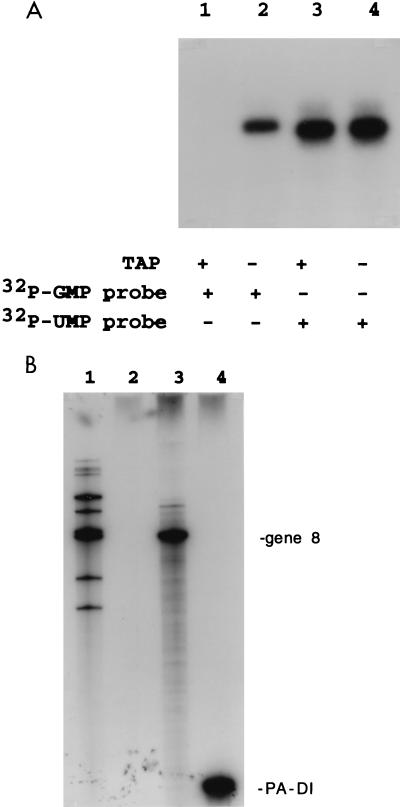
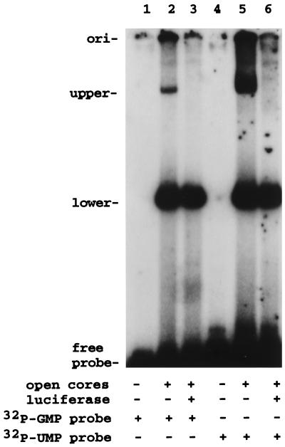
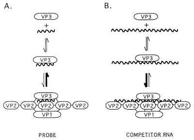
Guanylyltransferases are members of the nucleotidyltransferase family and function in mRNA capping by transferring GMP to the phosphate end of nascent RNAs. Although numerous guanylyltransferases have been identified, studies which define the nature of the interaction between the capping enzymes of any origin and their RNA substrates have been limited. Here, we have characterized the RNA-binding activity of VP3, a minor protein component of the core of rotavirions that has been proposed to function as the viral guanylyltransferase and to direct the capping of the 11 transcripts synthesized from the segmented double-stranded RNA (dsRNA) genome of these viruses. Gel shift analysis performed with disrupted (open) virion-derived cores and virus-specific RNA probes showed that VP3 has affinity for single-stranded RNA (ssRNA) but not for dsRNA. While the ssRNA-binding activity of VP3 was found to be sequence independent, the protein does exhibit preferential affinity for uncapped over capped RNA. Like the RNA-binding activity, RNA capping assays performed with open cores indicates that the guanylyltransferase activity of VP3 is nonspecific and is able to cap RNAs initiating with a G or an A residue. These data establish that all three rotavirus core proteins, VP1, the RNA polymerase; VP2, the core capsid protein; and VP3, the guanylyltransferase, have affinity for RNA but that only in the case of the RNA polymerase is the affinity sequence specific.


Figures

FIG. 1
FIG. 1
Protein composition of open cores and VP6-open cores. Samples of open cores (lanes 2 and 3), VP6-open cores (lane 4), and double-shelled particles (lane 5), each containing 5 μg of VP2, were analyzed by SDS–12% PAGE followed by staining with Coomassie blue. VP6 (4 μg) recovered from CaCl2-treated double-shelled particles was resolved in lane 1. The cores that were resolved in lane 2 were incubated with [α-32P]GTP prior to electrophoresis. VP3-GMP complexes that formed in this sample were identified by autoradiography (lane 6) and were used to confirm the position of VP3 in the gel.
FIG. 2
FIG. 2
Replicase activity of open cores and VP6-open cores. The products of replicase assays containing open cores (oc) or VP6-open cores (oc/VP6) and no added mRNA (none) or 5 μg of gene 8 or 6 mRNA were resolved by SDS–12% PAGE and detected by autoradiography. Background bands corresponding to the 11 genome segments in core preparations are routinely seen in assays performed with no exogenous RNA and are the products of an undefined activity (4).
FIG. 3
FIG. 3
RNA-protein complexes formed by open cores and VP6-open cores. (A) 32P-labeled SP72-v3′40 (7.5 pmol, 0.17 μg) was incubated alone or with open cores or VP6-open cores, each containing 3.3 μg of VP2, or with 2 μg of VP6 eluted from double-shelled particles with CaCl2. Some reaction mixtures also included luciferase RNA (1.7 pmol, 1.0 μg). (B) 32P-labeled SP72-v3′40 (7.5 pmol, 0.17 μg) was incubated alone (lane 1) or with VP6-open cores containing 3.3 μg of VP2 in the absence of competitor RNA for 60 min (lane 2) or 30 min (lane 3) or in the presence of 1.7 μg of luciferase RNA for 60 min (lane 5). Lane 4 shows complexes formed when 32P-labeled SP72-v3′40 was preincubated with the VP6-open cores for 30 min and then, after addition of 1.7 μg of luciferase RNA, the incubation was continued for another 30 min. Probe-protein complexes were detected by electrophoresis on 8% polyacrylamide gels and by autoradiography. ori, origin.
FIG. 4
FIG. 4
Effect of competitor RNA on the interaction of core proteins with a viral 3′-specific RNA probe. 32P-labeled SP72-v3′40 (4.4 pmol, 0.1 μg) was incubated with open cores and with the indicated amount of rabbit liver tRNA. The ratio of tRNA to probe in the reaction mixtures is expressed in terms of mass and moles. (A) Probe-protein complexes were resolved by electrophoresis, and the lower and upper complexes were detected by autoradiography. (B) The intensity of the upper and lower bands was determined with a phosphorimager, and the values were adjusted relative to 100% [probe bound (%)] for the reaction mixture containing no competitor RNA. The percentage of probe bound was plotted versus the molar ratio of tRNA to probe in the reaction mixtures. ori, origin.
FIG. 5
FIG. 5
Size of the competitor RNA and its impact on formation of the upper protein-probe complex. 32P-labeled SP72-v3′40 (50 ng) was incubated with open cores and no competitor RNA or 50 ng of cold competitor RNAs of the indicated size. The competitor RNAs were produced by runoff transcription of linearized SP72. Upper and lower protein-probe complexes were resolved by electrophoresis, and the band intensities of the complexes detected on the gel were determined with a phosphorimager. The values were adjusted relative to 100% for the assay which contained probe and open cores but no competitor RNA.
FIG. 6
FIG. 6
Protein composition of the upper and lower probe-protein complexes. 32P-labeled SP72-v3′40 was incubated with 35S-labeled VP6-open cores (ocs) (left) or open cores (right), and the mixtures were resolved by electrophoresis on a nondenaturing 8% polyacrylamide gel. The positions of upper and lower complexes in the gel were identified by autoradiography, and portions of the gel containing the complexes were cut out, soaked in sample buffer, and loaded onto SDS–12% polyacrylamide gels. After electrophoresis, the 35S-labeled proteins were detected by fluorography (shown). The position of proteins in the gel was determined by coelectrophoresis of 35S-labeled VP6-open cores. To confirm the position of VP3, VP3-[32P]GMP complexes were formed by coincubation of cold VP6-open cores and [α-32P]GTP prior to electrophoresis.
FIG. 7
FIG. 7
Recombinant VP1 interacts with the viral 3′-specific RNA probe to form the lower complex. 32P-labeled SP72-v3′40 (50 ng) was incubated alone or with either a preparation of open cores, an extract from rBVg1-infected Sf9 cells containing rVP1, or an extract from mock-infected Sf9 cells. Luciferase RNA (0.5 μg) was included in some reaction mixtures. Probe-protein complexes were resolved by electrophoresis and detected by autoradiography. ori, origin.
FIG. 8
FIG. 8
VP3 lacks specificity for the 5′-terminal sequences of viral mRNA. (A) The locations of sequences in RNA probes with respect to the wild-type gene 8 mRNA are indicated. The nonviral sequence UUAUU was used to link the 5′ and 3′ gene 8-specific sequences of the probes. ORF, open reading frame. (B) Five picomoles of the RNA probes, g8 5′-3′SacII, g8 5′-3′Eco47III, g8 GG44-86/3′, v-3′60, and nv-60, was incubated alone or with 3.3 μg of VP6-open cores in the presence or absence of 1.7 pmol (1 μg) of luciferase RNA. The upper and lower probe-protein complexes were resolved by electrophoresis and detected by autoradiography. ori, origin.
FIG. 9
FIG. 9
VP3 lacks affinity for the dsRNA genome. 32P-labeled SP72-v3′40 (50 ng) was incubated alone or with open cores in the absence or presence of either virion-derived dsRNA or luciferase RNA. The reaction mixtures were analyzed by electrophoresis and autoradiography. A phosphorimager was used to quantitate the levels of VP3- and VP1-probe complexes, and the values were adjusted relative to 100% for the reaction mixture containing probe and open cores but no competitor RNA (lane 2). ori, origin.
FIG. 10
FIG. 10
The guanylyltransferase activity of VP3 is nonspecific. SP72-v3′40, wild-type gene 8 mRNA, and PA-DI RNA were incubated with [32P]GTP and open cores in a capping assay. (A) SP72-v3′40 RNA ([32P]GMP probe) recovered from the reaction was coelectrophoresed on an SDS–14% polyacrylamide gel (lane 2) with 32P-labeled SP72-v3′40 RNA ([32P]UMP probe) (lane 4) synthesized by runoff transcription in the presence of [32P]UTP (lane 4). Portions of the [32P]GMP and [32P]UMP probes were treated with tobacco acid pyrophosphatase (TAP) prior to electrophoresis (lanes 1 and 3). (B) The gene 8 (lane 3) and PA-DI (lane 4) RNAs were recovered from capping assays by phenol-chloroform extraction, resolved by electrophoresis on a polyacrylamide-urea gel, and detected by autoradiography. 32P-labeled mRNAs made by transcriptionally active double-shelled particles served as markers on the gel (lane 1) (6). Lane 2 shows the products of a capping assay performed in the absence of added RNA.
FIG. 11
FIG. 11
VP3 preferentially binds uncapped RNA. Capped radiolabeled probe was made by incubating SP72-v3′40 with [32P]GTP and open cores ([32P]GMP probe). Noncapped radiolabeled probe was made by runoff transcription of SP72-v3′40 with [32P]UTP ([32P]UMP probe). Purified probes (0.12 μg) were incubated with 3.3 μg of VP6-open cores in the presence or absence of 1 μg of luciferase RNA. Probe-protein complexes were detected by electrophoresis and autoradiography. ori, origin.
FIG. 12
FIG. 12
Model for the effect that RNA size has on the formation of VP3-probe complexes.

References

    1. Bican P, Cohen J, Charpilienne A, Scherrer R. Purification and characterization of bovine rotavirus cores. J Virol. 1982;43:1113–1117. - PMC - PubMed
    1. Boyle J F, Holmes K V. RNA-binding proteins of bovine rotavirus. J Virol. 1986;51:561–568. - PMC - PubMed
    1. Chen D, Patton J T. Rotavirus RNA replication requires a single-stranded 3′ terminus for efficient minus-strand synthesis. J Virol. 1998;72:7387–7396. - PMC - PubMed
    1. Chen, D., and J. T. Patton. Unpublished data.
    1. Chen D Y, Zeng C Q-Y, Wentz M J, Gorziglia M, Estes M K, Ramig R F. Template-dependent, in vitro replication of rotavirus RNA. J Virol. 1994;68:7030–7039. - PMC - PubMed

LinkOut - more resources