. 1999 Feb;73(2):1438-46.
doi: 10.1128/JVI.73.2.1438-1446.1999.

Characterization and cell cycle regulation of the major Kaposi's sarcoma-associated herpesvirus (human herpesvirus 8) latent genes and their promoter

Affiliations

Characterization and cell cycle regulation of the major Kaposi's sarcoma-associated herpesvirus (human herpesvirus 8) latent genes and their promoter

R Sarid et al. J Virol. 1999 Feb.

Abstract

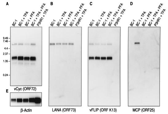
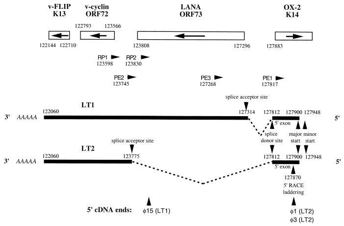
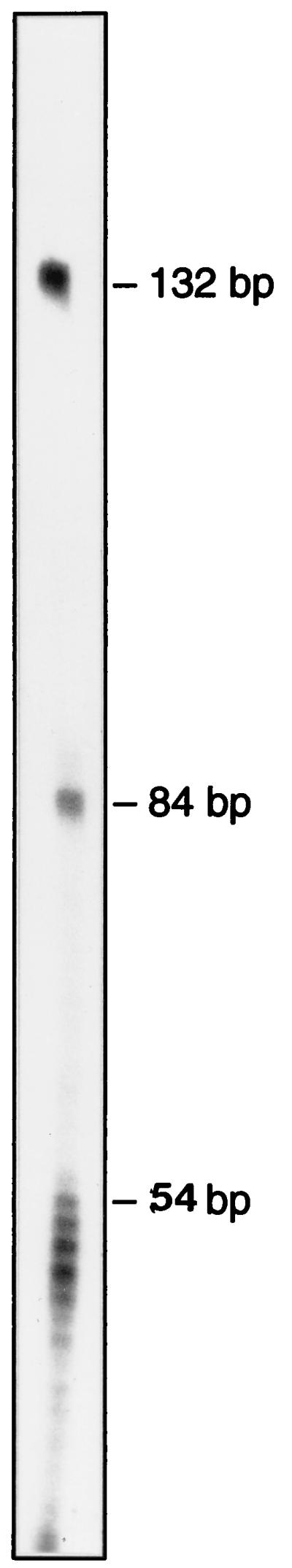
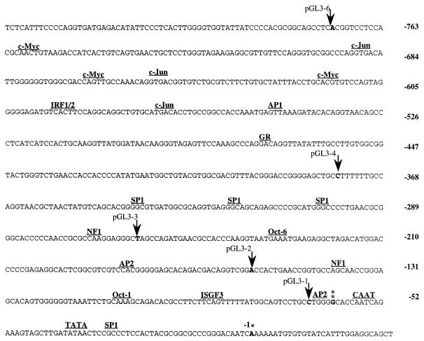
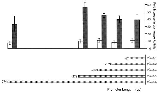
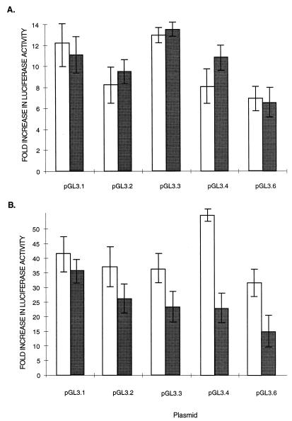
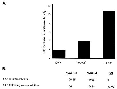
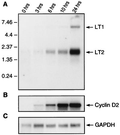
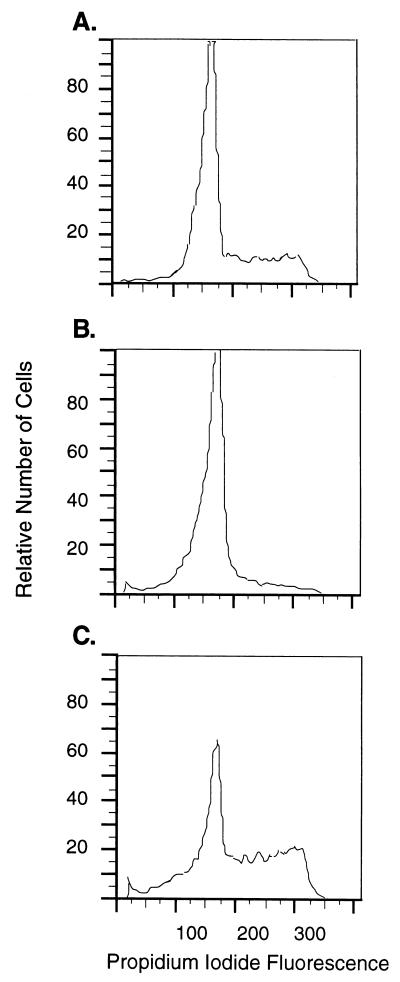
Retinoblastoma tumor suppressor protein (pRB) inhibition by tumor virus oncoproteins has been attributed to the need for these viruses to promote lytic viral nucleic acid synthesis by unscheduled entry into the S phase of the cell cycle. Kaposi's sarcoma-associated herpesvirus (KSHV or HHV8) encodes a functional cyclin (vCYC) which is expressed during latency and can direct phosphorylation of pRB. We mapped the two major latent transcripts encoding vCYC, latent transcript 1 (LT1) and LT2, by cDNA sequencing, 5' rapid amplification of cDNA ends, and primer extension analyses. Both LT1 and LT2 transcripts are spliced, originate from the same start site, and encode ORF K13 (vFLIP) as well as ORF72 (vCYC). The latency-associated nuclear antigen (LANA, ORF73) is encoded by LT1 but spliced from LT2. While differential expression of the two transcripts was not found, the promoter controlling LT1/LT2 transcription is regulated in a cell cycle-dependent manner. Activities of both KSHV LT1/LT2 and huCYC D1 luciferase promoter reporters transfected into NIH 3T3 cells increase 11- and 4-fold, respectively, after release from cell cycle arrest by serum starvation. Further, vCYC and huCYC D2 mRNA levels are low in naturally infected BCBL-1 cells arrested in late G1 with L-mimosine but increase in parallel during a 24-h period after release from cell cycle arrest. Cell cycle regulation of KSHV vCYC expression mimics cellular D cyclin regulation and may maintain infected cell cycling. This is consistent with an alternative hypothesis that tumor viruses have developed specific responses to innate cellular defenses against latent virus infection that include pRB-induced cell cycle arrest.


Figures

FIG. 1
FIG. 1
Northern hybridization of BC-1 mRNA with ORF72 (A), ORF73 (B), ORF K13 (C), and ORF 25 (D) probes. Probe hybridizations for mRNA from uninduced BC-1 cells (lane 1), BC-1 cells treated with 20-ng/ml TPA for 48 h (lane 2), BC-1 cells treated with 0.5 mM PFA (lane 3), BC-1 cells treated with both 20-ng/ml TPA and 0.5 mM PFA (lane 4), and KSHV-negative, EBV-infected P3HR1 cells treated with 20-ng/ml TPA (lane 5) are shown. ORF72 (vCYC) (panel A) and ORF K13 (vFLIP) (panel C) probes hybridize to both the 6.0-kb LT1 and the 2.0-kb LT2 bands while only the 6.0-kb LT1 band hybridizes with the ORF73 probe (panel B). A low-abundance 1-kb band is detected with the ORF72 probe alone (panel A). LT1 and LT2 bands are not induced by TPA and are not inhibited by PFA treatment, consistent with their designation as class I or latent viral transcripts (40). P3HR1 mRNA does not cross-hybridize to any of the KSHV probes. (E) The same as the blot shown in panel A except it has been stripped and reprobed with a β-actin probe to control for equal loading.
FIG. 2
FIG. 2
Depiction of the transcript mapping results for LT1 and LT2 based on cDNA sequencing, 5′ RACE, and primer extension studies. The arrangement and orientation of genes for this region of the KSHV BC-1 genome are shown with nucleotide positions designated by Russo et al. (39). Black bars represent the LT1 and LT2 transcripts, introns are represented as dashed lines, and nucleotide positions for polyadenylation sites, splice junctions, and start sites are indicated. The positions of 5′ truncation for cDNA phages φ1, φ3, and φ15 are shown. Primer positions and orientations used for 5′ RACE (RP1, RP2, and RP3) and primer extension (PE1, PE2, and PE3) analyses are shown.
FIG. 3
FIG. 3
Primer extension results obtained with the PE1 primer. Two start sites are present at 84 bp (nt 127,900) and 132 bp (nt 127,948) from the PE1 primer. A laddering pattern consistent with premature reverse transcription termination is present at 47 to 54 bp from the PE1 primer (nt 127,863 to 127,870), corresponding to the start sites determined by cDNA sequencing and 5′ RACE analyses.
FIG. 4
FIG. 4
Nucleotide sequence of the latent promoter region (LP1/2) for LT1-2. The first nucleotide of the presumed major transcription start site at nt 127,900 is marked by a single asterisk and is indicated in boldface. The second start site is marked by a double asterisk, and potential transcription factor binding sites in the promoter region are indicated. The locations of promoter deletions used for reporter studies indicated by arrows.
FIG. 5
FIG. 5
Luciferase expression levels for LP1/2-luciferase reporter constructs in HeLa (white bars) and BJAB (grey bars) cells. HeLa cells were transfected with 1 mg of reporter gene and 1 mg of control plasmid pcDNA3.1/lacZ. BJAB cells were transfected with 10 mg of reporter gene and 10 μg of control plasmid pcDNA3.1/lacZ. Mean promoter activities obtained from three transfections after normalization for β-galactosidase expression are indicated relative to the pGL3-luc basic reporter plasmid lacking a promoter sequence.
FIG. 6
FIG. 6
Effect of TPA treatment on LP1/2-luciferase promoter reporter constructs. HeLa (A) and BJAB (B) cells cultured in the presence (grey bars) or the absence (white bars) of 20-ng/ml TPA were transfected with LP1/2-luciferase constructs as described in the legend for Fig. 4 and assayed for luciferase activity. No evidence for TPA inducibility was found for either cell type.
FIG. 7
FIG. 7
(A) Cell cycle-dependent induction of the pGL3.6 LP1/2-luciferase reporter. NIH 3T3 cells were transfected with the indicated constructs and cultured in low serum (0.1% FCS) for 60 h. Bars indicate the levels of induction of luciferase activity 14 h after readdition of 20% FCS compared to pretreatment luciferase activity for the constitutively active CMV, the human cyclin D1, and the KSHV LP1/2 promoters. (B) Cell cycle distributions as determined by FACS analyses at 0 and 14 h after serum readdition. Serum starvation for 60 h arrested 90% of NIH 3T3 cells in G0/G1, whereas 32% of cells were entering S phase 14 h after serum stimulation.
FIG. 8
FIG. 8
Time course of KSHV vCYC (A) and cellular huCYC D2 (B) mRNA expression in BCBL-1 cells after release from l-mimosine G1 arrest. GAPDH mRNA expression (C) was used as a control for equal mRNA loading. Cells were arrested for 20 h by treatment with 200 mM l-mimosine and released from cell cycle arrest by washing in fresh medium. Cell cultures were harvested at 0, 3, 6, 10, and 24 h after l-mimosine washout and prepared for mRNA extraction. Expression of KSHV LT1 and LT2 mRNA mirrors expression of cellular CYC D2, which is expressed early in G1 phase of the cell cycle.
FIG. 9
FIG. 9
FACS analysis demonstrating that BCBL-1 cells are reversibly arrested by l-mimosine treatment. (A) Cell cycle distribution of exponentially growing BCBL-1 prior to l-mimosine arrest. (B) G1 arrest of BCBL-1 cells at 0 h as shown in Fig. 8, after treatment with 200 nM l-mimosine for 20 h. (C) Cell cycle progression of BCBL-1 cells 24 h after l-mimosine washout, as shown in Fig. 8.

References

    1. Albanese C, Johnson J, Watanabe G, Eklund N, Vu D, Arnold A, Pestell R G. Transforming p21ras mutants and c-Ets-2 activate the cyclin D1 promoter through distinguishable regions. J Biol Chem. 1995;270:23589–23597. - PubMed
    1. Bates S, Phillips A C, Clark P A, Stott F, Peters G, Ludwig R L, Vousden K H. p14ARF links the tumour suppressors RB and p53. Nature. 1998;395:124–125. - PubMed
    1. Bertin J, Armstrong R C, Ottilie S, Martin D A, Wang Y, Banks S, Wang G H, Senkevich T G, Alnemri E S, Moss B, Lenardo M J, Tomaselli K J, Cohen J I. Death effector domain-containing herpesvirus and poxvirus proteins inhibit both Fas- and TNFR1-induced apoptosis. Proc Natl Acad Sci USA. 1997;94:1172–1176. - PMC - PubMed
    1. Bodescot M, Perricaudet M. Epstein-Barr virus mRNAs produced by alternative splicing. Nucleic Acids Res. 1986;14:7103–7114. - PMC - PubMed
    1. Bulfone-Paus S, Dempsey L A, Maizels N. Host factors LR1 and Sp1 regulate the Fp promoter of Epstein-Barr virus. Proc Natl Acad Sci USA. 1995;92:8293–8297. - PMC - PubMed

Publication types