

Review
doi: 10.1128/JB.181.2.359-367.1999.
Processive antitermination
Affiliations
- PMID: 9882646
- PMCID: PMC93386
- DOI: 10.1128/JB.181.2.359-367.1999
Item in Clipboard
Review
Processive antitermination
J Bacteriol.
1999 Jan.
No abstract available
Figures
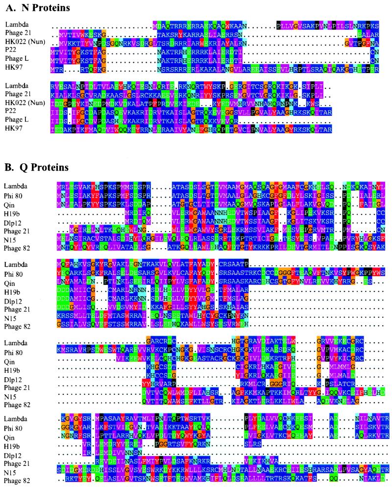
(A) Alignment of phage N proteins and the HK022 Nun protein. The color groupings reflect the frequency of amino acid substitutions in evolutionarily related protein domains: an amino acid is more likely to be replaced by one in the same color group than by one in a different color group in related proteins (34). The amino-proximal ARM regions were aligned by eye and according to the structures of the P22 and λ ARMs complexed to their cognate nut sites (see text and Fig. 2), and the remainder of the proteins was aligned by ClustalW (38). The dots indicate gaps introduced to improve the alignment. Aside from the ARM regions, the proteins fall into three very distantly related (or unrelated) families: (i) λ and phage 21; (ii) P22, phage L, and HK97; and (iii) HK022 Nun. The divergence of Nun from the N proteins is unsurprising because of their different functions. The sequence database was searched for additional N homologs with the PSI-BLAST program (3), using each of the listed sequences as a query, but none were found. Two N proteins were omitted from the alignment: that of phage H19b, because it differs by only three conservative substitutions from N of HK97 (E60D, K80E, and R100K) (3), and that of lambdoid phage φ80 (Phi 80), because it shows no resemblance to any of the other N proteins, lacking even an ARM (42, 69). (B) Alignment of phage Q proteins. The alignments were generated by ClustalW, and the database was searched for Q homologs as described above. These proteins fall into three very distantly related (or unrelated) families: (i) λ and Qin; (ii) H19b, Dlp12, and phage 21; and (iii) N15 and phage 82. Qin and Dlp12 are defective lambdoid prophages of E. coli, but it is likely that their Q proteins are active (see reference 16). The Q proteins of phages HK022 and P22 were omitted from the alignment because of their close similarity to that of λ. A putative and possibly defective Q, encoded by a sequence located upstream of Shiga-like toxin I genes in an E. coli isolate (72) and found by a BLAST search of the translated nucleotide sequence database, was omitted from the alignment because of its close similarity to the Q of phage H19B (61).
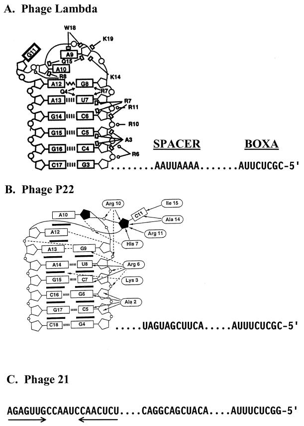
BOXA and BOXB RNAs and their interaction with the ARM of their cognate N proteins. The amino acid-nucleotide interactions are shown to the left except for BOXB of phage 21, for which the structure of the complex is unknown. The sequences of BOXA and BOXA-BOXB spacer are shown to the right. The dots to the left and right of the spacer sequences are for alignment. (A) λ N-ARM-BOXB complex (adapted from reference with permission of the publisher). Open circles, pentagons, and rectangles represent phosphates, riboses, and bases, respectively. Watson-Crick base pairs (||||) are indicated. The zigzag line denotes a sheared G · A base pair. Open circles, open rectangles, and arrowheads depict ionic, hydrophobic, and hydrogen-bonding interactions, respectively. Guanine-11, indicated by a bold rectangle, is extruded from the BOXB loop (see text). (B) P22 N-ARM-BOXB complex (adapted from reference with permission of the publisher). Open circles, pentagons, rectangles, and ovals represent phosphates, riboses, bases, and amino acids, respectively. The solid pentagons indicate riboses with a C2′-endo pucker. Base stacking ( ), intermolecular hydrogen bonding or electrostatic interactions (<-----), intermolecular hydrophobic or van der Waals interactions (←), intramolecular hydrogen bonds (––––) and Watson-Crick base pairs (|||||) are indicated. Cytosine-11 is extruded from the loop (see text). Note that the amino-terminal amino acid residue in the complex corresponds to Asn-14 in the complete protein (Fig. 1), and the displayed amino acids are numbered accordingly. (C) NUTL site of phage 21. The arrows indicate the inverted sequence repeats of BOXB.
), intermolecular hydrogen bonding or electrostatic interactions (<-----), intermolecular hydrophobic or van der Waals interactions (←), intramolecular hydrogen bonds (––––) and Watson-Crick base pairs (|||||) are indicated. Cytosine-11 is extruded from the loop (see text). Note that the amino-terminal amino acid residue in the complex corresponds to Asn-14 in the complete protein (Fig. 1), and the displayed amino acids are numbered accordingly. (C) NUTL site of phage 21. The arrows indicate the inverted sequence repeats of BOXB.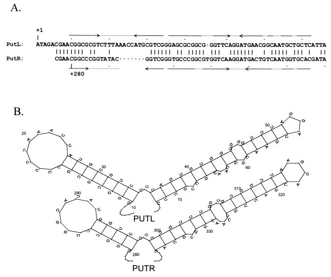
HK022 put sites and folded PUT RNAs. (A) Alignment of putL and putR (43). The numbers give distances from the start sites of the PL and PR promoters, respectively, and the pairs of arrows indicate inverted sequence repeats. (B) Folded PUTL and PUTR RNAs. The structures, which were generated by energy minimization as described (43), have been partially confirmed by genetic and biochemical studies (7, 43).
References
-
- Albrechtsen B, Squires C L, Li S, Squires C. Antitermination of characterized transcriptional terminators by the Escherichia coli rrnG leader region. J Mol Biol. 1990;213:123–134. - PubMed
-
- Atkinson B L, Gottesman M E. The Escherichia coli rpoB60 mutation blocks antitermination by coliphage HK022 Q-function. J Mol Biol. 1992;227:29–37. - PubMed
-
- Bailey M J, Hughes C, Koronakis V. Increased distal gene transcription by the elongation factor RfaH, a specialized homologue of NusG. Mol Microbiol. 1996;22:729–737. - PubMed
Publication types
MeSH terms
Substances
LinkOut - more resources
Full Text Sources
Other Literature Sources
Molecular Biology Databases
