. 1999 Jan;181(2):382-8.
doi: 10.1128/JB.181.2.382-388.1999.

Assembly of XcpR in the cytoplasmic membrane is required for extracellular protein secretion in Pseudomonas aeruginosa

Affiliations

Assembly of XcpR in the cytoplasmic membrane is required for extracellular protein secretion in Pseudomonas aeruginosa

G Ball et al. J Bacteriol. 1999 Jan.

Abstract

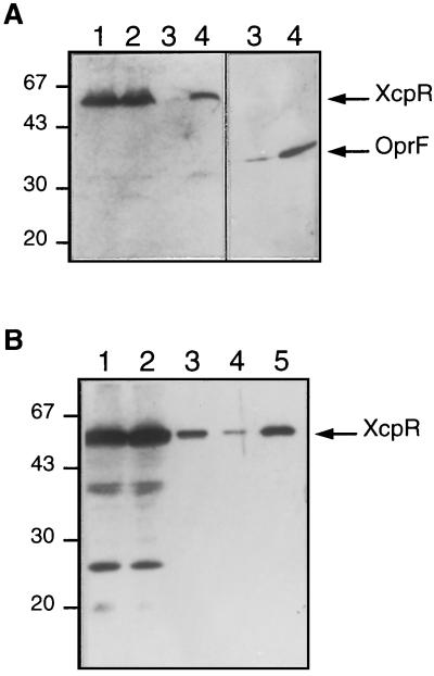
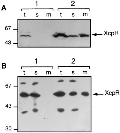
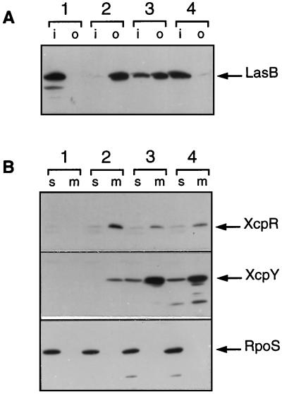
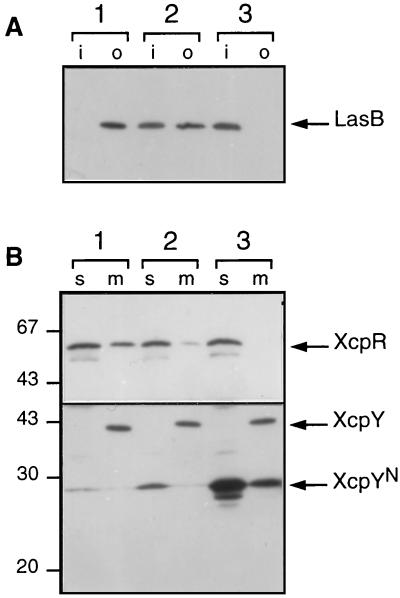
A broad range of extracellular proteins secreted by Pseudomonas aeruginosa use the type II or general secretory pathway (GSP) to reach the medium. This pathway requires the expression of at least 12 xcp gene products. XcpR, a putative nucleotide-binding protein, is essential for the secretion process across the outer membrane even though the protein contains no hydrophobic sequence that could target or anchor it to the bacterial envelope. For a better understanding of the relationship between XcpR and the other Xcp proteins which are located in the envelope, we have studied its subcellular localization. In a wild-type P. aeruginosa strain, XcpR was found associated with the cytoplasmic membrane. This association depends on the presence of the XcpY protein, which also appears to be necessary for XcpR stability. Functional complementation of an xcpY mutant required the XcpY protein to be expressed at a low level. Higher expression precluded the complementing activity of XcpY, although membrane association of XcpR was restored. This behavior suggested that an excess of free XcpY might interfere with the secretion by formation of inactive XcpR-XcpY complexes which cannot properly interact with their natural partners in the secretion machinery. These data show that a precise stoichiometric ratio between several components may be crucial for the functioning of the GSP.


Figures

FIG. 1
FIG. 1
Genetic organization and restriction map of the P. aeruginosa xcp region at 40 min. The 10.3-kb DNA fragment carrying the xcpP to -Z genes is shown by a double line; the 8.5-kb chromosomal sequence deleted in strain DZQ40 is shown by a dotted line; locations of plasmid subclones are indicated by bold lines. Restriction sites: A, AsuII; B, BamHI; Ba, BalI; E, EcoRI; N, NotI; S, SalI; Sc, ScaI; Sp, SphI; P, PstI. Only the relevant positions are indicated for the AsuII, PstI, and NotI restriction sites.
FIG. 2
FIG. 2
Subcellular localization of XcpR in P. aeruginosa. Soluble and membrane fractions were prepared as described in Materials and Methods. Proteins were separated by SDS-PAGE, transferred to nitrocellulose, and immunodetected with anti-XcpR serum. (A) Fractionation of wild-type PAO1 cells. Lanes: 1, total cells; 2, cell lysate; 3, soluble fraction; 4, membrane fraction. Immunodetection of the outer membrane protein OprF is shown as a fractionation control (right panel). Positions of protein molecular mass markers are indicated on the left in kilodaltons. (B) Fractionation of DZQ40/pMR1(xcpR) cells grown in the presence of 2 mM IPTG. The membrane pellet was extracted with 2% Triton X-100 in the presence of 1 mM MgCl2 and centrifuged again. Lanes: 1, total cell lysate; 2, soluble fraction; 3, membrane fraction; 4, soluble material after Triton X-100 extraction of the membrane fraction; 5, insoluble material after Triton X-100 extraction. Positions of molecular mass markers are indicated on the left in kilodaltons.
FIG. 3
FIG. 3
Membrane association of XcpR in the presence of XcpY. (A) Expression of XcpR or of XcpR and XcpY in P. aeruginosa. Lane sets: 1, strain DZQ40/pLFR4(xcpR)/pMMB67HE; 2, strain DZQ40/pLFR4(xcpR)/pSB31(xcpY). (B) Expression of XcpR in E. coli in the absence or presence of XcpY. Lane sets: 1, strains TG1/pMR1(xcpR)/pYZ4; 2, TG1/pMR1(xcpR)/pSB34(xcpY). Cells were grown in the presence of IPTG (2 mM for P. aeruginosa; 0.1 mM for E. coli) for specific expression of plasmid-encoded genes, and total cells (t) were fractionated into soluble (s) and membrane (m) fractions. Samples were analyzed by SDS-PAGE and immunoblotting with anti-XcpR serum. The band of lower molecular size corresponds to the pMR1-encoded LacI repressor (38.6 kDa) which is recognized by the serum. Positions of molecular mass markers are indicated on the left in kilodaltons.
FIG. 4
FIG. 4
Dosage-dependent complementation and interference in a P. aeruginosa xcpY51 mutant. Strain KS910-503 (xcpY51) containing plasmid pMMB67HE (control; lane set 1) or pSB31(xcpY) (lane sets 2 to 4) was grown in TSB in the absence (lane sets 1 and 2) or presence of 0.1 (lane set 3) or 2 (lane set 4) mM IPTG. Cells and extracellular medium were separated by centrifugation. (A) Whole cells (i) and proteins in culture supernatants (o) were analyzed by SDS-PAGE and immunoblotting with anti-LasB serum. (B) Cells were disrupted by sonication and fractionated by centrifugation as described in Materials and Methods. Samples of soluble (s) and membrane (m) fractions corresponding to equivalent amounts of cells were analyzed by SDS-PAGE followed by immunoblotting with anti-XcpR and anti-XcpY sera. Immunodetection of the cytosolic RpoS sigma factor was performed as a fractionation control.
FIG. 5
FIG. 5
Phenotypic suppression of interference by XcpR overexpression. Wild-type strain PAO1 containing plasmid pSB31 (xcpY; lane sets 1 and 2) or pMYR (xcpR xcpY; lane sets 3 and 4) was grown in the absence (lane sets 1 and 3) or presence (lane sets 2 and 4) of 2 mM IPTG. (A) Proteins in cell lysates (i) or culture supernatants (o) were separated by SDS-PAGE, blotted, and probed with anti-LasB serum. (B) Equivalent amounts of cellular proteins were analyzed by SDS-PAGE and detected with anti-XcpY (left) or anti-XcpR (right) serum. Positions of molecular mass markers are indicated on the left in kilodaltons.
FIG. 6
FIG. 6
Dominant negative effect of truncated XcpY protein. Strain PAO1 carrying pMYS (xcpY′) was grown in TSB in the absence (lanes 1) or presence of 10 (lane set 2) or 100 μM (lane set 3) IPTG. (A) Same as for Fig. 5A. (B) Cellular proteins were separated by SDS-PAGE and immunodetected with anti-XcpR (top) or anti-XcpY (bottom) serum. The truncated form of XcpY (XcpYN) is indicated. Positions of molecular mass markers are indicated on the left in kilodaltons.

Similar articles

Cited by

References

    1. Akrim M, Bally M, Ball G, Tommassen J, Teerink H, Filloux A, Lazdunski A. Xcp-mediated protein secretion in Pseudomonas aeruginosa: identification of two additional genes and evidence for regulation of xcp gene expression. Mol Microbiol. 1993;10:431–443. - PubMed
    1. Bagdasarian M M, Amann E, Lurz R, Rückert B, Bagdasarian M. Activity of the hybrid trp-lac (tac) promoter of Escherichia coli in Pseudomonas putida. Construction of broad-host-range, controlled-expression vectors. Gene. 1983;26:273–282. - PubMed
    1. Ball, G., and M. Bally. Unpublished data.
    1. Bally M, Ball G, Badère A, Lazdunski A. Protein secretion in Pseudomonas aeruginosa: the xcpA gene encodes an integral inner membrane protein homologous to Klebsiella pneumoniae secretion function PulO. J Bacteriol. 1991;173:479–486. - PMC - PubMed
    1. Bally M, Filloux A, Akrim M, Ball G, Lazdunski A, Tommassen J. Protein secretion in Pseudomonas aeruginosa: characterization of seven xcp genes and processing of secretory apparatus components by prepilin peptidase. Mol Microbiol. 1992;6:1121–1131. - PubMed

Publication types

MeSH terms

Substances

LinkOut - more resources