. 1999 Jan;181(2):401-10.
doi: 10.1128/JB.181.2.401-410.1999.

Heat-induced synthesis of sigma32 in Escherichia coli: structural and functional dissection of rpoH mRNA secondary structure

Affiliations

Heat-induced synthesis of sigma32 in Escherichia coli: structural and functional dissection of rpoH mRNA secondary structure

M Morita et al. J Bacteriol. 1999 Jan.

Abstract

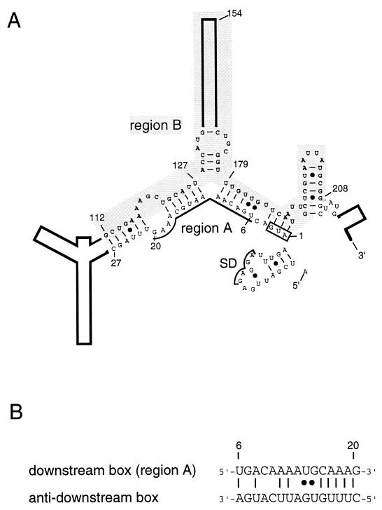
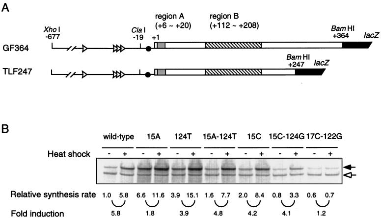
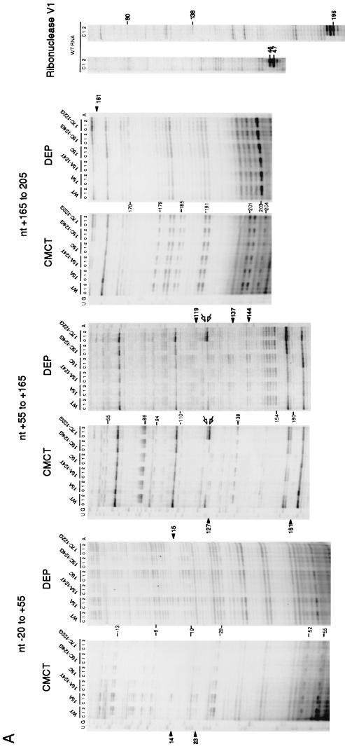
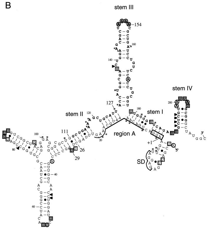
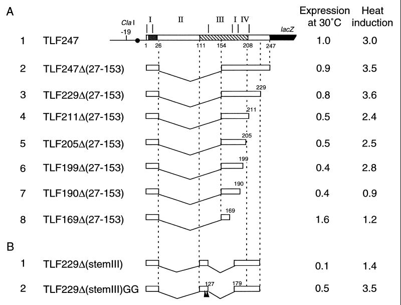
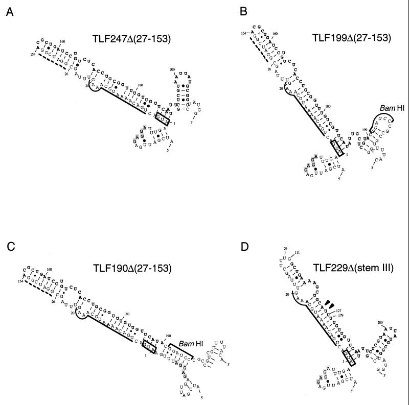
The heat shock response in Escherichia coli depends primarily on the increased synthesis and stabilization of otherwise scarce and unstable sigma32 (rpoH gene product), which is required for the transcription of heat shock genes. The heat-induced synthesis of sigma32 occurs at the level of translation, and genetic evidence has suggested the involvement of a secondary structure at the 5' portion (nucleotides -19 to +247) of rpoH mRNA in regulation. We now present evidence for the mRNA secondary structure model by means of structure probing of RNA with chemical and enzymatic probes. A similar analysis of several mutant RNAs with a mutation predicted to alter a base pairing or with two compensatory mutations revealed altered secondary structures consistent with the expression and heat inducibility of the corresponding fusion constructs observed in vivo. These findings led us to assess the possible roles of each of the stem-loop structures by analyzing an additional set of deletions and base substitutions. The results indicated not only the primary importance of base pairings between the translation initiation region of ca. 20 nucleotides (the AUG initiation codon plus the "downstream box") and the internal region of rpoH mRNA but also the requirement of appropriate stability of mRNA secondary structures for characteristic thermoregulation, i.e., repression at a low temperature and induction upon a temperature upshift.


Figures

FIG. 1
FIG. 1
Schematic representation of the 5′ portion (nt −19 to +247) of E. coli rpoH mRNA as predicted by use of Mulfold (14). (A) Secondary structure thought to be involved in modulating heat-induced synthesis of ς32 (21). Region A (nt +6 to 20), the initiation codon, and the Shine-Dalgarno (SD) sequence are indicated. Region B (nt +112 to 208) is shaded. Numbers refer to the nucleotides of the coding sequence. (B) Putative base pairing between the downstream box (region A) of rpoH and the “anti-downstream box” of 16S rRNA (spanning nt 1469 to 1483). •, G-U pairs.
FIG. 2
FIG. 2
Structure and expression of an rpoH-lacZ gene fusion (TLF247). (A) Schematic diagrams of the TLF247 fusion construct and GF364, studied previously (21). The locations of regions A and B are indicated. (B) SDS-PAGE patterns of fusion proteins expressed from the wild-type and mutant forms of TLF247. Cells were grown at 30°C and shifted to 42°C. Pulse-labeling with [35S]methionine was done for 2 min before or 3 min after the temperature shift. The labeled cells were disrupted, and immunoprecipitates obtained with anti–β-galactosidase serum were analyzed by SDS-PAGE as described in Materials and Methods. Closed and open arrows indicate fusion proteins and β-galactosidase ω protein (internal reference), respectively.
FIG. 3
FIG. 3
Structure probing of rpoH mRNA. (A) Urea-PAGE patterns of primer extension products. A 5′ segment (nt −60 to +247) of wild-type (WT) or mutant RNAs prepared in vitro was treated with CMCT, DEP, or RNase V1, and modified bases were identified by reverse transcription analysis as described in Materials and Methods. Treatment with CMCT was carried out at 30°C for 0 min (lanes c), 10 min (lanes 1), or 30 min (lanes 2), whereas treatment with DEP was done at 30°C for 0 min (lanes c), 5 min (lanes 1), or 10 min (lanes 2). Digestion with RNase V1 was done at 0°C for 30 min with 0 U (lanes c), 0.0005 U (lanes 1), or 0.001 U (lanes 2) of enzyme. Only some of the bases that were clearly modified by each treatment are indicated by nucleotide numbers. Relevant sequence ladders (wild type) are shown to the side as a reference. When comparing the observed bands with the sequence ladders, one should note that cDNA synthesis stops one residue before the modified base. Arrowheads indicate some of the modifications uniquely found in mutant RNA(s), whereas arrows indicate positions at which reverse transcriptase was arrested. Only the data for wild-type RNA are shown for experiments with RNase V1. (B) Modified bases identified with wild-type RNA in panel A are indicated on the mRNA secondary structure. The bases modified by CMCT or DEP are represented by squares or circles, respectively. The relative extents of modification are indicated by outlined stippled, outlined open, and nonoutlined shaded symbols for strong, modest, and weak reactivities, respectively. Arrowheads indicate sites cleaved by RNase V1. C-53 was fortuitously modified by DEP. Stems I, II, III, and IV are indicated, as are the initiation codon, region A, and the Shine-Dalgarno (SD) sequence. Region B (nt +112 to 208) is shown in boldface letters.
FIG. 3
FIG. 3
Structure probing of rpoH mRNA. (A) Urea-PAGE patterns of primer extension products. A 5′ segment (nt −60 to +247) of wild-type (WT) or mutant RNAs prepared in vitro was treated with CMCT, DEP, or RNase V1, and modified bases were identified by reverse transcription analysis as described in Materials and Methods. Treatment with CMCT was carried out at 30°C for 0 min (lanes c), 10 min (lanes 1), or 30 min (lanes 2), whereas treatment with DEP was done at 30°C for 0 min (lanes c), 5 min (lanes 1), or 10 min (lanes 2). Digestion with RNase V1 was done at 0°C for 30 min with 0 U (lanes c), 0.0005 U (lanes 1), or 0.001 U (lanes 2) of enzyme. Only some of the bases that were clearly modified by each treatment are indicated by nucleotide numbers. Relevant sequence ladders (wild type) are shown to the side as a reference. When comparing the observed bands with the sequence ladders, one should note that cDNA synthesis stops one residue before the modified base. Arrowheads indicate some of the modifications uniquely found in mutant RNA(s), whereas arrows indicate positions at which reverse transcriptase was arrested. Only the data for wild-type RNA are shown for experiments with RNase V1. (B) Modified bases identified with wild-type RNA in panel A are indicated on the mRNA secondary structure. The bases modified by CMCT or DEP are represented by squares or circles, respectively. The relative extents of modification are indicated by outlined stippled, outlined open, and nonoutlined shaded symbols for strong, modest, and weak reactivities, respectively. Arrowheads indicate sites cleaved by RNase V1. C-53 was fortuitously modified by DEP. Stems I, II, III, and IV are indicated, as are the initiation codon, region A, and the Shine-Dalgarno (SD) sequence. Region B (nt +112 to 208) is shown in boldface letters.
FIG. 4
FIG. 4
Structure and expression of fusion proteins from deletion derivatives of TLF247. (A) A series of 3′ deletions derived from the internal deletion TLF247Δ(27–153). Segments of mRNA that correspond to each of the stem structures (I to IV) are shown above the diagram, and nucleotide numbers are shown below. Regions A and B are indicated by stippled and hatched boxes, respectively. (B) A pair of deletions lacking stem III. Arrowheads indicate the positions of two G’s derived from the Δ(27–153) deletion. Cells were grown at 30°C and shifted to 42°C. Samples taken at time 0 (30°C) and 3 min after the shift were pulse-labeled with [35S]methionine for 1 min, followed by a 3-min chase. The labeled proteins were analyzed by immunoprecipitation, followed by SDS-PAGE as described in the legend to Fig. 2. Synthesis rates were normalized to that for the wild type (TLF247) labeled at 30°C. The apparently lower extents of induction observed here compared to those shown in Fig. 2B were due to slightly different procedures and not to instability of the fusion proteins examined (data not shown).
FIG. 5
FIG. 5
Predicted mRNA secondary structures for some of the deletion derivatives used. Structures predicted for RNA of 150 nt (starting from nt −19 for each; the sequences may include a BamHI junction and part of lacZ) and that have minimum free energy are shown for some representative constructs examined in Fig. 4. Only relevant portions are presented. The initiation codon, region A, and region B are indicated as described in the legend to Fig. 3B. The Shine-Dalgarno sequence is shown by shaded letters. Arrowheads indicate the positions where two G’s were replaced in constructing TLF229Δ(stemIII)GG (Fig. 4B). The broken line indicates extra bases inserted during construction (21).

References

    1. Bukau B. Regulation of the Escherichia coli heat shock response. Mol Microbiol. 1993;9:671–680. - PubMed
    1. Casadaban M J. Transposition and fusion of the lac genes to selected promoters in Escherichia coli using bacteriophage lambda and Mu. J Mol Biol. 1976;104:541–555. - PubMed
    1. Christiansen J, Egebjerg J, Larsen N, Garrett R. Analysis of rRNA structure: experimental and theoretical considerations. In: Spedding G, editor. Ribosome and protein synthesis: a practical approach. Oxford, England: IRL Press; 1991. pp. 229–252.
    1. Craig E A, Gross C A. Is hsp70 the cellular thermometer? Trends Biochem Sci. 1991;16:135–140. - PubMed
    1. de Smit M H, van Duin J. Secondary structure of the ribosome binding site determines translational efficiency: a quantitative analysis. Proc Natl Acad Sci USA. 1990;87:7668–7672. - PMC - PubMed

Publication types

MeSH terms

LinkOut - more resources