. 1999 Jan;181(2):685-8.
doi: 10.1128/JB.181.2.685-688.1999.

Subunit II of Bacillus subtilis cytochrome c oxidase is a lipoprotein

Affiliations

Subunit II of Bacillus subtilis cytochrome c oxidase is a lipoprotein

J Bengtsson et al. J Bacteriol. 1999 Jan.

Abstract

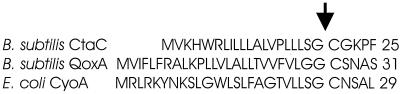
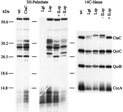
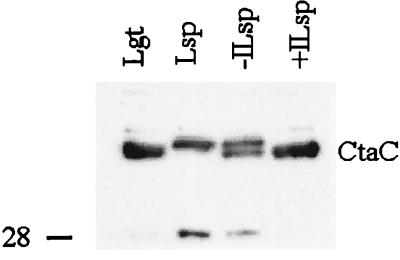
The sequence of the N-terminal end of the deduced ctaC gene product of Bacillus species has the features of a bacterial lipoprotein. CtaC is the subunit II of cytochrome caa3, which is a cytochrome c oxidase. Using Bacillus subtilis mutants blocked in lipoprotein synthesis, we show that CtaC is a lipoprotein and that synthesis of the membrane-bound protein and covalent binding of heme to the cytochrome c domain is not dependent on processing at the N-terminal part of the protein. Mutants blocked in prolipoprotein diacylglyceryl transferase (Lgt) or signal peptidase type II (Lsp) are, however, deficient in cytochrome caa3 enzyme activity. Removal of the signal peptide from the CtaC polypeptide, but not lipid modification, is seemingly required for formation of functional enzyme.


Figures

FIG. 1
FIG. 1
Deduced N-terminal sequence of subunit II polypeptides of three oxidases. CtaC, QoxA, and CyoA are subunits of cytochrome caa3, cytochrome aa3, and cytochrome bo3, respectively, of the specified bacteria. The signal peptidase type II signal cleavage site, indicated with an arrow, is supported by experimental data obtained for Bacillus sp. strain PS3 (6), Bacillus subtilis W23 (10), and E. coli (11).
FIG. 2
FIG. 2
Analysis of B. subtilis strains for membrane-bound lipoproteins and covalently bound heme by [3H]palmitate and [14C]ALA (heme) labeling, respectively. Fluorographs (2) of SDS gels (16% [wt/vol] acrylamide) (18) are shown. Growth conditions and preparation of extracts were as described in the text. Forty micrograms of membrane protein was loaded in each lane. The samples were as follows: wt, strain 168A; CtaC, LUH15; Lgt, LUH104; Lsp, LUH102; ILsp, LUH103. + and − indicate that the extract was prepared from cells grown in the presence or absence, respectively, of 1 mM IPTG. The numbers on the left indicate the positions of size markers. The specific 14C label in heme in the Lgt- and Lsp-deficient mutants was much lower than in the wild type for reasons that are discussed in the text. The fluorographs shown are overexposed to demonstrate that lipoproteins are absent from strain LUH104 and to more clearly show the CtaC polypeptide(s) in mutants defective in lipoprotein modification. The amount of CtaC polypeptide in LUH102 and LUH104 was about 60% of that in the wild type, as calculated from the [14C]heme in CtaC relative to that in QcrC; i.e., heme in QcrC was used as an internal standard with the assumption that the amount of QcrC polypeptide was the same in membranes from all strains.
FIG. 3
FIG. 3
Immunoblot analysis for CtaC polypeptide in membranes. The samples are the same as those analyzed for covalently bound heme in Fig. 2. SDS-PAGE was carried out as described for Fig. 2 except that about 35 μg of protein was loaded in each lane. This gel did not resolve the two forms of CtaC in the Lgt-deficient mutant. Proteins were electroblotted onto a polyvinylidene difluoride membrane under semidry conditions with Tris-glycine buffer with 20% methanol. The anti-CtaC serum, used at a 500-fold dilution, had been obtained by immunizing a rabbit with the peptide MLNALTEKRTRGC, corresponding to the C-terminal end of B. subtilis CtaC, conjugated to bovine serum albumin. The ECL Western blot system (Amersham) was used to detect antigens. The position of a 28-kDa size marker is indicated on the left.

References

    1. Calhoun M W, Thomas J W, Gennis R B. The cytochrome oxidase superfamily of redox-driven proton pumps. Trends Biochem Sci. 1994;19:325–330. - PubMed
    1. Chamberlain J P. Fluorographic detection of radioactivity in polyacrylamide gels with the water-soluble fluor, sodium salicylate. Anal Biochem. 1979;98:132–135. - PubMed
    1. Dartois V, Djavakhishvili T, Hoch J A. KapB is a lipoprotein required for KinB signal transduction and activation of the phosphorelay to sporulation in Bacillus subtilis. Mol Microbiol. 1997;26:1097–1108. - PubMed
    1. Hayashi S, Wu H C. Identification and characterization of lipid-modified proteins in bacteria. In: Hooper N M, Turner A J, editors. Lipid modification of proteins. Oxford, England: IRL Press; 1992. pp. 261–285.
    1. Hederstedt L. Molecular properties, genetics, and biosynthesis of Bacillus subtilis succinate dehydrogenase complex. Methods Enzymol. 1986;126:399–414. - PubMed

Publication types

MeSH terms

LinkOut - more resources