. 1999 Feb 1;514 ( Pt 3)(Pt 3):747-58.
doi: 10.1111/j.1469-7793.1999.747ad.x.

Contribution of Ca2+-activated K+ channels and non-selective cation channels to membrane potential of pulmonary arterial smooth muscle cells of the rabbit

Affiliations

Contribution of Ca2+-activated K+ channels and non-selective cation channels to membrane potential of pulmonary arterial smooth muscle cells of the rabbit

Y M Bae et al. J Physiol. .

Abstract

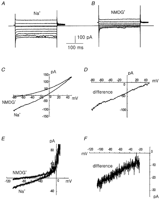
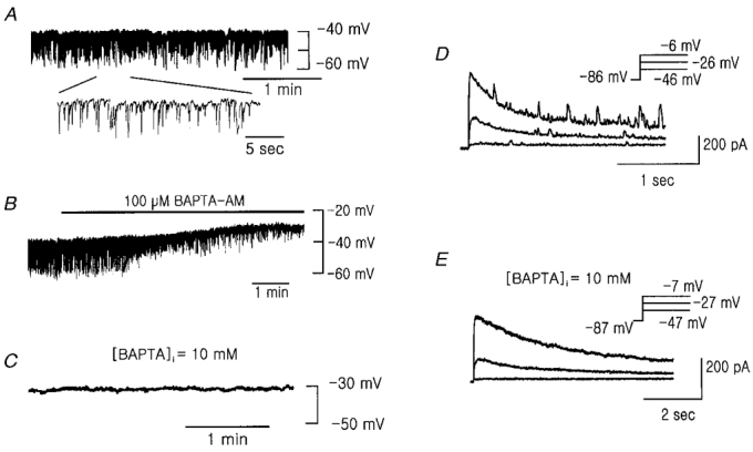
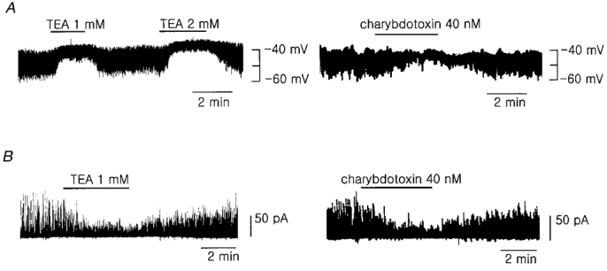
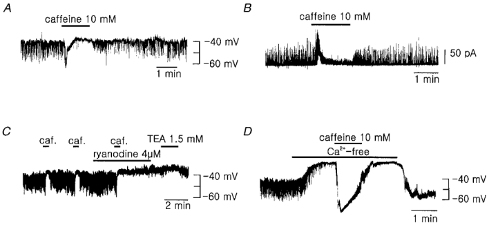
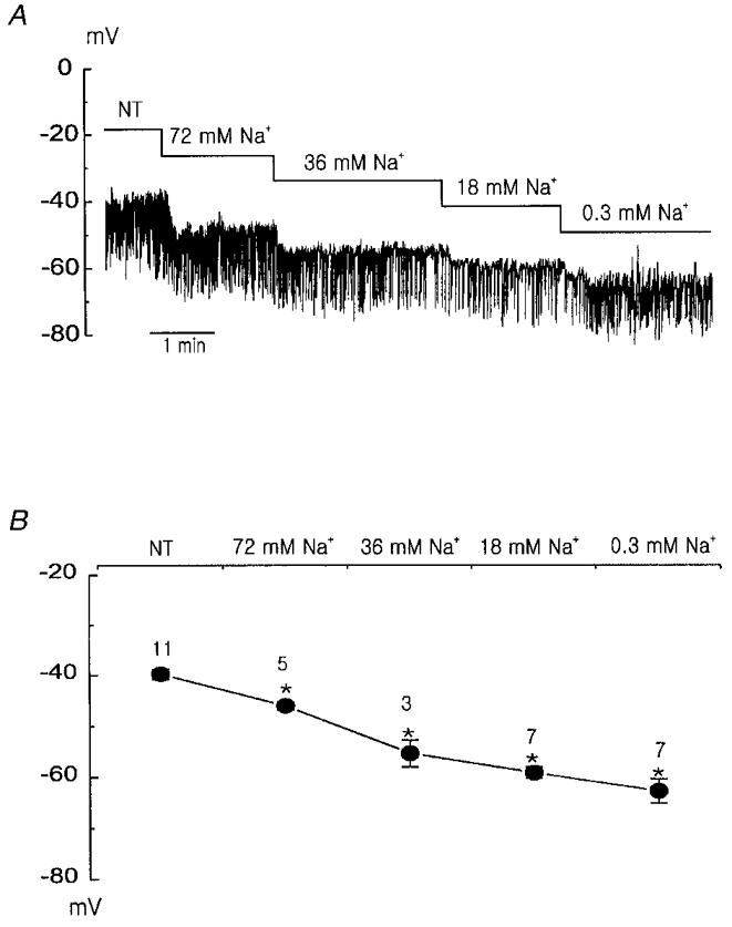
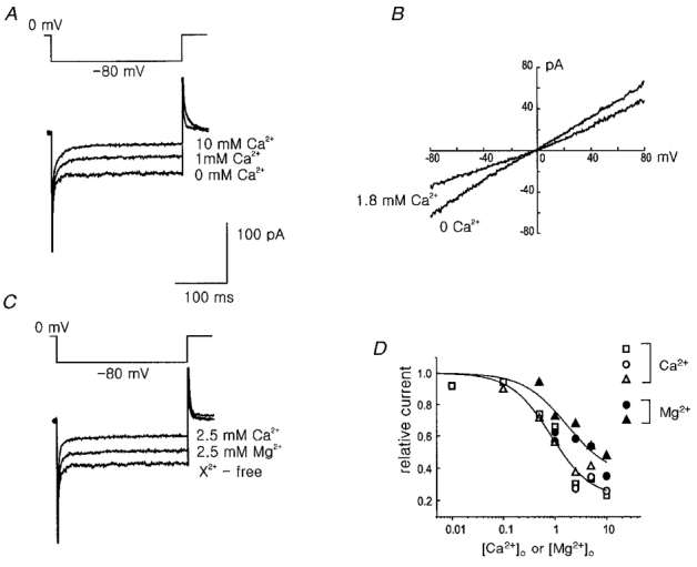
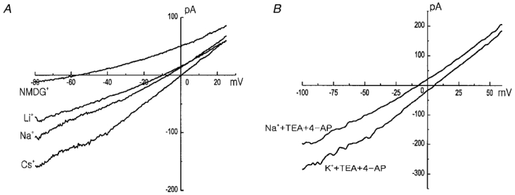
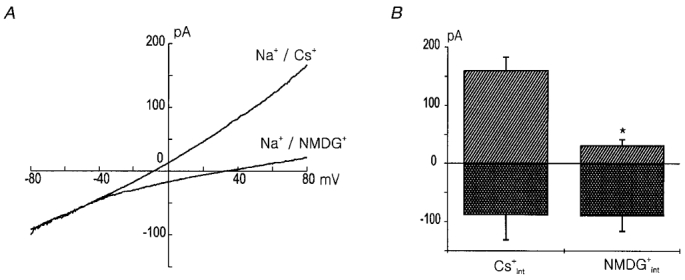
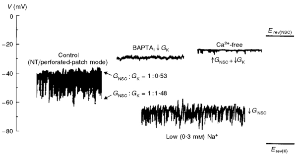
1. Using the perforated patch-clamp or whole-cell clamp technique, we investigated the contribution of Ca2+-activated K+ current (IK(Ca)) and non-selective cation currents (INSC) to the membrane potential in small pulmonary arterial smooth muscle cells of the rabbit. 2. The resting membrane potential (Vm) was -39.2 +/- 0.9 mV (n = 72). It did not stay at a constant level, but hyperpolarized irregularly, showing spontaneous transient hyperpolarizations (STHPs). The mean frequency and amplitude of the STHPs was 5.6 +/- 1. 1 Hz and -7.7 +/- 0.7 mV (n = 12), respectively. In the voltage-clamp mode, spontaneous transient outward currents (STOCs) were recorded with similar frequency and irregularity. 3. Intracellular application of BAPTA or extracellular application of TEA or charybdotoxin suppressed both the STHPs and STOCs. The depletion of intracellular Ca2+ stores by caffeine or ryanodine, and the removal of extracellular Ca2+ also abolished STHPs and STOCs. 4. Replacement of extracellular Na+ with NMDG+ caused hyperpolarization Vm of without affecting STHPs. Removal of extracellular Ca2+ induced a marked depolarization of Vm along with the disappearance of STHPs. 5. The ionic nature of the background inward current was identified. The permeability ratio of K+ : Cs+ : Na+ : Li+ was 1.7 : 1.3 : 1 : 0. 9, indicating that it is a non-selective cation current (INSC). The reversal potential of this current in control conditions was calculated to be -13.9 mV. The current was blocked by millimolar concentrations of extracellular Ca2+ and Mg2+. 6. From these results, it was concluded that (i) hyperpolarizing currents are mainly contributed by Ca2+-activated K+ (KCa) channels, and thus STOCs result in transient membrane hyperpolarization, and (ii) depolarizing currents are carried through NSC channels.


Figures

Figure 5
Figure 5. Isolation of the background current
A, currents measured in 148 mM Na+ solution in response to voltage steps between -110 mV and +30 mV from the holding potential of 0 mV. B, same as A but for extracellular NMDG+ instead of Na+. The horizontal bar indicates zero current level. C, current-voltage relation in 148 mM Na+ and NMDG+ solution in response to voltage ramp. D, current-voltage relation obtained by subtracting current measured in Na+-free (NMDG+) solution from that in 148 mM Na+-containing solution. Traces A-D were recorded with CsCl pipette solution and divalent cation-free bath solution in the ruptured whole-cell mode. Similar results were obtained in 16 other cells. E, current-voltage relation measured in the normal Tyrode solution and NMDG+ Tyrode (0.3 mM Na+) solution in the perforated patch-clamp mode. F, current-voltage relation of the difference current between the normal Tyrode and NMDG+ Tyrode solution. Similar results in 3 other cells.
Figure 1
Figure 1. Recording of the resting membrane potential and membrane currents in the PASMCs
A, representative trace of membrane potential. B, effect of bath application of 100 μM BAPTA AM on the membrane potential. A and B were recorded in the perforated patch-clamp mode. C, membrane potential recorded in ruptured whole-cell clamp mode with strong Ca2+ buffer (10 mM BAPTA) in the pipette. D and E, membrane currents elicited by step depolarizations to the indicated voltage from a holding potential of -86 or -87 mV in perforated patch-clamp mode (D) and whole-cell clamp mode (E), respectively.
Figure 2
Figure 2. Effects of KCa channel blockers on membrane potentials and membrane currents
A, effect of TEA (left) and charybdotoxin (right) on the membrane potential. B, effect of TEA and charybdotoxin on the STOCs recorded at the membrane potential of -31 mV in voltage-clamp mode. Perforated patch-clamp mode was used in A and B. Similar results were obtained in 4-9 other cells.
Figure 3
Figure 3. Regulation of the membrane potential by caffeine, ryanodine, and Cao2+
A and B, effect of bath application of 10 mM caffeine on the membrane potential (A) and membrane currents (B). The holding potential was -31 mV in trace B. C, effect of caffeine and ryanodine. TEA had little effect on the membrane potential while the STHPs were suppressed by the treatment with caffeine and ryanodine. D, effect of the extracellular Ca2+ removal (nominally Ca2+-free) on the membrane potential. Caffeine still evoked a marked hyperpolarization of the membrane potential while the STHPs were inhibited by the removal of the external Ca2+. Similar results were obtained in 3-7 other cells.
Figure 4
Figure 4. The effects of extracellular Na+ reduction on the membrane potential
A, hyperpolarization of the membrane potential caused by a reduction in extracellular Na+ concentration. B, summary of the effects of extracellular Na+ reduction on the membrane potential. The numbers on the symbols represent the number of tested cells. * indicates that the mean values are significantly different (P < 0.05) from the value measured in 143 mM Na+ normal Tyrode (NT) solution.
Figure 8
Figure 8. The blockade of the INSC by extracellular Ca2+ and Mg2+
The concentrations of Ca2+ and Mg2+ are nominal. A, extracellular Ca2+ blocks the INSC in a dose-dependent manner. The currents were elicited by step voltage to -80 mV from the holding potential of 0 mV. B, current-voltage relations measured in the presence and absence of extracellular 1.8 mM Ca2+. C, blockade of the INSC by external Mg2+ and Ca2+. D, comparison of the blockade of the INSC at the various concentrations of extracellular Ca2+ and Mg2+. The current values were measured at -80 mV and expressed relative to the currents measured in the divalent cation-free bath solution. Open symbols indicate the blockade by Ca2+ and filled symbols by Mg2+. Each cell used is represented by a different shape of symbol. CsCl pipette solution was used for the traces in A and C and NaCl pipette solution was used for B.
Figure 6
Figure 6. Cation selectivity of the background current
A, current-voltage relations measured in 148 mM CsCl, NaCl, LiCl, and NMDG-Cl bath solution. B, to obtain the K+ selectivity of the Na+-dependent background current, 10 mM TEA and 4-AP were added to the bath. CsCl pipette solution was used in both A and B.
Figure 7
Figure 7. The outward component of the INSC is cation dependent
A, current-voltage relation measured in the NaCl bath solution with Cs+ and NMDG+ in the pipette. Note the difference in the amplitude of the outward currents. The current traces are means of the currents obtained from the 3 different cells for each condition. B, the amplitudes of the currents measured at a voltage of ± 80 mV in A are compared. * indicates that they are significantly different (P < 0.05).
Figure 9
Figure 9. The relative contribution of the NSC and K+ selective channels
Erev(NSC) indicates the reversal potential of the background INSC and Erev(K) the reversal potential of the K+ channel, or the equilibrium potential for the K+ ion assuming the K+ selectivity of the K+ channels is complete. ↑GNSC indicates the increase of NSC channel conductance, ↓GNSC the decrease of NSC channel conductance, and ↓GK the decrease of K+ channel conductance. NT, normal Tyrode solution.

References

    1. Benham CD, Bolton TB. Spontaneous transient outward currents in single visceral and vascular smooth muscle cells of rabbit. The Journal of Physiology. 1986;381:385–406. - PMC - PubMed
    1. Casteels R, Kitamura K, Kuriyama H, Suzuki H. The membrane properties of the smooth muscle cells of the rabbit main pulmonary artery. The Journal of Physiology. 1977;271:41–61. - PMC - PubMed
    1. Daut J, Standen NB, Nelson MT. The role of the membrane potential of endothelial and smooth muscle cells in the regulation of coronary blood flow. Journal of Cardiovascular Electrophysiology. 1994;5:154–181. - PubMed
    1. Evans AM, Osipenko ON, Gurney AM. Properties of a novel K+ current that is active at resting potential in rabbit pulmonary artery smooth muscle cells. The Journal of Physiology. 1996;496:407–420. - PMC - PubMed
    1. Franco-Obregon A, Urena J, Lopez-Barneo J. Oxygen-sensitive calcium channels in vascular smooth muscle and their possible role in hypoxic arterial relaxation. Proceedings of the National Academy of Sciences of the USA. 1995;92:4715–4719. - PMC - PubMed

Publication types

MeSH terms

Substances

LinkOut - more resources