. 1999 Jan;103(1):47-54.
doi: 10.1172/JCI3756.

Decreased angiogenesis and arthritic disease in rabbits treated with an alphavbeta3 antagonist

Affiliations

Decreased angiogenesis and arthritic disease in rabbits treated with an alphavbeta3 antagonist

C M Storgard et al. J Clin Invest. 1999 Jan.

Abstract

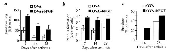
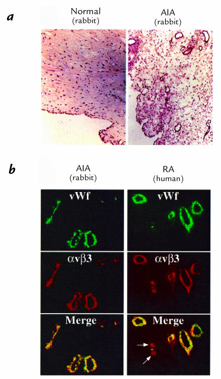
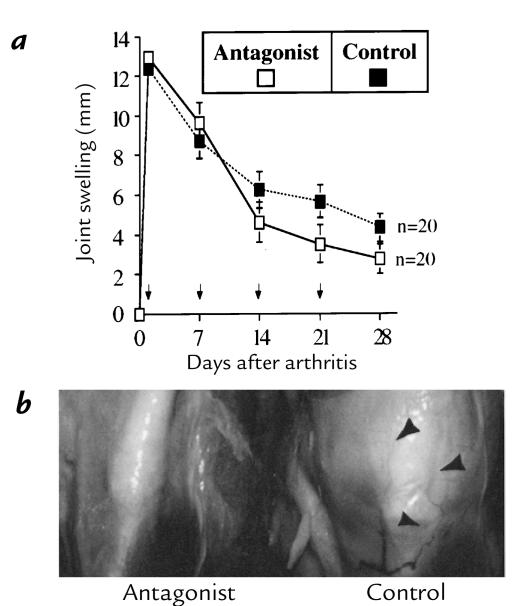
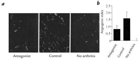
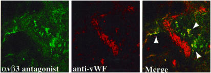
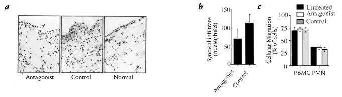
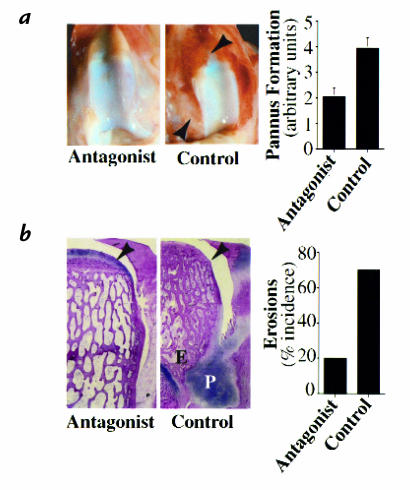
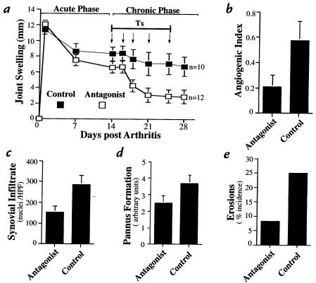
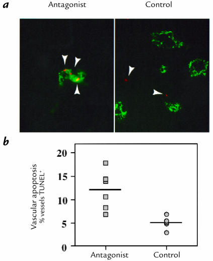
Rheumatoid arthritis (RA) is an inflammatory disease associated with intense angiogenesis and vascular expression of integrin alphavbeta3. Intra-articular administration of a cyclic peptide antagonist of integrin alphavbeta3 to rabbits with antigen-induced arthritis early in disease resulted in inhibition of synovial angiogenesis and reduced synovial cell infiltrate, pannus formation, and cartilage erosions. These effects were not associated with lymphopenia or impairment of leukocyte function. Furthermore, when administered in chronic, preexisting disease, the alphavbeta3 antagonist effectively diminished arthritis severity and was associated with a quantitative increase in apoptosis of the angiogenic blood vessels. Therefore, angiogenesis appears to be a central factor in the initiation and persistence of arthritic disease, and antagonists of integrin alphavbeta3 may represent a novel therapeutic strategy for RA.


Figures

Figure 1
Figure 1
The proangiogenic cytokine bFGF intensifies arthritis. (a) The addition of bFGF during induction of AIA enhanced arthritis severity compared with OVA alone, with increased joint swelling, greater pannus formation (b), and earlier and more frequent erosive disease (c). Pannus development was graded on a relative scale 0–5 (0 = normal to 5 = macroscopic cartilage erosion) (ref. 23). All data are expressed as mean ± SE (n = 8). AIA, antigen-induced arthritis; bFGF, basic fibroblast growth factor; OVA, ovalbumin.
Figure 2
Figure 2
AIA and human RA exhibit αvβ3 expression on angiogenic blood vessels. (a) Cryosections of infrapatellar tissue in AIA stained with H&E demonstrate marked neovascularization, synovial hypertrophy, and dense synovial inflammatory infiltrate relative to synovium from nonarthritic tissue (×100). (b) Immunofluorescent detection of blood vessels by vWf-specific antibody (green) and MAB LM609 directed to integrin αvβ3 (red) demonstrates colocalization of αvβ3 on the synovial endothelium (yellow merge) in rabbit AIA, as in human RA (×400). Note that microvascular angiogenic sprouts express only αvβ3 (arrows). H&E, hematoxylin and eosin; MAB, monoclonal antibody; RA, rheumatoid arthritis; vWf, von Willebrand factor.
Figure 3
Figure 3
Joint swelling is reduced after αvβ3 antagonist administration in AIA. (a) Rabbits with AIA were treated with bilateral intra-articular αvβ3 antagonist (EMD 66203; 0.5 ml, 2 mg/ml) or control peptide (EMD 69601) beginning 24 h after arthritis onset, and weekly thereafter for 4 weeks (arrows). Joint swelling, defined as the increase in knee diameter from normal (nonarthritic), was significantly reduced by the αvβ3 antagonist (days 14–28, P < 0.05, ANOVA) compared with control peptide. (b) Periarticular vascularization (arrowheads) was prominent in control-treated AIA compared with antagonist-treated groups.
Figure 4
Figure 4
Synovial vascularity is decreased after αvβ3 antagonist treatment. (a) Cryosections of the infrapatellar tissue obtained 28 days after arthritis onset in animals treated with the αvβ3 antagonist or control peptide (days 1, 7, 14, and 21) were stained for vWf as a marker of blood vessels, detected with FITC-labeled secondary antibody, and were digitally imaged (×200). (b) The relative increase in area of fluorescent pixels per field vs. normal was computed to determine the angiogenic index as described in Methods. Synovial neovascularization was significantly inhibited by the αvβ3 antagonist (P < 0.050, Student's t test). Data are expressed as mean ± SE (n = 20).
Figure 5
Figure 5
Fluorescent integrin antagonist colocalizes with synovial angiogenic microvessels. An FITC-conjugated αvβ3 antagonist peptide, EMD 80838 (cyclic-Arg-Gly-Asp-D-Phe-Lys-[fluoresceincarboxylic acid]; 0.5 mg/500 μl), was injected intra-articularly in rabbits with AIA. After 24 h, cryostat sections (5 μm) of synovial tissue were examined by confocal microscopy for the presence of peptide (green). Blood vessels were identified by antisera to vWF as detected with TRITC-labeled secondary antibody (red). Colocalization of αvβ3 antagonist is observed on angiogenic microvessels after signal merge (yellow), but not with mature vWF+ vessels (×630). TRITC, tetrarhodamine isothiocyanate.
Figure 6
Figure 6
Integrin αvβ3 antagonist reduces synovial inflammatory infiltrate in AIA without impairing leukocyte migration. (a) H&E stain of cryosections of the infrapatellar fat-pad 28 days after arthritis onset reveals a marked decrease in synovial cellular infiltrate in animals treated with the αvβ3 antagonist (days 1, 7, 14, and 21) compared with control. (b) Digital assessment of nuclei present in each field (three per joint, ×400) demonstrates that αvβ3 antagonist treatment resulted in a significant reduction in the cellular infiltrate (P < 0.05, Student's t test). Data are expressed as mean ± SE (n = 20). (c) In vitro chemotaxis on type II collagen toward synovial fluid by PBMCs or PMNs isolated from peripheral blood from rabbits with AIA. Data are expressed as mean ± SE of triplicate determinations. No effect of the αvβ3 antagonist on the migratory capacity of leukocytes was observed. PBMCs, peripheral blood mononuclear cells; PMNs, polymorphonuclear neutrophils.
Figure 7
Figure 7
Blockade of integrin αvβ3 decreases pannus formation and cartilage erosions. (a) Pannus development (arrowheads) was graded on a relative scale 0–5 (0 = normal to 5 = macroscopic erosion). Treatment with the αvβ3 antagonist significantly decreased pannus formation (P < 0.05, Student's t test) relative to control peptide. (b) Frontal sections of decalcified femoral condyle stained by H&E illustrate the protective effect of αvβ3 antagonist treatment on a representative sample of erosive disease (×10). (P indicates pannus, E indicates erosion, and the arrowhead indicates preservation of the articular cartilage in the antagonist-treated group.) Macroscopic cartilage erosions were decreased with αvβ3 antagonist treatment (P < 0.01, Student's t test). All data are expressed as mean ± SE (n = 20).
Figure 8
Figure 8
Chronic arthritis is ameliorated by an αvβ3 antagonist. (a) Administration of the antagonist (arrows) beginning 2 weeks after arthritis onset resulted in decreased joint swelling compared with controls (untreated) (P < 0.05, ANOVA). (b) Angiogenesis, as depicted by the angiogenic index (see Methods), was inhibited by αvβ3 antagonist treatment (P < 0.05, Student's t test). (c) Fewer infiltrating cells were observed in the synovium of αvβ3 antagonist–treated animals (P < 0.05, Student's t test), as assessed by digital computerized counting of nuclei. (d) Pannus was assessed as described in Methods. (e) A decrease in both pannus formation and cartilage erosion was observed in antagonist-treated animals relative to control. Data are expressed as mean ± SE (n = 12, antagonist; n = 10, control). HPF, high-power field; Tx, treatment.
Figure 9
Figure 9
Vascular cell apoptosis is associated with αvβ3 antagonist treatment. (a)Synovium from rabbits in the chronic AIA model were stained with TUNEL immunostaining (red) as an indicator of apoptosis, (arrowheads)and anti-vWF (green) as a marker of blood vessels (×400). (b) Apoptosis associated with blood vessels was detected and quantified blindly as percent blood vessels containing apoptotic cells in 20 fields (×400) per specimen. Apoptosis was increased specifically in the vasculature of arthritic synovium after αvβ3 antagonist treatment (P < 0.01, Student's t test). TUNEL, terminal deoxynucleotidyl transferase–mediated dUTP nick end-labeling.

Comment in

References

    1. Zvaifler NJ, Firestein GS. Pannus and pannocytes: alternative models of joint destruction in rheumatoid arthritis. Arthritis Rheum. 1994;37:783–789. - PubMed
    1. Kimball ES, Gross JL. Angiogenesis in pannus formation. Agents Actions. 1991;34:329–331. - PubMed
    1. Colville-Nash PR, Scott DL. Angiogenesis and rheumatoid arthritis: pathogenic and therapeutic implications. Ann Rheum Dis. 1992;51:919–925. - PMC - PubMed
    1. Paleolog E. Target effector role of vascular endothelium in the inflammatory response: insights from the clinical trial of anti-TNF alpha antibody in rheumatoid arthritis. Mol Pathol. 1997;50:225–233. - PMC - PubMed
    1. Qu Z, et al. Expression of basic fibroblast growth factor in synovial tissue from patients with rheumatoid arthritis and degenerative joint disease. Lab Invest. 1995;73:339–346. - PubMed

Publication types

MeSH terms

Substances