Case Reports
. 1999 Jan;103(1):97-106.
doi: 10.1172/JCI3312.

A novel leukocyte adhesion deficiency caused by expressed but nonfunctional beta2 integrins Mac-1 and LFA-1

Affiliations
Case Reports

A novel leukocyte adhesion deficiency caused by expressed but nonfunctional beta2 integrins Mac-1 and LFA-1

N Hogg et al. J Clin Invest. 1999 Jan.

Abstract

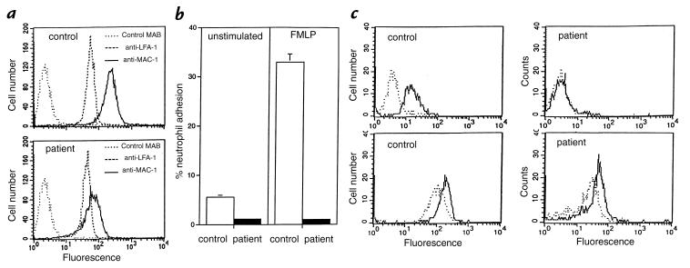
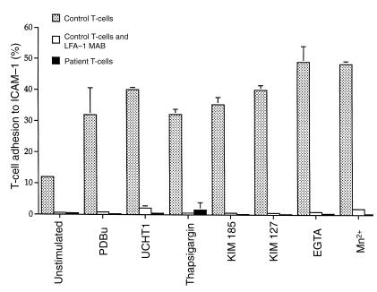
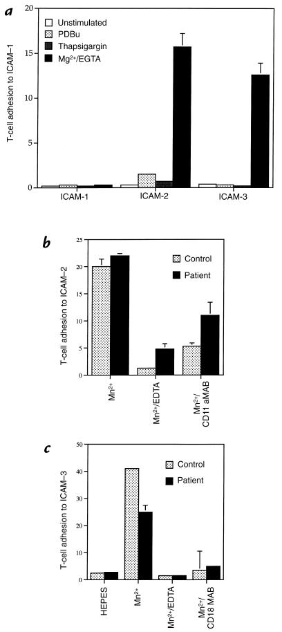
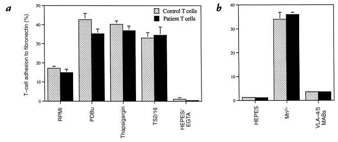
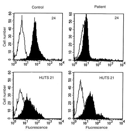
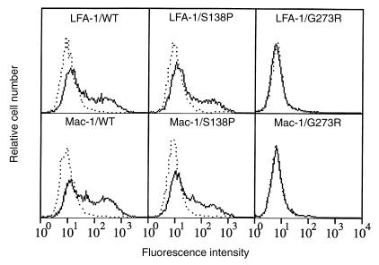
In the leukocyte adhesion deficiency (LAD)-1 syndrome, there is diminished expression of beta2(CD18) integrins. This is caused by lesions in the beta2-subunit gene and gives rise to recurrent bacterial infections, impaired pus formation, and poor wound healing. We describe a patient with clinical features compatible with a moderately severe phenotype of LAD-1 but who expresses the beta2 integrins lymphocyte function- associated molecule (LFA)-1 and Mac-1 at 40%-60% of normal levels. This level of expression should be adequate for normal integrin function, but both the patient's Mac-1 on neutrophils and LFA-1 on T cells failed to bind ligands such as fibrinogen and intercellular adhesion molecule (ICAM)-1, respectively, or to display a beta2-integrin activation epitope after adhesion-inducing stimuli. Unexpectedly, divalent cation treatment induced the patient's T cells to bind to ICAM-2 and ICAM-3. Sequencing of the patient's two CD18 alleles revealed the mutations S138P and G273R. Both mutations are in the beta2-subunit conserved domain, with S138P a putative divalent cation coordinating residue in the metal ion-dependent adhesion site (MIDAS) motif. After K562 cell transfection with alpha subunits, the mutated S138P beta subunit was coexpressed but did not support function, whereas the G273R mutant was not expressed. In summary, the patient described here exhibits failure of the beta2 integrins to function despite adequate levels of cell-surface expression.


Figures

Figure 1
Figure 1
Comparison of patient and control neutrophil β2 integrins. (a) Expression of cell membrane levels of Mac-1 (CD11b/CD18) and LFA-1 (CD11a/CD18). (b) Assay of neutrophil Mac-1 function: Ability of control (white bars) and patient (black bars) neutrophils to bind to immobilized ligand fibrinogen with and without exposure to 0.1 μM FMLP. (c) Ability of patient and control neutrophils to respond to external stimuli. Control, but not patient, neutrophils are able to express the β2-integrin activation reporter epitope recognized by MAB 24, indicative of active β2 integrins (top panels). Both patient and control neutrophils can upregulate Mac-1 (bottom panels) after exposure to 0.1 μM FMLP (dotted lines, –FMLP; solid lines, +FMLP), indicating that the patient cells have a normal response to external stimuli. LFA, lymphocyte function-associated molecule; MAB, monoclonal antibody.
Figure 2
Figure 2
The ability of patient (black bars) and control (dotted bars) T-cell LFA-1 to adhere to immobilized ICAM-1 after activating stimuli (see Methods). Several treatments were used that trigger LFA-1–mediated adhesion by intracellular signaling (PDBu, cross-linking the T-cell receptor with CD3 MAB UCHT1, Ca2+ mobilizer thapsigargin) or by acting extracellularly (Mg2+/EGTA, Mn2+, or β2-activating MABs KIM 127 and KIM 185). CD11a MAB 38 inhibited binding of control T cells under all conditions (white bars). ICAM, intercellular adhesion molecule; PDBu, phorbol-12,13 dibutyrate.
Figure 3
Figure 3
The distinctive recognition by patient T-cell LFA-1 of ligands ICAM-1, ICAM-2, and ICAM-3 after various integrin-activating protocols. (a) Ability of patient T cells to bind to immobilized ICAM-1, ICAM-2, and ICAM-3 either as untreated cells (white bars) or after treatment with PDBu (dotted bars), Ca2+ mobilizer thapsigargin (gray bars), and Mg2+/EGTA (black bars). Mg2+/EGTA is seen to induce patient T cells to bind to ICAM-2 and ICAM-3 but not to ICAM-1. (b) The binding of patient T cells (black bars) and control T cells (dotted bars) to ICAM-2 after treatment with 0.5 mM Mn2+ is prevented by chelation of divalent cations with EDTA and by CD11a function blocking MAB DF1524. (c) The binding of patient T cells (black bars) and control T cells (dotted bars) to ICAM-3 after 0.5 mM Mn2+ is prevented by chelation of divalent cations with EDTA and by CD18 function blocking MAB 60.3.
Figure 4
Figure 4
The ability of patient T cells and control T cells to adhere to immobilized fibronectin via β1 integrins. (a) Control (dotted bars) and patient (black bars) T cells were untreated (RPMI), or treated with stimuli (PDBu, thapsigargin), by the β1-activating MAB TS2/16 or by ablation of integrin-mediated adhesion with 1 mM EDTA. (b) The increased adhesion of control and patient T cells to fibronectin caused by divalent cation treatment with 0.5 mM Mn2+ could be blocked by a combination of VLA-4 (α4β1)– and VLA-5 (α5β1)–specific MABs, HP1/2 and SAM-1, respectively. RPMI, Roswell Park Memorial Institute medium.
Figure 5
Figure 5
Expression patterns of β2- and β1-integrin activation reporter epitopes on T cells detected by flow cytometry after exposure to 0.5 mM Mn2+ (black) or without Mn2+ (white) at 37°C for 25 min. Control, but not patient's, T cells express β2 activation reporter epitope detected by MAB 24 (top panels). Conversely, the patient's T cells and control T cells express equivalent levels of the β1 activation reporter epitope detected with MAB HUTS 21 (bottom panels).
Figure 6
Figure 6
LFA-1 and Mac-1 expression profiles of K562 transfectants. Wild-type CD18 cDNA or CD18 cDNA carrying the S138P or G273R mutations were transfected into K562 together with CD11a cDNA (top panels) or CD11b cDNA (bottom panels). Expression of LFA-1 and Mac-1 was detected with the MAB 1B4 (solid lines). Background fluorescence was observed with MAB OX34 (dotted lines).

References

    1. Butcher EC, Picker JL. Lymphocyte homing and homeostasis. Science. 1996;272:60–66. - PubMed
    1. Brown EJ. Adhesive interactions in the immune system. Trends Cell Biol. 1997;7:289–295. - PubMed
    1. Bachmann MF, et al. Distinct roles for LFA-1 and CD28 during activation of naive T cells: adhesion versus costimulation. Immunity. 1997;7:549–557. - PubMed
    1. Brown EJ. Complement receptors and phagocytosis. Curr Opin Immunol. 1991;3:76–82. - PubMed
    1. Ross GD, Reed W, Dalzell JG, Becker SE, Hogg N. Macrophage cytoskeleton association with CR3 and CR4 regulates receptor mobility and phagocytosis of iC3b-opsonized erythrocytes. J Leukoc Biol. 1992;51:109–117. - PubMed

Publication types

MeSH terms