Case Reports
. 1999 Jan;103(1):129-35.
doi: 10.1172/JCI4165.

Oxidative stress causes enhanced endothelial cell injury in human heme oxygenase-1 deficiency

Affiliations
Case Reports

Oxidative stress causes enhanced endothelial cell injury in human heme oxygenase-1 deficiency

A Yachie et al. J Clin Invest. 1999 Jan.

Abstract

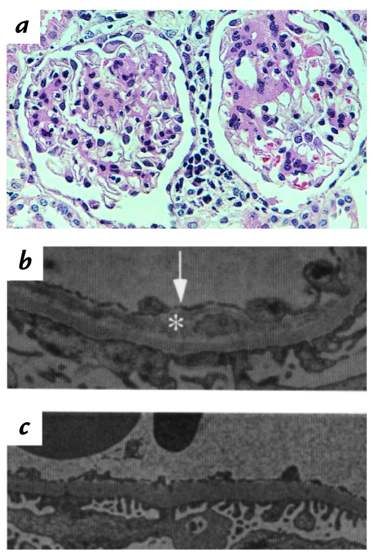
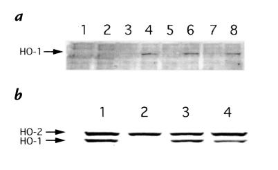
The first known human case of heme oxygenase-1 (HO-1) deficiency is presented in this report. The patient is a six-year-old boy with severe growth retardation. He has been suffering from persistent hemolytic anemia characterized by marked erythrocyte fragmentation and intravascular hemolysis, with paradoxical increase of serum haptoglobin and low bilirubin. An abnormal coagulation/fibrinolysis system, associated with elevated thrombomodulin and von Willebrand factor, indicated the presence of severe, persistent endothelial damage. Electron microscopy of renal glomeruli revealed detachment of endothelium, with subendothelial deposition of an unidentified material. Iron deposition was noted in renal and hepatic tissue. Immunohistochemistry of hepatic tissue and immunoblotting of a cadmium-stimulated Epstein-Barr virus-transformed lymphoblastoid cell line (LCL) revealed complete absence of HO-1 production. An LCL derived from the patient was extremely sensitive to hemin-induced cell injury. Sequence analysis of the patient's HO-1 gene revealed complete loss of exon-2 of the maternal allele and a two-nucleotide deletion within exon3 of the paternal allele. Growth retardation, anemia, iron deposition, and vulnerability to stressful injury are all characteristics observed in recently described HO-1 targeted mice. This study presents not only the first human case of HO-1 deficiency but may also provide clues to the key roles played by this important enzyme in vivo.


Figures

Figure 1
Figure 1
Plasma and peripheral blood smears. (a) Centrifuged peripheral venous blood from control (left) and patient (right). (b) Peripheral blood smear was stained by May-Grünwald and Giemsa. Arrows indicate fragmented erythrocytes.
Figure 2
Figure 2
Pathology of renal glomerular loop. (a) Hematoxylin and eosin staining of the renal glomeruli. (b) Electron microscopy of glomerular capillary of the patient. Arrow indicates the detached endothelium and asterisk indicates subendothelial deposit. (c) Electron microscopy of control glomerular capillary.
Figure 3
Figure 3
Iron staining of the kidney and liver. Paraffin-embedded specimens of the kidney and liver were stained for iron. (a) Kidney. ×200. (b) Liver. ×200. Asterisk indicates a glomerulus.
Figure 4
Figure 4
Immunohistochemical staining of HO-1 in the liver. Paraffin-embedded specimen of the liver was stained with anti–HO-1 antiserum. (a) Control. (b) Patient. Arrows indicate Kupffer cells. HO-1, heme oxygenase-1.
Figure 5
Figure 5
Immunoblotting of HO-1 induced in cadmium-stimulated cells. (a) PBMC from patient (lanes 1 and 2) and control 1 (lanes 3 and 4). Control 2 (lanes 5 and 6), and control 3 (lanes 7 and 8) were cultured alone (lanes 1, 3, 5, and 7) or with cadmium (lanes 2, 4, 6, and 8). Lysate samples were prepared as shown in Methods. Blotted membrane was reacted simultaneously with anti–HO-1 antiserum and anti–HO-2 antiserum, followed by the reaction with HRP-conjugated anti–rabbit IgG. (b) LCLs from patient, parents, and control were cultured with cadmium and treated similarly. Lane 1, control; lane 2, patient; lane 3, father; and lane 4, mother. HRP, horseradish peroxidase; LCL, lymphoblastoid cell line, PBMC, peripheral blood mononuclear cells.
Figure 6
Figure 6
Cytotoxicity assay. LCLs from patient (closed circles) and control (open circles) were cultured for 24 h with different concentrations of hemin. Dead or apoptotic cells were identified by lowered forward light scatter and high annexin-V binding using a flow cytometer. Percent survival was determined compared with cells cultured with medium. Each datum represents the mean ± SD of five independent experiments. *P < 0.001.
Figure 7
Figure 7
Structural organization of HO-1 gene and construction of PCR primers for mutational analysis.
Figure 8
Figure 8
Mutational analysis of HO-1 gene. (a) Reverse transcriptase-PCR for HO-1 mRNA. M denotes molecular weight marker. Lane 1, control; lane 2, patient; lane 3, father; and lane 4, mother. (b) Sequence analysis of paternal allele and normal control allele. (c) Sequence analysis of maternal allele and normal control allele. (d) Mutation- specific PCR of genomic DNA. Data from control (lane 1), patient (lane 2), father (lane 3), and mother (lane 4) are shown. (e) Control PCR.

References

    1. Maines MD. Heme oxygenase: function, multiplicity, regulatory mechanism, and clinical applications. FASEB J. 1988;2:2557–2568. - PubMed
    1. Maines MD. The heme oxygenase system: a regulator of second messenger gases. Annu Rev Pharmacol Toxicol. 1997;37:517–554. - PubMed
    1. Okinaga S, et al. Regulation of human heme oxygenase-1 gene expression under thermal stress. Blood. 1996;87:5074–5084. - PubMed
    1. Cantoni L, Rossi C, Rizzardini M, Gardina M, Ghezzi P. Interleukin-1 and tumor necrosis factor induce hepatic haeme oxygenase. Biochem J. 1991;279:891–894. - PMC - PubMed
    1. Willis D, Moore AR, Frederick R, Willoughby DA. Heme oxygenase: a novel target for the modulation of the inflammatory response. Nat Med. 1996;2:87–90. - PubMed

Publication types

MeSH terms