. 1999 Jan 11;144(1):83-98.
doi: 10.1083/jcb.144.1.83.

Coronin promotes the rapid assembly and cross-linking of actin filaments and may link the actin and microtubule cytoskeletons in yeast

Affiliations

Coronin promotes the rapid assembly and cross-linking of actin filaments and may link the actin and microtubule cytoskeletons in yeast

B L Goode et al. J Cell Biol. .

Abstract

Coronin is a highly conserved actin-associated protein that until now has had unknown biochemical activities. Using microtubule affinity chromatography, we coisolated actin and a homologue of coronin, Crn1p, from Saccharomyces cerevisiae cell extracts. Crn1p is an abundant component of the cortical actin cytoskeleton and binds to F-actin with high affinity (Kd 6 x 10(-9) M). Crn1p promotes the rapid barbed-end assembly of actin filaments and cross-links filaments into bundles and more complex networks, but does not stabilize them. Genetic analyses with a crn1Delta deletion mutation also are consistent with Crn1p regulating filament assembly rather than stability. Filament cross-linking depends on the coiled coil domain of Crn1p, suggesting a requirement for Crn1p dimerization. Assembly-promoting activity is independent of cross-linking and could be due to nucleation and/or accelerated polymerization. Crn1p also binds to microtubules in vitro, and microtubule binding is enhanced by the presence of actin filaments. Microtubule binding is mediated by a region of Crn1p that contains sequences (not found in other coronins) homologous to the microtubule binding region of MAP1B. These activities, considered with microtubule defects observed in crn1Delta cells and in cells overexpressing Crn1p, suggest that Crn1p may provide a functional link between the actin and microtubule cytoskeletons in yeast.


Figures

Figure 3
Figure 3
Crn1p bundles actin filaments and can cross-link actin filaments and microtubules. (A) Low speed pelleting of Crn1p–F-actin complexes. Crn1p (0.5 μM) or Crn1p (Δ600–651; 0.5 μM) was mixed with preassembled yeast actin filaments (2 μM), incubated for 15 min at 25°C, and centrifuged for 3 min at low speed (13,000 g). The pellets and supernatants were analyzed by SDS-PAGE and Coomassie staining. (B) Crn1p decreases the apparent viscosity of actin filament solutions in a concentration-dependent manner. Variable concentrations of Crn1p or yeast fimbrin/Sac6p (0.15–2.5 μM) were added to preassembled actin filaments (7 μM), and apparent viscosity was measured by the falling ball assay. (C) Electron micrographs of Crn1p-actin filament bundles and fimbrin/Sac6p-actin filament bundles. Crn1p or Sac6p (0.5 μM) was added to preassembled yeast actin filaments (5 μM), incubated for 30 min at 25°C, then negatively stained and examined by electron microscopy. (D) Cosedimentation of microtubules with Crn1p-F-actin bundles. 4 μM taxol-stabilized microtubules, 4 μM preassembled F-actin, 2 μM Crn1p, and 2 μM Sac6p were mixed in the combinations indicated in the figure and incubated for 30 min at 25°C. The reactions then were centrifuged at low speed (13,000 g) for 3 min, and the pellets and supernatants were analyzed by SDS-PAGE gels and Coomassie staining. Bar, 200 nm.
Figure 4
Figure 4
Crn1p promotes the rapid barbed-end assembly and cross-linking of actin filaments into branched networks. (A) Effects of Crn1p on assembly kinetics of yeast actin by light scattering assay. Monomeric yeast actin (5 μm) in G-buffer was mixed with variable concentrations of Crn1p and assembly initiation salts at time 0. Actin filament assembly was monitored by change in light scattering at 400 nm in a spectrophotometer. Steady state actin filament assembly was reached by 20 min in all reactions, and at this point reactions contained similar polymer levels (see Materials and Methods). (B) Effects of Crn1p on the assembly kinetics of pyrene-actin. Variable concentrations of Crn1p (0, 0.5, or 1 μM) were added to 5 μM actin (4 μM yeast actin and 1 μM pyrene-labeled rabbit muscle actin), and actin filament assembly was monitored by increase in pyrene fluorescence (excitation 365 nm/excitation 407 nm). Fluorescence units are arbitrary. (C) Electron micrographs of actin assembly reactions containing 5 μM yeast actin in the absence or presence of 0.5 μM Crn1p. Samples were removed from the assembly reactions at 2 min and at steady state (30 min), fixed in 0.5% glutaraldehyde, negatively stained, and examined by electron microscopy. (D) Crn1p causes a concentration-dependent increase in apparent viscosity when coassembled with actin. Variable concentrations of Crn1p (0.2–1.5 μM) were added to monomeric yeast G-actin (7 μM), incubated for 20 min at 25°C, and apparent viscosity was measured by the falling ball assay. (E) Crn1p induces barbed-end actin filament assembly. The assembly of 5 μM actin (4 μM yeast actin and 1 μM pyrene-labeled rabbit muscle actin) was compared in the presence or absence of 100 nM cytochalasin D, which blocks barbed-end filament assembly, and in the presence or absence of 0.5 μM Crn1p. Assembly was nucleated with preassembled, sheared yeast actin filament seeds (0.5 μM). (F) Effects of Crn1p (Δ600–651) on the kinetics of yeast actin filament assembly measured by light scattering assay. The assembly of monomeric yeast actin (5 μM) was monitored by light scattering as above in the presence or absence of 0.5 μM Crn1p (Δ600–651), which lacks filament cross-linking activity. (G) Effects of Crn1p (Δ600– 651) on the kinetics of pyrene-actin assembly. Variable concentrations of Crn1p (Δ600–651; 0, 0.25, 0.5, or 1 μM) were added to 5 μM monomeric actin (4 μM yeast actin and 1 μM pyrene-labeled rabbit muscle actin), and actin filament assembly was monitored by increase in pyrene fluorescence as above. Bar, 200 nm.
Figure 4
Figure 4
Crn1p promotes the rapid barbed-end assembly and cross-linking of actin filaments into branched networks. (A) Effects of Crn1p on assembly kinetics of yeast actin by light scattering assay. Monomeric yeast actin (5 μm) in G-buffer was mixed with variable concentrations of Crn1p and assembly initiation salts at time 0. Actin filament assembly was monitored by change in light scattering at 400 nm in a spectrophotometer. Steady state actin filament assembly was reached by 20 min in all reactions, and at this point reactions contained similar polymer levels (see Materials and Methods). (B) Effects of Crn1p on the assembly kinetics of pyrene-actin. Variable concentrations of Crn1p (0, 0.5, or 1 μM) were added to 5 μM actin (4 μM yeast actin and 1 μM pyrene-labeled rabbit muscle actin), and actin filament assembly was monitored by increase in pyrene fluorescence (excitation 365 nm/excitation 407 nm). Fluorescence units are arbitrary. (C) Electron micrographs of actin assembly reactions containing 5 μM yeast actin in the absence or presence of 0.5 μM Crn1p. Samples were removed from the assembly reactions at 2 min and at steady state (30 min), fixed in 0.5% glutaraldehyde, negatively stained, and examined by electron microscopy. (D) Crn1p causes a concentration-dependent increase in apparent viscosity when coassembled with actin. Variable concentrations of Crn1p (0.2–1.5 μM) were added to monomeric yeast G-actin (7 μM), incubated for 20 min at 25°C, and apparent viscosity was measured by the falling ball assay. (E) Crn1p induces barbed-end actin filament assembly. The assembly of 5 μM actin (4 μM yeast actin and 1 μM pyrene-labeled rabbit muscle actin) was compared in the presence or absence of 100 nM cytochalasin D, which blocks barbed-end filament assembly, and in the presence or absence of 0.5 μM Crn1p. Assembly was nucleated with preassembled, sheared yeast actin filament seeds (0.5 μM). (F) Effects of Crn1p (Δ600–651) on the kinetics of yeast actin filament assembly measured by light scattering assay. The assembly of monomeric yeast actin (5 μM) was monitored by light scattering as above in the presence or absence of 0.5 μM Crn1p (Δ600–651), which lacks filament cross-linking activity. (G) Effects of Crn1p (Δ600– 651) on the kinetics of pyrene-actin assembly. Variable concentrations of Crn1p (Δ600–651; 0, 0.25, 0.5, or 1 μM) were added to 5 μM monomeric actin (4 μM yeast actin and 1 μM pyrene-labeled rabbit muscle actin), and actin filament assembly was monitored by increase in pyrene fluorescence as above. Bar, 200 nm.
Figure 1
Figure 1
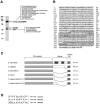
(A) Coisolation from yeast extracts of actin and Crn1p on a microtubule affinity column. Proteins were isolated from S. cerevisiae extracts using an affinity column containing taxol-stabilized bovine microtubules (see Materials and Methods). Shown is a Coomassie-stained gel of proteins eluted from the column by 0.5 M KCl. The proteins in this fraction were digested with trypsin, and the resulting peptides were identified by LC/MS/MS tandem mass spectrometry and matched to predicted gene products in the S. cerevisiae genome database. All of the yeast peptides identified in the column fraction shown were derived from actin or the product of the YLR429w gene. The peptide sequences are shown adjacent to the protein bands from which they were derived. (B) Predicted amino acid sequence of the YLR429w (CRN1) gene product. The unique region is boxed, and contains two sequences (underlined) that share homology with the microtubule binding region of MAP1B. A single consensus target sequence for Cdc28p kinase is shown in bold. (C) Alignment of the domain structures of S. cerevisiae, C. elegans, D. discoideum, B. taurus, and H. sapiens coronins. Each protein contains a conserved WD repeat region and COOH-terminal coiled-coil domain (shaded regions). In addition, each coronin has a COOH-terminal unique region (not shaded). The unique regions vary greatly in length and sequence. The unique region of yeast coronin (CRN1) is distinct from other coronins and contains two sequences (hatched boxes) with homology to the microtubule binding region of MAP1B. (D) Alignment of a sequence in MAP1B that contains a KKE/D microtubule binding motif with a similar sequence found in the unique region of S. cerevisiae coronin (Crn1p).
Figure 2
Figure 2
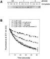
Crn1p binds to actin filaments and microtubules. (A) Immunoblot of total wild-type (WT) and crn1Δ cell proteins probed with coronin antibodies. (B) Crn1p binds to actin filaments with high affinity. Crn1p (0.5 nM) was mixed with variable concentrations of phalloidin-stabilized yeast actin filaments. After incubation, the actin filaments were pelleted, and the pellets and supernatants were analyzed by SDS-PAGE and immunoblotting with Crn1p antibodies. (C) Crn1p binds to microtubules with weak affinity. Full-length Crn1p (0.5 μM) was mixed with variable concentrations of taxol-stabilized bovine brain microtubules. After incubation, the microtubules were pelleted, and the pellets (P) and supernatants (S) were analyzed by SDS-PAGE and Coomassie staining.
Figure 5
Figure 5
Crn1p has no significant effects on actin critical concentration (actin Cc) at steady state or the rate of actin filament depolymerization. (A) Crn1p does not affect significantly the actin Cc at steady state. Three different concentrations of preassembled yeast actin filaments (0.5, 1, or 2 μM) were mixed with four different molar ratios of Crn1p to actin (0, 1:10, 1:3, 1:1), incubated for 20 min, and the actin filaments in the reactions were pelleted by high speed centrifugation. Levels of unpolymerized actin (actin in the supernatant) were compared by SDS-PAGE and Coomassie staining. (B) Crn1p does not affect the rate of actin filament depolymerization. 5 μM preassembled F-actin (4 μM yeast actin and 1 μM pyrene-labeled rabbit muscle actin) was added to 0.5 μM Crn1p, 1 μM Crn1p, 1 μM Sac6p/fimbrin, or 100 nM cytochalasin D. After incubation for 15 min at 25°C, filament disassembly was induced by the addition of 40 μM latrunculin A at time 0, and depolymerization was monitored by change in pyrene fluorescence as described in Fig. 4 b.
Figure 6
Figure 6
Activities of Crn1p domains. (A) Coomassie-stained SDS-PAGE gel of Crn1p and Crn1p subfragments expressed and purified from E. coli. A = full-length (amino acids 1–651), B = 1–400, C = 400–651, and D = 1–599. (B) Biochemical activities of Crn1p polypeptides. Each polypeptide was tested for microtubule and F-actin binding by cosedimentation assay, ability to promote F-actin assembly by light scattering assay, and actin filament bundling by low speed pelleting assay and electron microscopy.
Figure 6
Figure 6
Activities of Crn1p domains. (A) Coomassie-stained SDS-PAGE gel of Crn1p and Crn1p subfragments expressed and purified from E. coli. A = full-length (amino acids 1–651), B = 1–400, C = 400–651, and D = 1–599. (B) Biochemical activities of Crn1p polypeptides. Each polypeptide was tested for microtubule and F-actin binding by cosedimentation assay, ability to promote F-actin assembly by light scattering assay, and actin filament bundling by low speed pelleting assay and electron microscopy.
Figure 7
Figure 7
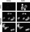
Localization of Crn1p in yeast cells. Actin and Crn1p localization in wild-type (DDY1088) and crn1Δ (DDY1521) strains was examined by double-label immunofluorescence microscopy using Crn1p and actin antibodies.
Figure 8
Figure 8
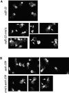
Synthetic defects in actin organization in crn1Δ cof1-22 and crn1Δ act1-159 cells. Haploid segregants from crn1Δ × cof1-22 and crn1Δ × act1-159 genetic crosses were grown to log phase at 20°C, and actin organization was examined by rhodamine phalloidin staining. crn1Δ cells and wild-type cells had normal actin staining (not shown). Actin staining is shown for cof1-22 and cof1-22 crn1Δ cells (A) and for act1-159 and act1-159 crn1Δ cells (B). Arrows mark defects in actin organization specific to the double mutants.
Figure 9
Figure 9
Deletion of the CRN1 gene causes pronounced defects in cytoplasmic microtubules at a low frequency. Wild-type (DDY1519) and crn1Δ (DDY1518) cells were grown at 20°C to OD600 = 0.5 and examined by immunofluorescence microscopy using tubulin antibodies. Approximately 5% of the medium to large budded crn1Δ cells had abnormally long cytoplasmic microtubules (marked by arrows) that emanate from the microtubule organizing center in the bud and extend back towards or through the bud neck into the mother cell. Similar defects were never seen in the wild-type cells (n > 1,000 cells).
Figure 10
Figure 10
Overexpression of GST-Crn1p causes severe defects in cell growth and microtubule and actin organization. Wild-type cells (DDY130) carrying the pEGKT-CRN1 plasmid were grown to log phase at 25°C in minimal selective medium (lacking uracil and leucine) with glucose. Cells were washed and transferred to minimal selective medium with 2% raffinose for 12 h of growth at 25°C. Then 2% galactose was added to induce the expression of GST-Crn1p. After 4 h growth at 25°C, the cells were examined by double label immunofluorescence microscopy using actin and tubulin antibodies. Bar, 10 μM.

References

    1. Ayscough, K.R., and D.G. Drubin. 1997. Immunofluorescence microscopy of yeast cells. In Cell Biology, A Laboratory Handbook. Academic Press. San Diego, CA.
    1. Ayscough KR. In vivo functions of actin-binding proteins. Curr Opin Cell Biol. 1998;10:102–111. - PubMed
    1. Ayscough KR, Stryker J, Pokala N, Sanders M, Crews P, Drubin DG. High rates of actin filament turnover in budding yeast and roles for actin in establishment and maintenance of cell polarity revealed using the actin inhibitor latrunculin A. J Cell Biol. 1997;137:399–416. - PMC - PubMed
    1. Barnes G, Andrea K, Louie, Botstein D. Yeast proteins associated with microtubules in vitro and in vivo. Mol Biol Cell. 1992;3:29–47. - PMC - PubMed
    1. Belmont L, Drubin DG. The yeast V159N actin mutant reveals roles for actin dynamics in vivo. J Cell Biol. 1998;142:1289–1299. - PMC - PubMed

Publication types

MeSH terms