. 1999 Jan 11;144(1):161-73.
doi: 10.1083/jcb.144.1.161.

Role of CDMP-1 in skeletal morphogenesis: promotion of mesenchymal cell recruitment and chondrocyte differentiation

Affiliations

Role of CDMP-1 in skeletal morphogenesis: promotion of mesenchymal cell recruitment and chondrocyte differentiation

N Tsumaki et al. J Cell Biol. .

Abstract

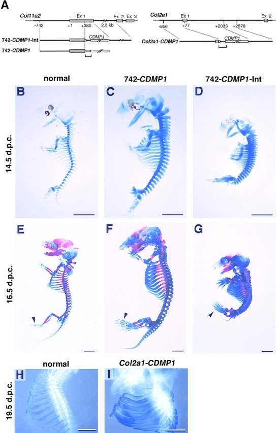
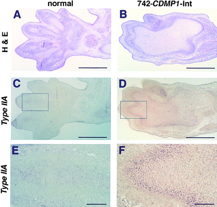
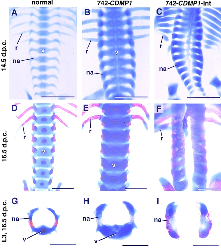
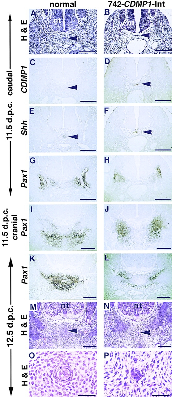
Cartilage provides the template for endochondral ossification and is crucial for determining the length and width of the skeleton. Transgenic mice with targeted expression of recombinant cartilage-derived morphogenetic protein-1 (CDMP-1), a member of the bone morphogenetic protein family, were created to investigate the role of CDMP-1 in skeletal formation. The mice exhibited chondrodysplasia with expanded cartilage, which consists of the enlarged hypertrophic zone and the reduced proliferating chondrocyte zone. Histologically, CDMP-1 increased the number of chondroprogenitor cells and accelerated chondrocyte differentiation to hypertrophy. Expression of CDMP-1 in the notochord inhibited vertebral body formation by blocking migration of sclerotome cells to the notochord. These results indicate that CDMP-1 antagonizes the ventralization signals from the notochord. Our study suggests a molecular mechanism by which CDMP-1 regulates the formation, growth, and differentiation of the skeletal elements.


Figures

Figure 1
Figure 1
Transgene constructs and skeletal abnormalities of CDMP-1 transgenic mice. (A) Diagrams of the DNA constructs used to generate Col11a2-CDMP1 (left) and Col2a1-CDMP1 (right) transgenic mice. The gene structures of Col11a2 and Col2a1 are shown at the top of each panel. Boxes indicate the coding regions and solid lines denote noncoding sequences. The transgenes are shown below the genomic map. Brackets indicate intron cassettes. (B–I) Skeletons of CDMP-1 transgenic mice. Entire skeletons of normal (B and E), 742- CDMP1 transgenic (C and F), and 742- CDMP1-Int transgenic (D and G) embryos were stained with alcian blue (B–D) and alcian blue plus alizarin red (E–G). The rib cages of a normal mouse (H) and a Col2a1-CDMP1 transgenic mouse (I) were stained with alcian blue and shown in dark field. (B–D) Embryos at 14.5 d.p.c.; (E–G) 16.5 d.p.c.; (H and I), 19.5 d.p.c. 742- CDMP1 transgenic mice were large, whereas 742-CDMP1-Int and Col2a1-CDMP1 transgenic mice were small. The skeletal components were thick in all transgenic mice. In 742-CDMP1-Int and Col2a1 transgenic mice, the ribs were so thick that intercostal spaces were almost eliminated. Ossification had just started at metatarsals in normal and transgenic mice (E–G, arrowheads). Scale bars, 2 mm.
Figure 2
Figure 2
Examination of the skeleton of the forelimbs and ribs of CDMP-1 transgenic mice. (A–C) Skeletal components of the forelimb at 16.5 d.p.c. stained with alcian blue and alizarin red. In the 742-CDMP1 (B) and 742- CDMP1-Int (C) transgenic mice, the distal components (brackets), such as the carpals, metacarpals, and phalanges, were larger than those of normal mice (A), whereas the proximal component such as the scapula of the transgenic mice was not so enlarged. The extent of deformity of 742-CDMP1-Int transgenic mice was more severe than that of 742-CDMP1 transgenic mice. (D and E) Skeletal components of the forelimb at 19.5 d.p.c. stained with alcian blue shown in the dark field. In the Col2a1-CDMP1 transgenic mouse (E), the skeleton of the paws was much enlarged compared with the normal mouse (D). Note the difference in the scale between D and E. 742-CDMP1- Int and Col2a1-CDMP1 transgenic mice had fused joints, whereas 742- CDMP1 transgenic mice did not. The humerus, radius, and ulna of 742- CDMP1-Int and Col2a1-CDMP1 transgenic mice were united by bone. (F–H) Ribs at 16.5 d.p.c. stained with alcian blue and alizarin red. The ribs of 742-CDMP1 (G) and 742-CDMP1-Int (H) transgenic mice were thicker than those of normal mice (F) at 16.5 d.p.c. (I and J) Ribs at 19.5 d.p.c. stained with alcian blue shown in the dark field. The Col2a1-CDMP1 transgenic mouse (I) also had much thicker ribs than normal mice (J). The degree of the thickness of the skeleton and the enlargement of the paw in 742- CDMP1-Int transgenic mice were consistently more severe than those of 742-CDMP1 transgenic mice. Although stages were different, the degree of deformity in the Col2a1-CDMP1 transgenic mouse appeared to be more severe than that of 742-CDMP1-Int transgenic mice. s, scapula; h, humerus; r, radius; u, ulna. Brackets in A–E indicate skeletal components of paws including carpals, metacarpals, and phalanges. Scale bars, 1 mm.
Figure 3
Figure 3
Gene expression of CDMP-1 transgenic mice. (A) Northern blot analysis using total RNAs extracted from limb buds of normal (lane 1), 742- CDMP1 (lane 2), and 742-CDMP1-Int (lane 3) transgenic mice at 14.5 d.p.c. For each lane, 20 μg RNA was loaded, transferred to the nylon membrane, and hybridized with corresponding probes indicated at the left of the panels. The bottom panel shows the ethidium bromide–stained gel before transfer. Transgenic CDMP-1 mRNA was expressed strongly compared with endogenous Gdf5/CDMP1 mRNA (arrowhead). The expression levels of Ihh and Col10a1 mRNAs, markers for chondrocyte differentiation, were elevated in the transgenic mice. (B) Distribution of CDMP1 mRNA in the forelimb of normal (left), 742-CDMP1 transgenic (middle), and 742-CDMP1-Int transgenic (right) mice at 14.5 d.p.c. (Top) Staining with hematoxylin and eosin. (Bottom) In situ hybridization of semiserial sections using CDMP1 antisense probe. In the transgenic mice, CDMP-1 mRNA was distributed in proliferating chondrocytes, but not in hypertrophic chondrocytes. Note that the height of the hypertrophic zone was increased in transgenic mice. (C) CDMP1 and Col11a2 expression in the rib of normal (left) and 742- CDMP1-Int transgenic (right) mice at 16.5 d.p.c. (Top) Staining with hematoxylin and eosin. (Middle) In situ hybridization of CDMP1 antisense probe of semiserial sections. (Bottom) Col11a2 antisense probe. In the transgenic mice, CDMP-1 mRNA was distributed in proliferating chondrocytes, but not in hypertrophic chondrocytes. The localization of CDMP-1 mRNA was identical to that of Col11a2 mRNA. (D) Immunohistochemical analysis of the primordial cartilage of the tarsals of normal (left) and 742- CDMP1-Int transgenic (right) mice at 14.5 d.p.c. (Top) Staining with hematoxylin and eosin. (Bottom) Immunostaining of anti–CDMP-1 polyclonal antibody showed transgenic cartilage contained a significant amount of CDMP-1. r, resting chondrocytes; p, proliferating chondrocytes; h, hypertrophic chondrocytes. Scale bars, B, 500 μm; C, 200 μm; D, 50 μm.
Figure 4
Figure 4
Altered endochondral bone formation in Col11a2-CDMP1 transgenic mice. (A) Histological sections of the humerus of normal (left) and 742- CDMP1-Int transgenic mice (right) at 14.5 d.p.c. (Top) Staining with Safranin O-fast green-iron hematoxylin of entire humerus. The transgenic mice (right) had the increased height of a zone of hypertrophic chondrocytes (h) compared to normal mice (left). In the transgenic mice, the primordial cartilage was wide and elbow joints were fused. (Bottom) Magnifications of the boxed regions of the distal part of the humerus in the top panels. The height of zones of proliferating chondrocytes was reduced in the transgenic mice. Note that arrays of chondrocytes at the bottom (asterisk) were different from those of chondrocytes located above in the transgenic mice. They were chondrocytes of the proximal part of the ulna. Hematoxylin and eosin staining. (B) Spatial expression of marker genes for endochondral bone formation in the forelimb of normal (left) and 742- CDMP1-Int transgenic (right) mice at 16.5 d.p.c. (Top row) Staining with Safranin O-fast green-iron hematoxylin. The skeletal components consist of proliferating and hypertrophic zones of chondrocytes and bone. In the transgenic mice, the carpals and distal part of the radius and ulna were completely fused and underwent endochondral bone formation as a single component. Semiserial sections were hybridized with cRNA probes of corresponding genes indicated on the left. Areas expressing Ihh (fourth row) and type X collagen (fifth row) mRNAs were remarkably enlarged in transgenic mice. r, resting chondrocytes; p, proliferating chondrocytes; h, hypertrophic chondrocytes. Scale bars, 100 μm.
Figure 5
Figure 5
Mesenchymal condensation and perichondrium of CDMP-1 transgenic mice. (A, C, and E) Normal mice. (B, D, and F) 742-CDMP1-Int transgenic mice. (A and B) Axial sections of mesenchymal condensation in the forelimb bud at 12.5 d.p.c. Mesenchymal condensation was already expanded in transgenic mice. Hematoxylin and eosin staining. (C and D) AgNORs in mesenchyme of boxed portions in top panels. In normal mice (C), peripheral cells (arrowheads) located between the central part of mesenchymal condensation (c) and surrounding mesenchymal cells (m) contained large number of AgNORs, indicative of proliferative activities. In transgenic mice (D), the number of peripheral cells was increased and formed multiple layers. (E and F) Perichondrium (arrows) of epiphyseal cartilage (e) of the distal ulna at 14.5 d.p.c. Compared with normal mice, transgenic mice showed thick perichondrium consisting of multiple cell layers. Scale bars, 50 μm.
Figure 6
Figure 6
Expression of type IIA collagen mRNA in the forelimb at 13.5 d.p.c. (A and B) Hematoxylin and eosin staining. In the transgenic mice, the phalanges, carpals, and distal part of the radius and ulna were completely fused and formed single thick cartilage anlage. (C–F) In situ hybridization with the antisense type IIA collagen mRNA probe of semiserial sections (C and D) and magnification of the boxed regions (E and F). Type IIA mRNA-positive cells surrounded the cartilage anlage in transgenic mice, whereas only faint signals were detected in normal mice. Scale bars, A–D, 500 μm; E and F, 100 μm.
Figure 7
Figure 7
Spinal columns stained with alcian blue and alizarin red. Normal (A, D, and G), 742-CDMP1 transgenic (B, E, and H), and 742-CDMP1-Int transgenic (C, F, and I) mice. (A–F) The spinal columns with the ribs were viewed from the ventral side at 14.5 d.p.c. (A–C) and at 16.5 d.p.c. (D–F). 742-CDMP1 transgenic mice had thick skeletal components including the vertebral bodies (v), neural arches (na), and ribs (r) (B and E). In 742-CDMP1- Int transgenic mice, the vertebral bodies were absent (C and F). (G–I) Axial view of the third lumber vertebrae of mice at 16.5 d.p.c. The vertebral body was enlarged in 742-CDMP1 transgenic mice (H) and absent in 742- CDMP1-Int transgenic mice (I). v, vertebral bodies; na, neural arch; r, rib. Scale bars, 1 μm.
Figure 8
Figure 8
Histology and expression analysis of spine of 742- CDMP1-Int transgenic mice. (A–H) Axial semiserial sections at caudal level (in tail region) of the spine of normal (A, C, E, and G) and 742-CDMP1-Int transgenic (B, D, F, and H) embryos at 11.5 d.p.c. Staining with hematoxylin and eosin (A and B). In situ hybridization of CDMP1 (C and D), Shh (E and F), and Pax1 (G and H) antisense probes. In transgenic mice, CDMP-1 mRNA was localized in the notochord. (I and J) In situ hybridization of sections at the cranial level (thoracic region) of the spine of normal (I) and 742-CDMP1-Int transgenic (J) embryos at 11.5 d.p.c. using Pax1 antisense probe. (K–N) Axial semiserial sections of spine in the thoracic region in normal (K and M) and 742- CDMP1-Int transgenic (L and N) embryos at 12.5 d.p.c. In situ hybridization of Pax1 (K and L) and staining with hematoxylin and eosin (M and N). In normal mice, Pax1-expressing sclerotome cells migrated toward the notochord to form aggregation as the developmental stage progressed (G, I, and K). In transgenic mice, those cells remained laterally (H, J, and L). (O and P) Magnifications of the region around the notochord in M and N, respectively. In 742-CDMP1-Int transgenic mice, mesenchymal cells around the notochord were disorganized, and the sheath of the notochord was not clear. Arrowhead, notochord; nt, neural tube. Scale bars, A–N, 200 μm; O and P, 50 μm.
Figure 9
Figure 9
Schematic drawing of the action of CDMP-1 on cartilage formation and differentiation. (Left) Skeletogenesis is initiated with commitment of mesenchymal condensation to the chondrocytic lineage and followed by a series of differentiation process of chondrocytes and replacement by bone in endochondral bone formation. These temporal events are represented spatially with zone structures named as resting (r), proliferative (p), and hypertrophic zones (h) which are arrayed from end to center of the primordial cartilage. (Right) We propose that CDMP-1 promotes the recruitment of mesenchymal cells into the chondrogenic lineage, resulting in the radial expansion of the primordial cartilage. CDMP-1 affects endochondral ossification by accelerating differentiation of chondrocytes to hypertrophy, resulting in the expansion of bones. Through these mechanisms, CDMP-1 regulates the width and length of the skeletal components.

References

    1. Chang SC, Hoang B, Thomas JT, Vukicevic S, Luyten FP, Ryba NJ, Kozak CA, Reddi AH, Moos M., Jr Cartilage-derived morphogenetic proteins. New members of the transforming growth factor-β superfamily predominantly expressed in long bones during human embryonic development. J Biol Chem. 1994;269:28227–28234. - PubMed
    1. Christ B, Wilting J. From somites to vertebral column. Anat Anz. 1992;174:23–32. - PubMed
    1. Crocker J, Nar P. Nuclear organizer regions in lymphomas. J Pathol. 1987;151:111–118. - PubMed
    1. Deutsch U, Dressler GR, Gruss P. Pax 1, a member of a paired box homologous murine gene family, is expressed in segmented structures during development. Cell. 1988;53:617–625. - PubMed
    1. Dewulf N, Verschueren K, Lonnoy O, Morén A, Grimsby S, Vande K, Spiegle, Miyazono K, Huylebroeck D, Ten P, Dijke Distinct spatial and temporal expression patterns of two type I receptors for bone morphogenetic proteins during mouse embryogenesis. Endocrinology. 1995;136:2652–2663. - PubMed

Substances