. 1999 Jan 1;13(1):64-75.
doi: 10.1101/gad.13.1.64.

Functional role of p35srj, a novel p300/CBP binding protein, during transactivation by HIF-1

Affiliations

Functional role of p35srj, a novel p300/CBP binding protein, during transactivation by HIF-1

S Bhattacharya et al. Genes Dev. .

Abstract

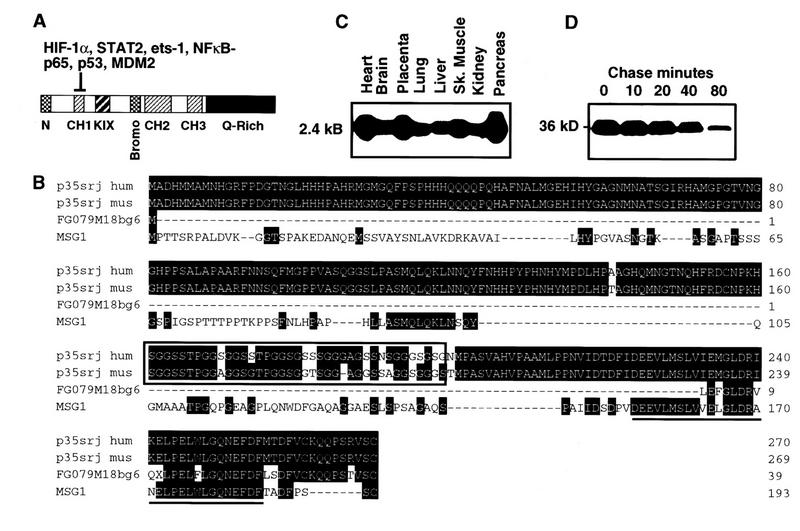
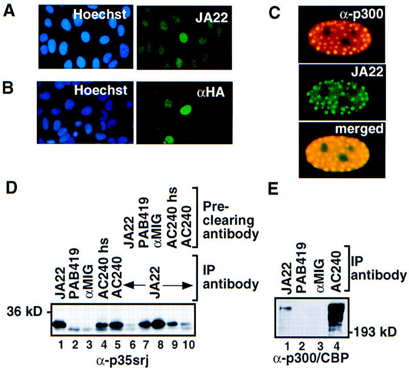
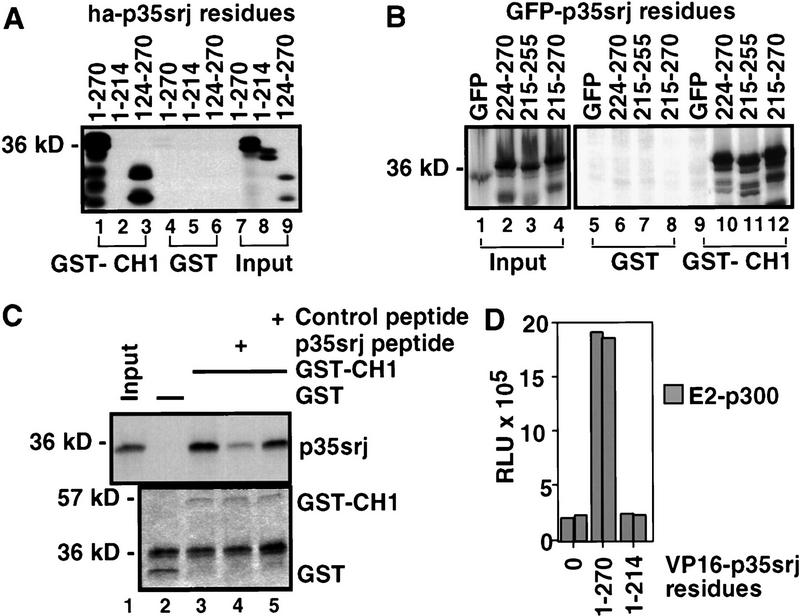
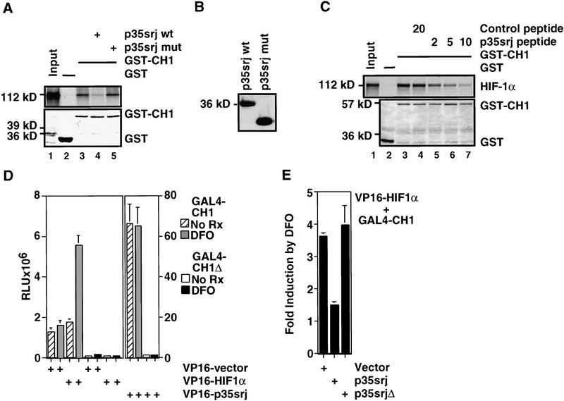
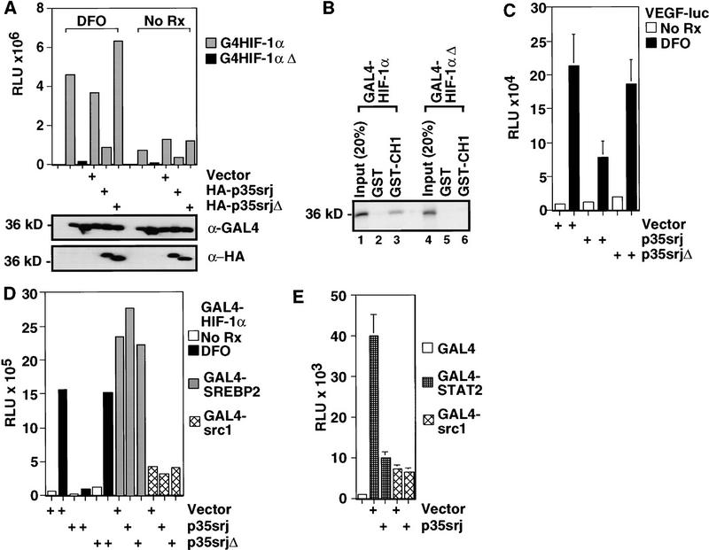
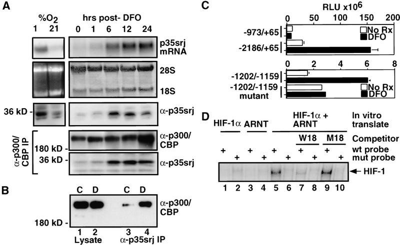
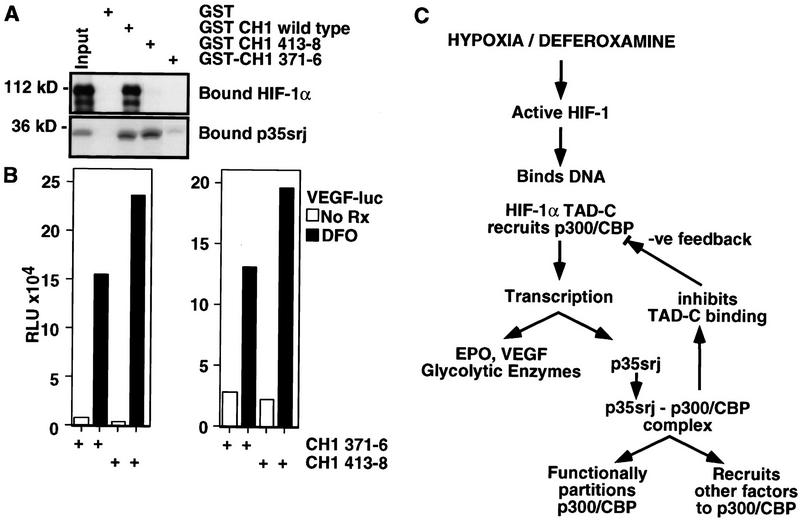
Recruitment of p300/CBP by the hypoxia-inducible factor, HIF-1, is essential for the transcriptional response to hypoxia and requires an interaction between the p300/CBP CH1 region and HIF-1alpha. A new p300-CH1 interacting protein, p35srj, has been identified and cloned. p35srj is an alternatively spliced isoform of MRG1, a human protein of unknown function. Virtually all endogenous p35srj is bound to p300/CBP in vivo, and it inhibits HIF-1 transactivation by blocking the HIF-1alpha/p300 CH1 interaction. p35srj did not affect transactivation by transcription factors that bind p300/CBP outside the CH1 region. Endogenous p35srj is up-regulated markedly by the HIF-1 activators hypoxia or deferoxamine, suggesting that it could operate in a negative-feedback loop. In keeping with this notion, a p300 CH1 mutant domain, defective in HIF-1 but not p35srj binding, enhanced endogenous HIF-1 function. In hypoxic cells, p35srj may regulate HIF-1 transactivation by controlling access of HIF-1alpha to p300/CBP, and may keep a significant portion of p300/CBP available for interaction with other transcription factors by partially sequestering and functionally compartmentalizing cellular p300/CBP.


Figures

Figure 1
Figure 1
Cloning, expression, and stability of p35srj. (A) p300 and CBP contain multiple, conserved regions. These include three cysteine–histidine rich regions (CH1–3), the KIX domain (which binds CREB), a bromodomain, and a glutamine-rich region. The p300–CH1 domain, fused to GST bearing a protein kinase A phosphorylation site, was labeled with 32P and used as a probe to clone p35srj. (B) p35srj peptide sequence and homology. The serine–glycine rich junction is conserved in murine p35srj, and is boxed. The minimal p300-binding domain (see Fig. 3) is underlined. FG079M18bg6 is a fragment from the Fugu genome project, detected by database search. MSG1 is a homologous human gene also identified by database searches. (C) Expression of p35srj, as reflected the results of a multiple human tissue Northern blot (Clontech) probed with human p35srj cDNA. (D) The stability of p35srj was analyzed by pulse-chase analysis performed in exponentially growing C2C12 cells. 35S-labeled p35srj was immunoprecipitated at the indicated time points.
Figure 2
Figure 2
p35srj is a nuclear protein bound to p300/CBP. (A) Cellular localization of endogenous p35srj. U2-OS cells were stained with Hoechst to demonstrate cell nuclei, and with anti-p35srj monoclonal antibody JA22, to demonstrate endogenous p35srj. (B) U2-OS cells were transfected with HA–p35srj, cell nuclei stained with Hoechst, and immunostained with anti-HA monoclonal antibody. (C) Colocalization of endogenous p35srj with p300. U2-OS cells were transfected with HA–p300 and stained with anti-HA polyclonal antibody to reveal typical p300 dot-like structures characteristic of p300-overproducing cells. These cells were reacted simultaneously with anti-p35srj monoclonal antibody, JA22, to show the colocalization of endogenous p35srj in the p300 dots. The merged exposure confirms that the dots colocalize. (D) Efficiency of endogenous p35srj coimmunoprecipitation with anti-p300/CBP antibodies. Anti-p35srj Western blot of immunoprecipitates (IP) from U2-OS cell lysates. Immunoprecipitations were performed with monoclonal antibodies to p35srj (JA22, lane 1), control antibodies (PAB419 and rabbit anti-mouse IgG, αMIG, lanes 2,3), and antibody to p300/CBP (AC240) (lanes 4,5). The AC240hs immunoprecipitation (lane 4) was performed in 300 mm NaCl. The supernatants from the above immunoprecipitates were reprecipitated with JA22 (lanes 6–10) to assess the relative levels of free p35srj. (E) Anti-p300/CBP Western blot of immunoprecipitates (IP) from U2-OS cell lysates.
Figure 3
Figure 3
p35srj carboxy-terminal residues are required for CH1 binding. (A) Binding of 35S-labeled, in vitro translated p35srj derivatives to GST or GST–p300CH1 fusion proteins (lanes 1–6). Twenty percent of the in vitro-translated input was loaded in lanes 7–9. (B) The indicated residues from the carboxyl terminus of p35srj, fused to green fluorescent protein (GFP), as carrier, were synthesized as 35S-labeled peptides by in vitro translation (lanes 1–4), and were assayed for ability to bind GST or GST–p300CH1 (lanes 5–12). (C) Effect of p35srj 224–255 peptide on in vitro binding of p35srj to p300–CH1. (Top) The binding of 35S-labeled p35srj, generated by in vitro translation, to GST–p300CH1 was tested in the presence of the p35srj peptide (20 μg, lane 4) and compared to either a control peptide (20 μg, lane 5) or no peptide (lane 3). Lane 1 shows 20% of the input in vitro translate. (Bottom) Coomassie stain of the gel demonstrating relative amounts of the GST and GST–p300CH1 proteins. (D) Mammalian two-hybrid assay in U2-OS cells transfected with a 3× E2–luciferase reporter (50 ng), E2–p300 (20 ng), expression vectors encoding the indicated p35srj derivatives fused to VP16 (25 ng), and CMV–lacZ (50 ng). The results of a representative experiment, performed in duplicate, are shown. E2–p300 contains the DNA-binding domain of BPV E2, fused to p300.
Figure 4
Figure 4
p35srj competes with HIF-1α for binding to p300–CH1 in vitro and in vivo. (A) Effect of baculoviral p35srj on in vitro binding of HIF-1α to p300–CH1. (Top) The binding of 35S-labeled HIF-1α (lane 1) generated by in vitro translation to bacterially produced GST (lane 2) or GST–p300CH1 (lane 3) immobilized on glutathione–Sepharose beads was tested. The binding of HIF-1α to GST–p300CH1 was also tested in the presence of baculovirally expressed wild-type (lane 4) or mutant p35srj (expressing p35srj residues 1–160) as control (lane 5). (Bottom) Coomassie stain of the gel demonstrating relative amounts of the GST and GST–p300CH1 proteins. (B) Anti-p35srj Western blot of SF21 cell lysates used in A. (C) Effect of p35srj 224–255 peptide on in vitro binding of HIF-1α to p300–CH1. (Top) The binding of HIF-1α to GST–p300CH1 was tested in the presence of increasing concentrations of the wild-type peptide (lanes 5–8) and compared to either a control, irrelevant peptide (lane 4) or no peptide (lane 3).Amounts of peptide used are shown in micrograms. Twenty percent of the HIF-1α input was loaded in lane 1. (Bottom) Coomassie stain of the gel demonstrating relative amounts of the GST and GST–p300CH1 proteins. (D) Effect of DFO on a mammalian two-hybrid interaction between GAL4–CH1 and VP16–HIF1α 723–826 (left) or VP16–p35srj (right). Hep3B cells were cotransfected with the indicated GAL4 and VP16 fusion plasmids (40 ng each), 3× GAL4–luc reporter (100 ng), and CMV–lacZ (100 ng). GAL4–CH1 contains p300 residues 300–528. GAL4–CH1Δ lacks p300 residues 346–410, and served as a control. Results are presented as relative luciferase units (RLU, mean of three independent experiments ± s.e.m.). (E) Effect of p35srj on the two-hybrid interaction between GAL4–p300CH1 and VP16–HIF1α. Hep3B cells were cotransfected with VP16HIF1α and GAL4–CH1 (40 ng each), and either vector control or p35srj expression plasmids (80 ng each), and CMV–lacZ (100 ng). p35srjΔ lacks residues 215–270. Results (mean of three independent experiments ± s.e.m.) are presented as fold induction of luciferase activity by DFO. A fold induction of 1 represents absence of induction.
Figure 5
Figure 5
p35srj inhibits HIF-1 transactivation by specifically affecting CH1 function. (A) Effect of p35srj on the activation of a GAL4–luciferase reporter by GAL4–HIF-1α (723–826). (Top) Hep3B cells were transiently cotransfected with a 3× GAL4–luc reporter (0.2 μg), and the indicated GAL4–HIF1α (0.2 μg), and HA–p35srj expression plasmids (0.2 μg), and CMV–lacZ (0.1 μg). GAL4–HIF1αΔ lacks the carboxy-terminal 13 residues of HIF-1α. Results are presented as RLU. The differences noted were statistically significant in three independent experiments. (Middle; bottom) The levels of GAL4–HIF1α and HA-tagged p35srj proteins in the above cell lysates were assayed by Western blotting with anti-GAL4 and anti-HA antibodies, respectively. The p35srjΔ mutation deletes residues 215–270, i.e., the p300–CH1 binding domain. (B) Effect of mutating the HIF-1α carboxyl terminus on its binding to p300–CH1. 35S-labeled proteins encoded by the indicated GAL4–HIF-1α plasmids were generated by in vitro translation, and their ability to bind GST–p300CH1 (lanes 3,6) or GST (as control, lanes 2,5) was assayed. Twenty percent of the in vitro-translated input was loaded as control in each case (lanes 1,4). (C) Effect of p35srj on the activation of a VEGF promoter–luciferase reporter by DFO. Hep3B cells were transiently cotransfected with a VEGF–luc reporter (40 ng), the indicated p35srj expression plasmids (300 ng each) and CMV–lacZ (100 ng). Results are presented as RLU (mean of three independent experiments ± s.e.m.). (D) Effect of p35srj on HIF-1α, SREBP2, and src1 transactivation. The indicated GAL4 fusion proteins (2 ng), were cotransfected with p35srj expression plasmids (4 ng), GAL4–luc reporter (0.1 μg), and CMV–lacZ (0.2 μg) into Hep3B cells. Results are presented as RLU and are representative of several independent experiments. (E) Effect of p35srj on STAT2 and src1 transactivation. The indicated GAL4 fusion proteins (2 ng), were cotransfected with p35srj expression plasmid or vector control (10 ng), GAL4–luc reporter (0.1 μg), and CMV–lacZ (0.3 μg) into U2-OS cells. Results are presented as relative luciferase units (mean of four independent experiments ± s.e.m.).
Figure 6
Figure 6
p35srj is induced by deferoxamine and hypoxia. (A) Hep3B cells were grown to confluence and exposed to either 1 or 21% O2 for 6 hr or to 100 μm DFO for the indicated periods of time. Total RNA from these cells was Northern blotted and probed for p35srj (top). 28S and 18S rRNA are shown as loading controls. Whole-cell lysates (50 μg per lane) from these cells were Western blotted to detect p35srj, using JA22 mAb (α-p35srj). Cell lysates were also immunoprecipitated with anti-p300/CBP mAb, AC240 (α-p300/CBP IP), and Western blotted with anti-p300/CBP monoclonal antibodies (α-p300/CBP) and anti-p35srj monoclonal antibody JA22 (α-p35srj, bottom). (B) Hep3B cells were grown to confluence and exposed to 100 μm DFO for 5 hr. Cell lysates from control (C) cells or DFO treated (D) cells were immunoprecipitated with anti-p35srj monoclonal antibody JA22 (lanes 3 and 4, respectively), and Western blotted with anti-p300/CBP monoclonal antibodies. One-fortieth of the total cell lysate used for each immunoprecipitation was loaded in lanes 1 and 2, respectively. (C) Effect of DFO on the p35srj promoter. Hep3B cells were transfected with the indicated human p35srj promoter–luciferase construct (300 ng), and CMV–lacZ (100 ng). The −1202/−1159 wild-type reporter plasmid contains the sequence 5′-gtgtgcgcgtggtgccatacgggacgtgcagctacgtgcccacc, whereas the −1202/−1159 mutant contains the mutated sequence 5′-gtgtgcgaaaggtgccatacgggaaaagcagctaaaagcccacc, cloned upstream of TK–luciferase. The HIF-1 consensus sequences in the wild-type are underlined. Results are presented as RLU (mean of three independent experiments ± s.e.m.). (D) The p35srj promoter binds HIF-1 in vitro. A gel-shift assay using 32P-labeled double-stranded wild-type and mutant oligonucleotide probes, derived from the p35srj promoter (see B), was performed. The probes were incubated with HIF-1α, ARNT, or both proteins, which were generated by in vitro translation. Oligonucleotides, W18 and M18, are wild-type and mutant competitors for HIF-1 binding, and were introduced at 100 fold molar excess.
Figure 7
Figure 7
Sequestering p35srj enhances HIF-1 transactivation. (A) Effect of mutations in GST p300–CH1 on binding to HIF-1α and to p35srj. GST–CH1 wild type includes p300 amino acids 300–528. In GST–CH1–371–6, and CH1–413–8, the indicated residues were replaced with a NAAIRS sequence. The relative efficiency of binding of in vitro translated HIF-1α or p35srj to the wild-type and mutant GST–CH1 peptides was assayed. (B) Effect of mutant CH1 peptides on activation of VEGF promoter by deferoxamine. The mutants are the NAAIRS-substituted derivatives of CH1 (p300 residues 300–528) described in A. Hep3B cells were transiently cotransfected with a VEGF–luc reporter (40 ng), the indicated HA-tagged CH1 expression plasmids (300 ng each) and CMV–lacZ (100 ng). Results are presented as fold induction of the VEGF reporter by DFO relative to the uninduced activity. Two independent experiments are shown. The cells in the experiment represented at left were analyzed at a higher density than those at right. (C) Model: Putative roles of p35srj in HIF-1 transactivation.

References

    1. Arany Z, Newsome D, Oldread E, Livingston DM, Eckner R. A family of transcriptional adaptor proteins targeted by the E1A oncoprotein. Nature. 1995;374:81–84. - PubMed
    1. Arany Z, Huang LE, Eckner R, Bhattacharya S, Jiang C, Goldberg MA, Bunn HF, Livingston DM. An essential role for p300/CBP in the cellular response to hypoxia. Proc Natl Acad Sci. 1996;93:12969–12973. - PMC - PubMed
    1. Ausubel F, Brent R, Kingston RE, Moore DD, Seidman JG, Smith JA, Struhl K. Short protocols in molecular biology. New York, NY: John Wiley & Sons; 1995.
    1. Avantaggiati ML, Ogryzko V, Gardner K, Giordano A, Levine AS, Kelly K. Recruitment of p300/CBP in p53-dependent signal pathways. Cell. 1997;89:1175–1184. - PubMed
    1. Bhattacharya S, Eckner R, Grossman S, Oldread E, Arany Z, D’Andrea A, Livingston DM. Cooperation of Stat2 and p300/CBP in signalling induced by interferon-α. Nature. 1996;383:344–347. - PubMed

Publication types

MeSH terms

Associated data