. 1999 Jan 22;274(4):2126-31.
doi: 10.1074/jbc.274.4.2126.

Identification of a cytokine-induced repressor of interleukin-1 stimulated expression of stromelysin 1 (MMP-3)

Affiliations

Identification of a cytokine-induced repressor of interleukin-1 stimulated expression of stromelysin 1 (MMP-3)

R C Borghaei et al. J Biol Chem. .

Abstract

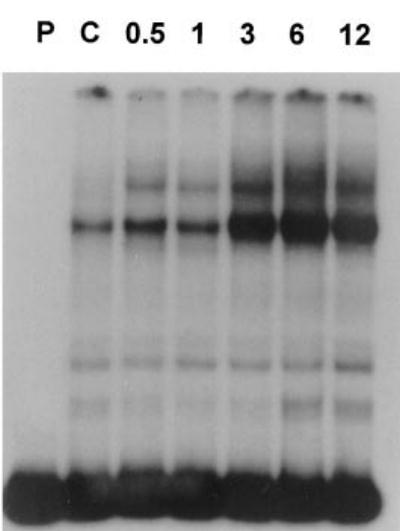
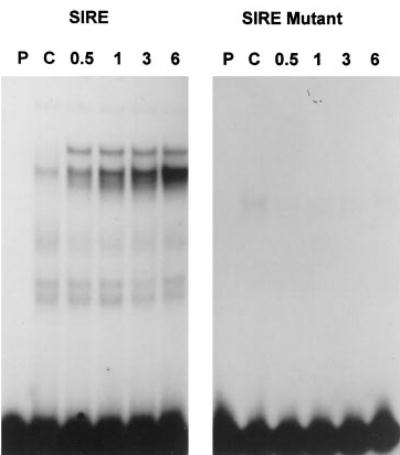
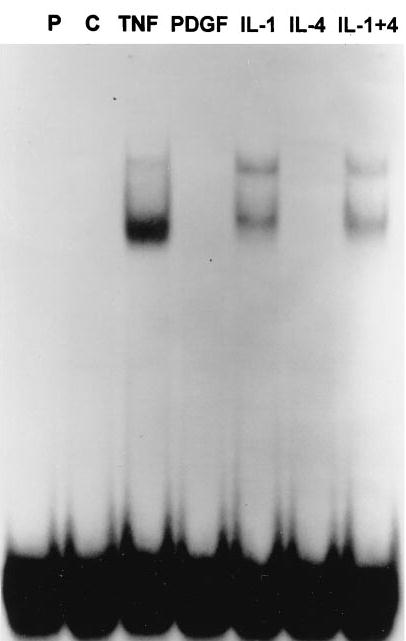
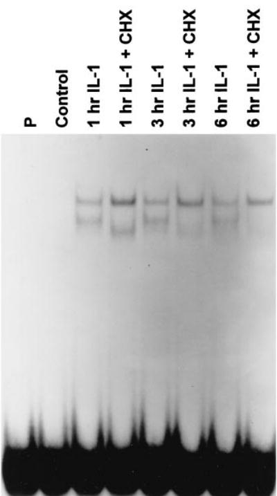
Stromelysin 1 (MMP-3) is a matrix metalloproteinase with broad substrate specificity that has been linked to joint and tissue destruction associated with chronic inflammatory diseases such as rheumatoid arthritis and periodontitis. Transcription of the stromelysin gene is induced by inflammatory cytokines such as interleukin 1 (IL-1) and tumor necrosis factor as well as a number of other cytokines and mitogens, but the exact mechanisms involved in its regulation are not fully understood. To identify transcription factors and cis elements potentially involved in the IL-1 induction of stromelysin, the human stromelysin 5'-flanking region was screened by electrophoretic mobility shift assay for IL-1-induced DNA-binding complexes in human synovial and gingival fibroblasts. Here we report the identification of such a complex binding to the region -1614 to -1595 (5'-G(T)TTTTTCCCCCCATCAAAG-3') termed the stromelysin IL-1 responsive element site. Binding to this site is also induced by tumor necrosis factor but not by platelet-derived growth factor or interleukin 4. UV cross-linking demonstrates that there are at least two DNA-binding proteins involved, of approximately 48 and 52 kDa. Transient transfection experiments in human foreskin fibroblasts demonstrate that proteins binding to this site act as a repressor of IL-1-induced expression of the stromelysin gene.


Figures

Fig. 1
Fig. 1. IL-1 induced binding to the region −1693 to −1575 of stromelysin 1
The region from −1693 to −1575 of the stromelysin 5′-flanking region was amplified by PCR and end-labeled with 32P. This probe was then incubated with nuclear extracts (10 μg) isolated from synovial fibroblast cultures at the indicated times after addition of IL-1 (100 ng/ml). Similar results were obtained with extracts isolated from three different individuals and also with extracts isolated from gingival fibroblasts from three different individuals. P, probe alone, no extract; C, control extract, no IL-1.
Fig. 2
Fig. 2. Identification of SIRE at −1614 to −1595
A, schematic for isolation of SIRE. Overlapping sets of oligodeoxynucleotide probes were made encompassing the region −1693 to −1575. The pattern of IL-1-induced binding to probe 4 and finally to 4B was identical to that seen with the larger probe. B, 10 μg of nuclear extract isolated from synovial fibroblasts treated for 6 h with IL-1 were incubated with 32P-labeled probe 4. Binding was competed with indicated molar excess of the unlabeled probe 4 but not by a 100-fold excess of probe 5. Identical results were obtained with extracts isolated from gingival fibroblasts. P, probe alone, no extract.
Fig. 3
Fig. 3. Mutation of two nucleotides in probe 4B abolishes DNA binding
The same nuclear extract (5 μg) isolated from human gingival fibroblasts at the indicated times after stimulation with IL-1 was incubated with both wild-type (5′-GTTTTTTCCCCCCATCAAAG-3′) and mutant (5′-GTTTTTTCTCACCATCAAAG-3′) 32P-labeled oligonucleotide probes as shown. P, probe alone, no extract; C, control extract, no IL-1.
Fig. 4
Fig. 4. Binding to SIRE is also induced by tumor necrosis factor but not by platelet-derived growth factor or IL-4
Nuclear extracts (5 μg) isolated 6 h after stimulation of gingival fibroblast cultures with the indicated cytokine (10 μg/ml) were incubated with a 32P-labeled probe containing the wild-type SIRE region. Similar results were observed with extracts isolated from synovial fibroblasts. P, probe alone, no extract; C, control extract, no cytokine; TNF, tumor necrosis factor; PDGF, platelet-derived growth factor.
Fig. 5
Fig. 5. De novo protein synthesis is not required for IL-1-induced binding to the SIRE site
5 μg of nuclear extract isolated at the indicated hours after stimulation with IL-1β (100 ng/ml) in the presence or absence of 100 μg/ml cycloheximide (CHX) were incubated with the 32P-labeled probe containing the SIRE binding site. P, probe alone, no extract.
Fig. 6
Fig. 6. UV cross-linking of two proteins to the SIRE site
40 μg of nuclear extract isolated from gingival fibroblast cultures 1 h after IL-1 stimulation in the presence and absence of cycloheximide (CHX) were incubated in EMSA with a bromodeoxyuridine-tagged, 32P-labeled probe corresponding to the SIRE site. The binding reaction was exposed to UV irradiation for 15 min before electrophoresis on 10% SDS-polyacrylamide gel electrophoresis along with molecular mass standards.
Fig. 7
Fig. 7. IL-1 induced binding to the SIRE site in HFF cells
Five μg of nuclear extract isolated from HFF cells at the indicated times after stimulation with IL-1 were incubated with the 32P-labeled probe corresponding to the SIRE binding site. P, probe alone, no extract; C, control extract, no IL-1.
Fig. 8
Fig. 8. Site-directed mutagenesis of the SIRE site increases IL-1-induced transcription from the stromelysin promoter
Subconfluent cultures of human foreskin fibroblasts were co-transfected with SVβ-gal along with a stromelysin luciferase reporter construct. pGLStro contains a 2-kilobase fragment of the wild-type stromelysin promoter; pGLΔSac is a deletion mutant missing the SIRE region, and pGLmStro is a mutant construct with the SIRE region altered as shown in Fig. 3. Results are from three independent experiments performed in triplicate and normalized with β-galactosidase for transfection efficiency and expressed as fold IL-1 induction. Basal activities were similar for all three constructs.

References

    1. Woessner JF., Jr FASEB J. 1991;5:2145–2154. - PubMed
    1. Suzuki K, Enghild JJ, Morodomi T, Salvesen G, Nagase H. Biochemistry. 1990;29:10261–10270. - PubMed
    1. Brinckerhoff CE, Suzuki K, Mitchell TI, Oram F, Coon CI, Palmiter RD, Nagase H. J Biol Chem. 1990;265:22262–22269. - PubMed
    1. Birkedal-Hansen H. J Periodontal Res. 1993;28:500–510. - PubMed
    1. Harris EJ. In: Textbook of Rheumatology. Kelley W, Harris ED Jr, Ruddy S, Sledge CB, editors. W. B. Saunders Co; Philadelphia: 1993. pp. 833–873.

Publication types