. 1999 Feb;19(2):967-78.
doi: 10.1128/MCB.19.2.967.

Isolation and functional characterization of two distinct sexual-stage-specific promoters of the human malaria parasite Plasmodium falciparum

Affiliations

Isolation and functional characterization of two distinct sexual-stage-specific promoters of the human malaria parasite Plasmodium falciparum

K J Dechering et al. Mol Cell Biol. 1999 Feb.

Abstract

Transmission of malaria depends on the successful development of the sexual stages of the parasite within the midgut of the mosquito vector. The differentiation process leading to the production of the sexual stages is delineated by several developmental switches. Arresting the progression through this sexual differentiation pathway would effectively block the spread of the disease. The successful development of such transmission-blocking agents is hampered by the lack of a detailed understanding of the program of gene expression that governs sexual differentiation of the parasite. Here we describe the isolation and functional characterization of the Plasmodium falciparum pfs16 and pfs25 promoters, whose activation marks the developmental switches executed during the sexual differentiation process. We have studied the differential activation of the pfs16 and pfs25 promoters during intraerythrocytic development by transfection of P. falciparum and during gametogenesis and early sporogonic development by transfection of the related malarial parasite P. gallinaceum. Our data indicate that the promoter of the pfs16 gene is activated at the onset of gametocytogenesis, while the activity of the pfs25 promoter is induced following the transition to the mosquito vector. Both promoters have unusual DNA compositions and are extremely A/T rich. We have identified the regions in the pfs16 and pfs25 promoters that are essential for high transcriptional activity. Furthermore, we have identified a DNA-binding protein, termed PAF-1, which activates pfs25 transcription in the mosquito midgut. The data presented here shed the first light on the details of processes of gene regulation in the important human pathogen P. falciparum.


Figures
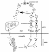
FIG. 1
FIG. 1
Life cycle of the human malaria parasite P. falciparum. Infection in humans begins with the introduction of sporozoites in the bloodstream by a bite of an infected Anopheles mosquito (a). The sporozoites are cleared by the liver (b), where cyclic asexual development initiates (c to g). Merozoites invade erythrocytes and transform into a ring-stage parasite (d). Subsequent trophic growth (e) and mitotic divisions lead to the production of up to 32 merozoites in a schizont (f), which bursts and releases new merozoites in the bloodstream. The merozoites can either reinitiate the erythrocytic asexual multiplication cycle (g) or commit to sexual differentiation (h). Initially, sexually committed parasites adapt the typical postinvasion ring-like shape (i) and cannot be discriminated from asexual parasites on a morphological basis. Sexual differentiation becomes morphologically apparent with the appearance of stage II gametocytes (j), which mature into female and male stage V gametocytes (k). The gametocytes are adapted to infect mosquitoes. Following the blood meal of a mosquito, the female gametocyte transforms in a macrogamete (l). The male gametocyte undergoes three rapid nuclear divisions and produces eight microgametes (m) that fertilize (n) the macrogamete, which then transforms in an invasive ookinete (o). This ookinete traverses the midgut epithelium and forms an oocyst at the side of the basal lamina. Sporogonic development (p) subsequently leads to the production of sporozoites that are released from the oocyst, accumulate in the salivary gland of the mosquito, and are infectious upon a new bite (a). DNA synthesis occurs at the transition from ring-stage parasite to schizont (d to f) (23) and at the transition from sexually committed ring-stage parasite to gametocyte (i and j) (24). Accordingly, both transitions are inhibited by pyrimethamine (9). Transcription of the pfs16 gene is induced in sexually committed ring stages, whereas the pfs25 gene is activated immediately following transmission (13).
FIG. 2
FIG. 2
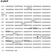
(A) Nucleotide sequence of the pfs16 upstream region. Numbering is with respect to the ATG translation initiation codon, which is italicized. The transcription start site as determined by RNase protection is indicated with an arrow. The starting positions of deletion mutants used in transfection studies are indicated in bold and above the sequence. Sequence elements with homology to the binding site of the yeast MATα2 homeobox transcription factor are in bold and overlined. (B) Nucleotide sequence of the pfs25 upstream region. Numbering is with respect to the ATG translation initiation codon (italicized). Arrows indicate positions of the transcription initiation sites. The starting positions of deletion mutants of plasmid pFS25LUC are indicated in bold and above the sequence. The AAGGAATA sequences that serve as the recognition sites for the PAF-1 transcription factor identified in this work are underlined and in bold. A CCAAT box is overlined.
FIG. 2
FIG. 2
(A) Nucleotide sequence of the pfs16 upstream region. Numbering is with respect to the ATG translation initiation codon, which is italicized. The transcription start site as determined by RNase protection is indicated with an arrow. The starting positions of deletion mutants used in transfection studies are indicated in bold and above the sequence. Sequence elements with homology to the binding site of the yeast MATα2 homeobox transcription factor are in bold and overlined. (B) Nucleotide sequence of the pfs25 upstream region. Numbering is with respect to the ATG translation initiation codon (italicized). Arrows indicate positions of the transcription initiation sites. The starting positions of deletion mutants of plasmid pFS25LUC are indicated in bold and above the sequence. The AAGGAATA sequences that serve as the recognition sites for the PAF-1 transcription factor identified in this work are underlined and in bold. A CCAAT box is overlined.
FIG. 3
FIG. 3
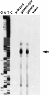
Analysis of the transcription initiation site of the pfs16 gene. RNase protection was performed on RNA of the different developmental stages of P. falciparum and on yeast RNA as indicated. An arrow indicates the position of the major protected fragment. The letters G, A, T, and C indicate the products of a sequencing reaction that was run along the protected fragments for size determination. Note that the sequence depicted in the figure does not contain a single cytosine base.
FIG. 4
FIG. 4
A. Transfections of mosquito- and blood-stage parasites with pfs16, pfs25, and hrp3 promoter-reporter constructs. CAT signals from P. gallinaceum mosquito- and P. falciparum blood-stage parasites transfected with the plasmids schematically depicted at the left side. Arrows indicate the transcriptional start sites of the pfs16 and pfs25 genes; open circles represent the transcription termination signals provided by the hrp2 sequences. The positions at which the unacetylated (N) and monoacetylated (1M and 3M) forms of [14C]chloramphenicol migrate are indicated. Plasmids p49.20 (P. gallinaceum mosquito-stage transfections) and pA0 (P. falciparum blood-stage transfections) were cotransfected to affirm the success of the transfection, and the numbers in parentheses indicate the relative luciferase activities induced by these plasmids. (B) Comparison of the transcriptional activities of the pfs16 and pfs25 promoters. P. gallinaceum mosquito-stage parasites were transfected with plasmids pCAT-L16.1ΔSX and pCAT25.1. CAT activities were normalized to the luciferase activity induced by cotransfected plasmid p49.20.
FIG. 4
FIG. 4
A. Transfections of mosquito- and blood-stage parasites with pfs16, pfs25, and hrp3 promoter-reporter constructs. CAT signals from P. gallinaceum mosquito- and P. falciparum blood-stage parasites transfected with the plasmids schematically depicted at the left side. Arrows indicate the transcriptional start sites of the pfs16 and pfs25 genes; open circles represent the transcription termination signals provided by the hrp2 sequences. The positions at which the unacetylated (N) and monoacetylated (1M and 3M) forms of [14C]chloramphenicol migrate are indicated. Plasmids p49.20 (P. gallinaceum mosquito-stage transfections) and pA0 (P. falciparum blood-stage transfections) were cotransfected to affirm the success of the transfection, and the numbers in parentheses indicate the relative luciferase activities induced by these plasmids. (B) Comparison of the transcriptional activities of the pfs16 and pfs25 promoters. P. gallinaceum mosquito-stage parasites were transfected with plasmids pCAT-L16.1ΔSX and pCAT25.1. CAT activities were normalized to the luciferase activity induced by cotransfected plasmid p49.20.
FIG. 5
FIG. 5
Activities of the pfs16 and hrp3 promoters during sexual differentiation of P. falciparum. (A) Asynchronous cultures of P. falciparum were transfected at day 0 with plasmids pHLH and pCAT-L16.1, and reporter activities were monitored over time. The upper graph shows the activities of the pfs16 and hrp3 promoters; the lower graph shows the parasitemia of the culture, discriminating between asexual parasites, gametocytes (gct), and sexually committed ring stages (cr). The number of sexually committed ring stages was inferred from the number of stage II gametocytes appearing 48 h later (7). (B) Cultures were transfected with plasmid pHLH or pLUC16.1 at day 0 and treated with pyrimethamine from days 4 to 7. Error bars indicate standard deviations calculated from three independent experiments.
FIG. 6
FIG. 6
GFP expression driven by the pfs16 promoter in P. gallinaceum mosquito midgut stages. Fluorescence (left) and light transmission (right) images are shown. Gametocytes ingested by a mosquito transform into gametes (A [female gamete]) that fertilize and develop into invasive ookinetes (C) via the intermediate retort stages (B). Parasites that had successfully been transfected with plasmid pGFP16.2 show a bright fluorescence signal (left).
FIG. 7
FIG. 7
Deletion mapping of the pfs16 and pfs25 promoters. P. gallinaceum mosquito-stage parasites were transfected with the constructs schematically depicted at the left side. (A) Deletion mapping of the pfs16 promoter. CAT activities were normalized to the luciferase activity induced by cotransfected plasmid p49.20. (B) Deletion mapping of the pfs25 promoter. Luciferase activities were normalized to the CAT activity of cotransfected pCAT-L16.1ΔSX.
FIG. 8
FIG. 8
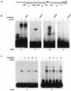
The pfs25 promoter is a target for DNA-binding proteins. (A) Schematic representation of locations of the probes used in EMSAs. (B) EMSAs with probes A through D and a nuclear extract derived from P. gallinaceum gametes. Competitions included molar excesses of unlabeled probes A through D as indicated. The competition with probe D accidentally was with a 10-fold instead of the intended 100-fold molar excess. (C) Cross-competition assays. Radiolabeled probes B and C were incubated with the nuclear extract. Competitions included 100-fold molar excesses of probes A through D as indicated.
FIG. 9
FIG. 9
The AAGGAATA elements in the pfs25 promoter recruit a mosquito stage-specific DNA-binding protein that activates transcription. (A) Oligonucleotide TFB25 (AATTCATAAGGAATATAG and its complement AATTCTATATTCCTTATG) was incubated with a nuclear extract derived from P. gallinaceum (g) or P. falciparum (Pfg) gametes or from an asynchronous P. falciparum blood-stage culture that contained both asexual parasites and gametocytes (Pfb). Competitions included either a 100-fold molar excess of the unlabeled oligonucleotide TFB25 or a 100-fold molar excess of an oligonucleotide containing a mutant version of the putative binding site (MUT; AATTCATAAGGCCGCTAG and its complement AATTCTAGCGGCCTTATG). (B) Control on the activity of the P. falciparum blood-stage nuclear extract. Radiolabeled oligonucleotide KAHRP (30) was incubated with a nuclear extract of the blood stages of P. falciparum parasites. The unlabeled oligonucleotide was added as a competitor DNA as indicated. (C) Radiolabeled probe C was incubated with a nuclear extract derived from P. gallinaceum gametes. A batch of nuclear extract different from that used in the experiments depicted in panel A was used, which resulted in the formation of an additional nonspecific complex, indicated by an asterisk. Binding reactions were supplemented with specific competitor DNAs as indicated. (D) Transfection of P. gallinaceum mosquito-stage parasites with plasmid pPFS25LUC and a version of the plasmid in which the two AAGGAATA elements were mutated to AAGGCCGC (pPFS25-LUC-MUT). Plasmids were cotransfected with pCAT-L16.1ΔSX, and luciferase activities were normalized to CAT activities. Values are from three transfections; error bars indicate standard deviations.
FIG. 9
FIG. 9
The AAGGAATA elements in the pfs25 promoter recruit a mosquito stage-specific DNA-binding protein that activates transcription. (A) Oligonucleotide TFB25 (AATTCATAAGGAATATAG and its complement AATTCTATATTCCTTATG) was incubated with a nuclear extract derived from P. gallinaceum (g) or P. falciparum (Pfg) gametes or from an asynchronous P. falciparum blood-stage culture that contained both asexual parasites and gametocytes (Pfb). Competitions included either a 100-fold molar excess of the unlabeled oligonucleotide TFB25 or a 100-fold molar excess of an oligonucleotide containing a mutant version of the putative binding site (MUT; AATTCATAAGGCCGCTAG and its complement AATTCTAGCGGCCTTATG). (B) Control on the activity of the P. falciparum blood-stage nuclear extract. Radiolabeled oligonucleotide KAHRP (30) was incubated with a nuclear extract of the blood stages of P. falciparum parasites. The unlabeled oligonucleotide was added as a competitor DNA as indicated. (C) Radiolabeled probe C was incubated with a nuclear extract derived from P. gallinaceum gametes. A batch of nuclear extract different from that used in the experiments depicted in panel A was used, which resulted in the formation of an additional nonspecific complex, indicated by an asterisk. Binding reactions were supplemented with specific competitor DNAs as indicated. (D) Transfection of P. gallinaceum mosquito-stage parasites with plasmid pPFS25LUC and a version of the plasmid in which the two AAGGAATA elements were mutated to AAGGCCGC (pPFS25-LUC-MUT). Plasmids were cotransfected with pCAT-L16.1ΔSX, and luciferase activities were normalized to CAT activities. Values are from three transfections; error bars indicate standard deviations.

Similar articles

Cited by

References

    1. Alano P, Carter R. Sexual differentiation in malaria parasites. Annu Rev Microbiol. 1990;44:429–449. - PubMed
    1. Baker D A, Daramola O, McCrossan M V, Harmer J, Targett G A T. Subcellular localization of Pfs16, a Plasmodium falciparum gametocyte antigen. Parasitology. 1994;108:129–137. - PubMed
    1. Billker O, Lindo V, Panico M, Etienne A E, Paxton T, Dell A, Rogers M, Sinden R E, Morris H R. Identification of xanthurenic acid as the putative inducer of malaria development in the mosquito. Nature. 1998;392:289–292. - PubMed
    1. Bohne W, Wirsing A, Gross U. Bradyzoite-specific gene expression in Toxoplasma gondii requires minimal genomic elements. Mol Biochem Parasitol. 1997;85:89–98. - PubMed
    1. Bruce M C, Alano P, Carter R. Commitment of the malaria parasite Plasmodium falciparum to sexual and asexual development. Parasitology. 1990;100:191–200. - PubMed

Publication types

MeSH terms

Associated data

LinkOut - more resources