. 1999 Feb;19(2):1068-80.
doi: 10.1128/MCB.19.2.1068.

Retinoblastoma protein contains a C-terminal motif that targets it for phosphorylation by cyclin-cdk complexes

Affiliations

Retinoblastoma protein contains a C-terminal motif that targets it for phosphorylation by cyclin-cdk complexes

P D Adams et al. Mol Cell Biol. 1999 Feb.

Abstract

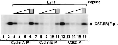
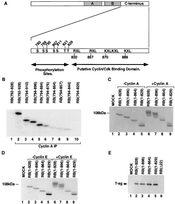
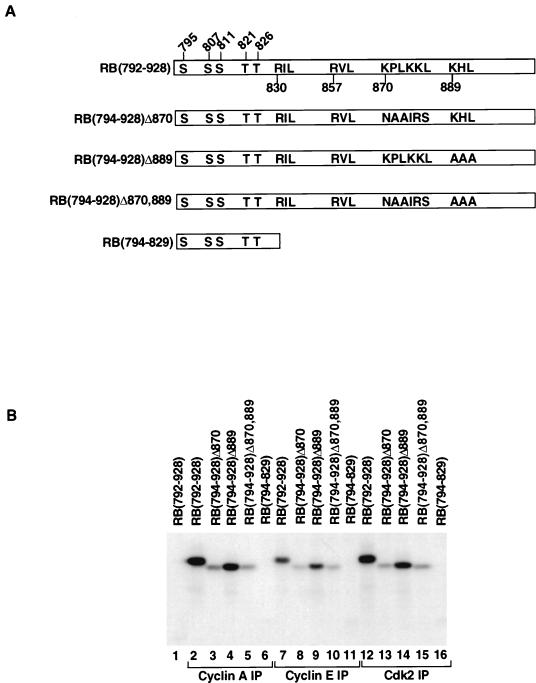
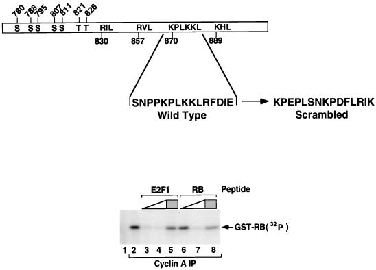
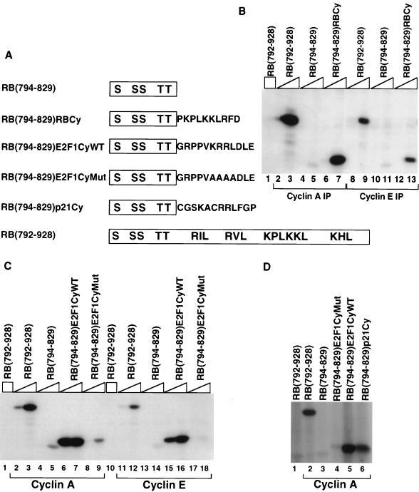
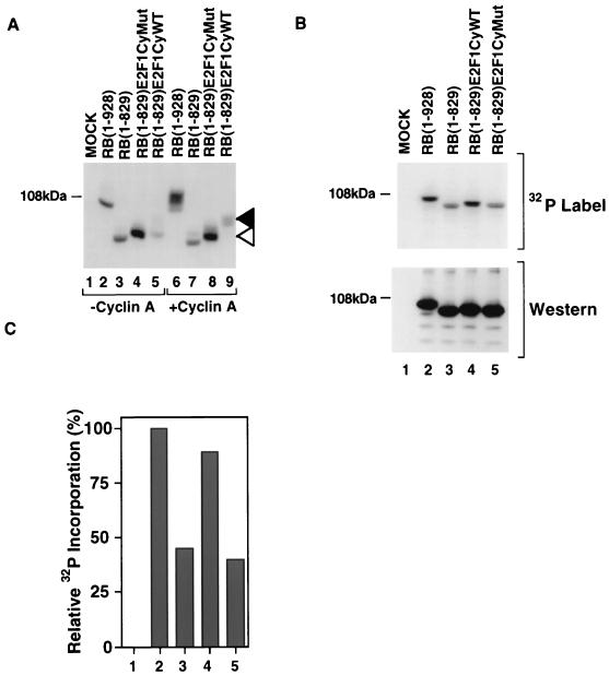
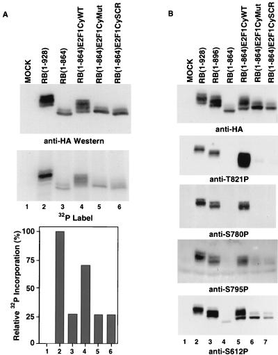
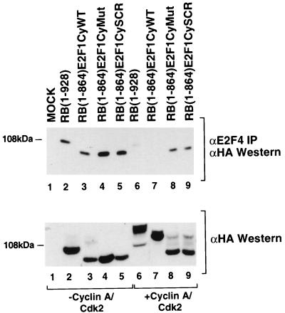
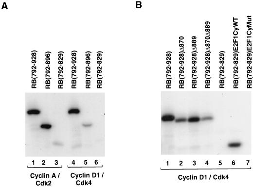
Stable association of certain proteins, such as E2F1 and p21, with cyclin-cdk2 complexes is dependent upon a conserved cyclin-cdk2 binding motif that contains the core sequence ZRXL, where Z and X are usually basic. In vitro phosphorylation of the retinoblastoma tumor suppressor protein, pRB, by cyclin A-cdk2 and cyclin E-cdk2 was inhibited by a short peptide spanning the cyclin-cdk2 binding motif present in E2F1. Examination of the pRB C terminus revealed that it contained sequence elements related to ZRXL. Site-directed mutagenesis of one of these sequences, beginning at residue 870, impaired the phosphorylation of pRB in vitro. A synthetic peptide spanning this sequence also inhibited the phosphorylation of pRB in vitro. pRB C-terminal truncation mutants lacking this sequence were hypophosphorylated in vitro and in vivo despite the presence of intact cyclin-cdk phosphoacceptor sites. Phosphorylation of such mutants was restored by fusion to the ZRXL-like motif derived from pRB or to the ZRXL motifs from E2F1 or p21. Phospho-site-specific antibodies revealed that certain phosphoacceptor sites strictly required a C-terminal ZRXL motif whereas at least one site did not. Furthermore, this residual phosphorylation was sufficient to inactivate pRB in vivo, implying that there are additional mechanisms for directing cyclin-cdk complexes to pRB. Thus, the C terminus of pRB contains a cyclin-cdk interaction motif of the type found in E2F1 and p21 that enables it to be recognized and phosphorylated by cyclin-cdk complexes.


Figures

FIG. 1
FIG. 1
Inhibition of cyclin-cdk2 by an E2F1-derived peptide. U2OS osteosarcoma cells were lysed and immunoprecipitated (IP) with anti-cyclin A, anti-cyclin E, anti-cdk2, or control (lane 1, anti-SV40 T-antigen) antibodies as indicated. In vitro kinase assays were performed with GST-RB(792–928) in the presence of no competitor peptide (lanes 1, 2, 7, and 12), 1.4, 14, or 140 μM wild-type E2F1-derived cyclin-cdk2 binding peptide (open triangles), or 140 μM scrambled version of the cyclin-cdk2 binding peptide (shaded squares) (lanes 6, 11, and 16). Phosphorylated proteins were resolved by SDS-polyacrylamide gel electrophoresis and detected by autoradiography.
FIG. 2
FIG. 2
Residues 864 to 884 are required for phosphorylation of pRB in vitro and in vivo. (A) Schematic of pRB protein and its C terminus. The top line shows the full-length pRB protein. Shaded boxes correspond to the A and B subdomains of the pRB viral oncoprotein binding domain (also referred to as the pRB pocket). The bottom line shows an expansion of the pRB C terminus. Indicated are the locations of the candidate S/T-P cyclin-cdk in vivo phosphorylation sites and sequences related to the previously defined cyclin binding sequence. The actual sequences in each case are as follows: at 830, RIL; at 857, RVL; at 870, KPLKKL; at 889, KHL. There are no RXL or KXL motifs between residues 780 and 829 and no S/T-P motifs downstream of residue 829. (B) U2OS osteosarcoma cells were lysed and immunoprecipitated with anti-cyclin A or control (lane 1, anti-SV40 T-antigen) antibodies as indicated. In vitro kinase assays were performed in the presence of 1 μg of the indicated GST-RB fusion proteins. Phosphorylated proteins were resolved by SDS-polyacrylamide gel electrophoresis, transferred to nitrocellulose, and detected by autoradiography. (C and D) SAOS2 RB−/− osteosarcoma cells were transiently transfected with plasmids encoding HA-tagged versions of the indicated pRB mutants in the presence or absence of plasmids encoding cyclin A (C) or cyclin E (D). Cell lysates were fractionated by SDS-polyacrylamide gel electrophoresis (6% polyacrylamide) and Western blotted with an anti-HA antibody. (E) Cells stably producing SV40 T antigen (COS cells) were transiently transfected with plasmids encoding HA-tagged versions of the indicated pRB mutants, lysed, and immunoprecipitated with an anti-HA antibody. Coimmunoprecipitation of SV40 T antigen was detected by anti-SV40 T-antigen Western blot analysis.
FIG. 3
FIG. 3
A consensus cyclin-cdk binding motif between residues 870 and 875 mediates efficient phosphorylation of pRB. (A) Schematic illustrating the mutants used in the experiment in this figure. (B) U2OS osteosarcoma cells were lysed and immunoprecipitated with anti-cyclin A, anti-cyclin E, anti-cdk2, or control (lane 1, anti-SV40 T-antigen) antibodies as indicated. In vitro kinase assays were performed in the presence of 1 μg of the indicated GST-RB fusion proteins. Phosphorylated proteins were resolved by SDS-polyacrylamide gel electrophoresis, transferred to nitrocellulose, and detected by autoradiography. Comparable levels of the GST-RB proteins were confirmed by anti-GST Western blot analysis (data not shown).
FIG. 4
FIG. 4
A synthetic peptide spanning the KPLKKL residues of pRB inhibits phosphorylation of pRB by cyclin A-cdk2. The schematic shows the amino acid sequences of the wild type and scrambled versions of the pRB-derived peptide (residues 866 to 880) tested as inhibitors of cyclin A-associated kinase activity. U2OS osteosarcoma cells were lysed and immunoprecipitated with anti-cyclin A or control (lane 1, anti-SV40 T-antigen) antibodies as indicated. In vitro kinase assays were performed with GST-RB(792–928) in the presence of no competitor peptide (lanes 1 and 2), 1.4 or 140 μM wild-type E2F1-derived cyclin-cdk2 binding peptide (open triangle) (lanes 3 and 4), 1.4 or 140 μM wild-type pRB peptide (residues 866 to 880) (open triangle) (lanes 6 and 7), or 140 μM scrambled versions of the E2F1 and pRB peptides (shaded squares) (lanes 5 and 8).
FIG. 5
FIG. 5
A minimal cyclin-cdk2 targeting sequence derived from pRB, E2F1, or p21 will promote pRB phosphorylation in vitro. (A) Schematic of the pRB mutants used in the experiment in this figure. (B to D) U2OS osteosarcoma cells were lysed and immunoprecipitated with anti-cyclin A, anti-cyclin E, or control (lane 1, anti-SV40 T-antigen) antibodies as indicated. In vitro kinase assays were performed in the presence of 1 μg of GST–RB(792–928) (open squares) or 0.1 or 1 μg of the indicated GST-RB fusion proteins (open triangles). Phosphorylated proteins were resolved by SDS-polyacrylamide gel electrophoresis and detected by autoradiography.
FIG. 6
FIG. 6
A minimal cyclin-cdk targeting sequence derived from pRB, E2F1, or p21 will promote pRB Phosphorylation in vivo. (A) SAOS2 RB−/− osteosarcoma cells were transiently transfected with plasmids encoding HA-tagged versions of the indicated pRB mutants in the presence or absence of a plasmid encoding cyclin A as indicated. The cells were lysed, and soluble proteins were fractionated by SDS-polyacrylamide gel electrophoresis (6% polyacrylamide) and Western blotted with an anti-HA antibody. The open arrowhead marks the position of unphosphorylated RB(1–829)CyE2F1Mut and RB(1–829)CyE2F1WT. The solid arrowhead marks the position of phosphorylated RB(1–829)CyE2F1WT. (B) U2OS RB+/+ osteosarcoma cells were transiently transfected with plasmids encoding HA-tagged versions of the indicated pRB mutants, metabolically labelled with [32P]orthophosphate, lysed, and immunoprecipitated with an anti-HA antibody. Bound proteins were resolved by SDS-polyacrylamide gel electrophoresis (10% polyacrylamide) and detected by autoradiography (top) and anti-HA Western blot analysis (bottom). Note that the characteristic pRB mobility shift following phosphorylation is not apparent under these electrophoretic conditions, in contrast to the conditions used in panel A. (C) 32P incorporation relative to wild-type pRB was determined by phosphorimager analysis of panel B.
FIG. 7
FIG. 7
Identification of pRB phosphosites that require a C-terminal cyclin-cdk2 binding site. (A). U2OS RB+/+ osteosarcoma cells were transiently transfected with plasmids encoding HA-tagged versions of the indicated pRB mutants, metabolically labelled with [32P]orthophosphate, lysed, and immunoprecipitated with an anti-HA antibody. Bound proteins were resolved by SDS-polyacrylamide gel electrophoresis (6% polyacrylamide) and detected by anti-HA Western blot analysis (top) and autoradiography (middle). 32P incorporation relative to wild-type pRB was determined by phosphorimager analysis (bottom). (B) U2OS RB+/+ osteosarcoma cells were transiently transfected with plasmids encoding HA-tagged versions of the indicated pRB mutants. Cell extracts were prepared and immunoblotted with the indicated antibodies.
FIG. 8
FIG. 8
Phosphorylation of pRB mediated by the heterologous cyclin-cdk2 binding sequence of E2F1 results in disruption of pRB-E2F4 complexes. Asynchronously growing SAOS2 cells were transfected with plasmids encoding the HA-tagged versions of the indicated pRB mutants in the presence or absence of cyclin A and cdk2, as indicated. The cells were lysed, and an aliquot of the soluble proteins (150 μg) was fractionated by SDS-polyacrylamide gel electrophoresis (6% polyacrylamide) and Western blotted with an anti-HA antibody (bottom). The remainder of the lysate was immunoprecipitated with an anti-E2F4 monoclonal antibody. The immunoprecipitates were fractionated by SDS-polyacrylamide gel electrophoresis (6% polyacrylamide) and Western blotted with the anti-HA antibody (top).
FIG. 9
FIG. 9
A cyclin-cdk2 binding motif can participate in substrate recognition by cyclin D1-cdk4. In vitro kinase reactions were performed with recombinant cyclin A-cdk2 or cyclin D1-cdk4 and the indicated GST-RB fusion proteins. Comparable amounts of each GST-RB fusion protein were added in each reaction, as determined by Bradford assay and confirmed by Coomassie blue staining. Phosphorylated proteins were resolved by SDS-polyacrylamide gel electrophoresis and detected by autoradiography.

References

    1. Adams P D, Kaelin W G. Transcriptional control by E2F. Semin Cancer Biol. 1995;6:99–108. - PubMed
    1. Adams P D, Sellers W R, Sharma S K, Wu A D, Nalin C M, Kaelin W G. Identification of a cyclin-cdk2 recognition motif present in substrates and p21-like cdk inhibitors. Mol Cell Biol. 1996;16:6623–6633. - PMC - PubMed
    1. Bartkova J, Lukas J, Muller H, Lutzhoft D, Strauss M, Bartek J. Cyclin D1 protein expression and function in human breast cancer. Int J Cancer. 1994;57:353–361. - PubMed
    1. Botz J, Zerfass-Thome K, Spitzovsky D, Delius H, Vogt B, Eilers M, Hatzigeorgiou A, Jansen-Durr P. Cell cycle regulation of the murine cyclin E gene depends on an E2F binding site in the promoter. Mol Cell Biol. 1996;16:3401–3409. - PMC - PubMed
    1. Cantley L C, Auger K R, Carpenter C, Duckworth B, Graziani A, Kapeller R, Soltoff S. Oncogenes and signal transduction. Cell. 1991;64:281–302. - PubMed

Publication types

MeSH terms

LinkOut - more resources