. 1999 Feb;19(2):1081-91.
doi: 10.1128/MCB.19.2.1081.

An SH2 domain-containing 5' inositolphosphatase inhibits insulin-induced GLUT4 translocation and growth factor-induced actin filament rearrangement

Affiliations

An SH2 domain-containing 5' inositolphosphatase inhibits insulin-induced GLUT4 translocation and growth factor-induced actin filament rearrangement

P Vollenweider et al. Mol Cell Biol. 1999 Feb.

Abstract

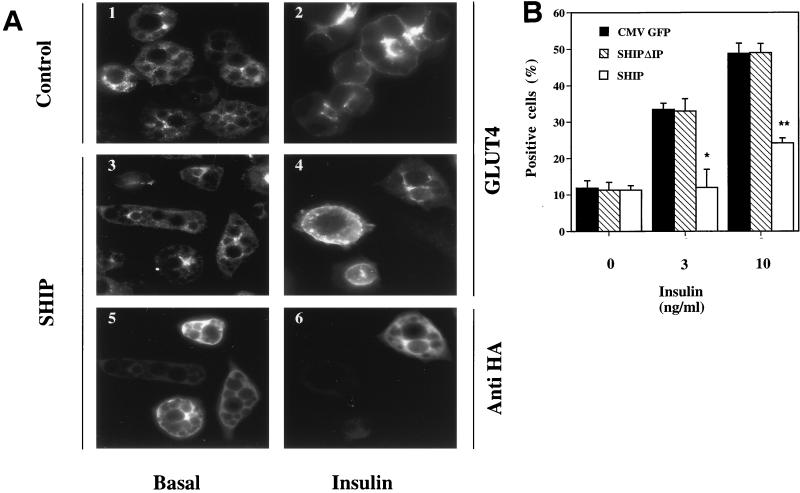
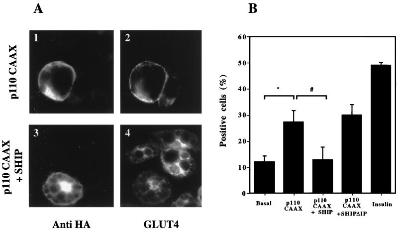
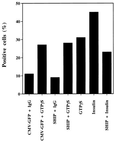
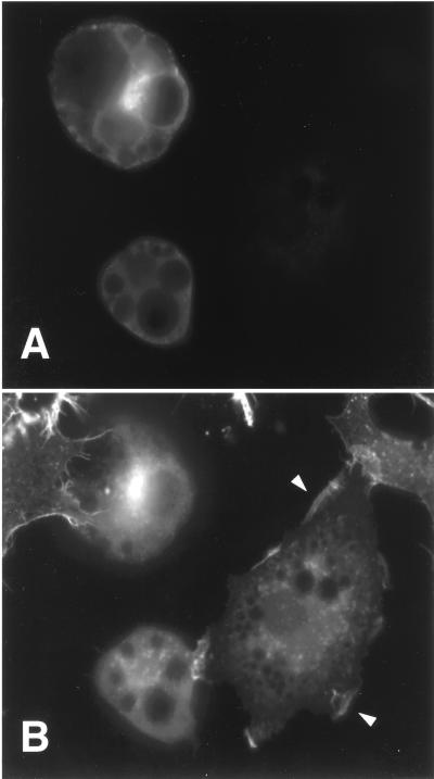
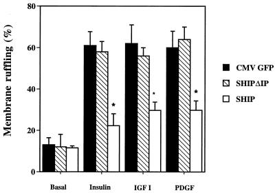
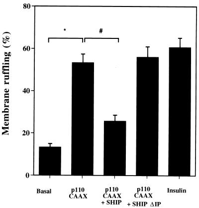
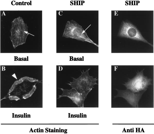
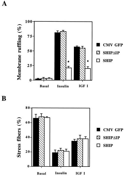
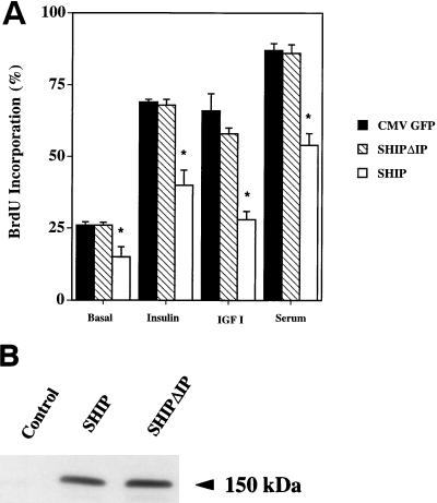
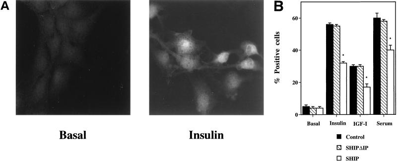
Tyrosine kinase receptors lead to rapid activation of phosphatidylinositol 3-kinase (PI3 kinase) and the subsequent formation of phosphatidylinositides (PtdIns) 3,4-P2 and PtdIns 3,4, 5-P3, which are thought to be involved in signaling for glucose transporter GLUT4 translocation, cytoskeletal rearrangement, and DNA synthesis. However, the specific role of each of these PtdIns in insulin and growth factor signaling is still mainly unknown. Therefore, we assessed, in the current study, the effect of SH2-containing inositol phosphatase (SHIP) expression on these biological effects. SHIP is a 5' phosphatase that decreases the intracellular levels of PtdIns 3,4,5-P3. Expression of SHIP after nuclear microinjection in 3T3-L1 adipocytes inhibited insulin-induced GLUT4 translocation by 100 +/- 21% (mean +/- the standard error) at submaximal (3 ng/ml) and 64 +/- 5% at maximal (10 ng/ml) insulin concentrations (P < 0.05 and P < 0.001, respectively). A catalytically inactive mutant of SHIP had no effect on insulin-induced GLUT4 translocation. Furthermore, SHIP also abolished GLUT4 translocation induced by a membrane-targeted catalytic subunit of PI3 kinase. In addition, insulin-, insulin-like growth factor I (IGF-I)-, and platelet-derived growth factor-induced cytoskeletal rearrangement, i.e., membrane ruffling, was significantly inhibited (78 +/- 10, 64 +/- 3, and 62 +/- 5%, respectively; P < 0.05 for all) in 3T3-L1 adipocytes. In a rat fibroblast cell line overexpressing the human insulin receptor (HIRc-B), SHIP inhibited membrane ruffling induced by insulin and IGF-I by 76 +/- 3% (P < 0.001) and 68 +/- 5% (P < 0.005), respectively. However, growth factor-induced stress fiber breakdown was not affected by SHIP expression. Finally, SHIP decreased significantly growth factor-induced mitogen-activated protein kinase activation and DNA synthesis. Expression of the catalytically inactive mutant had no effect on these cellular responses. In summary, our results show that expression of SHIP inhibits insulin-induced GLUT4 translocation, growth factor-induced membrane ruffling, and DNA synthesis, indicating that PtdIns 3,4,5-P3 is the key phospholipid product mediating these biological actions.


Figures

FIG. 1
FIG. 1
Effect of SHIP on insulin-induced GLUT4 translocation in 3T3-L1 adipocytes. 3T3-L1 adipocytes on coverslips were injected into the nucleus with CMV-GFP expression vector as a control, SHIP expression vector, or a catalytically inactive mutant of SHIP (SHIPΔIP) at a concentration of 0.1 mg/ml. Proteins were allowed to express for 20 to 24 h. Cells were then starved for 2 h in serum-free medium, stimulated without or with insulin (3 or 10 ng/ml) for 20 min, and fixed. Immunostaining was performed with rabbit polyclonal anti-GLUT4 (F349), mouse monoclonal anti-GFP, and mouse monoclonal anti-HA antibodies to allow detection of the tagged SHIP proteins. Cells positive for GFP, SHIP, or SHIPΔIP expression were counted for GLUT4 translocation. (A) Images of immunofluorescent GLUT4 staining in 3T3-L1 adipocytes. Panels 1 and 2 show typical staining in basal conditions and after insulin stimulation, respectively. Individual cells were injected into the nucleus with SHIP cDNA. Panels 5 and 6 show injected cells, and panels 3 and 4 represent GLUT4 staining of injected cells. (B) Summary data are given as bar graphs. Each bar represents mean ± SE of four to five experiments. Black bars represent values for control experiments, striped bars represent values for SHIPΔIP, and open bars represent SHIP-expressing cells. SHIP completely inhibited insulin-induced GLUT4 translocation at submaximal insulin concentrations, and by about 60% at maximal insulin concentrations (∗, P < 0.05; ∗∗, P < 0.01 versus control).
FIG. 2
FIG. 2
Effect of p110-CAAX and SHIP coinjection on GLUT4 translocation in 3T3-L1 adipocytes. 3T3-L1 adipocytes on coverslips were injected into the nucleus with CMV-GFP as a control, p110-CAAX with CMV-GFP, p110-CAAX with SHIP, or p110-CAAX with SHIPΔIP. All expression vectors were injected at a concentration of 0.1 mg/ml. After 20 to 24 h to allow for protein expression, cells were stimulated without or with insulin at 10 ng/ml as indicated and then fixed for staining. Cells positive for GFP or SHIP and SHIPΔIP (HA antibody) expression were scored for GLUT4 translocation. (A) Immunofluorescent GLUT4 staining. Individual cells were injected with p110-CAAX alone (1 and 2) or with p110-CAAX along with SHIP (3 and 4). The left panels show injected cells, and the right panels demonstrate GLUT4 staining. (B) Summary data are given as bar graphs. Each bar represents the mean ± SE for three experiments. p110-CAAX expression induced GLUT4 translocation, which was about half that of the insulin effect. Concomitant overexpression of SHIP inhibited the ability of p110-CAAX to induce GLUT4 translocation (∗, P < 0.05 versus basal; #, P < 0.05 versus p110-CAAX), whereas SHIPΔIP coexpression with p110-CAAX had no effect.
FIG. 3
FIG. 3
Effects of SHIP on GTPγS-induced GLUT4 translocation in 3T3-L1 adipocytes. 3T3-L1 adipocytes on scored glass coverslips were injected into the nucleus with either the expression vector for SHIP or CMV-GFP. Proteins were allowed to express for 20 to 24 h. After this time period, the same cells were serum starved for 2 h and injected into the cytoplasm with 5 mM GTPγS mixed with sheep IgG. After 30 min the cells were fixed and immunostained for GLUT4 as described above. Cells positive for SHIP or CMV-GFP expression and cytoplasmic sheep IgG were counted for GLUT4 translocation. Cytoplasmic GTPγS injection alone induced GLUT4 translocation in 31% of the cells. From the cells expressing either CMV-GFP or SHIP and injected with GTPγS, 27 and 28% were positive, respectively, for GLUT4 translocation. In the same experiment, SHIP expression inhibited insulin-induced GLUT4 translocation by about 50%.
FIG. 4
FIG. 4
Effects of SHIP on insulin-induced membrane ruffling in 3T3-L1 adipocytes. 3T3-L1 adipocytes on glass coverslips were injected into the nucleus with the expression vector for SHIP, and the protein was allowed to express for 24 h. After an overnight starvation, cells were then stimulated with 100 ng of insulin per ml for 20 min, fixed, and stained for expressed protein and actin filament rearrangement. (A) Photograph of 3T3-L1 adipocytes stained with anti-HA antibody demonstrating SHIP expression in the cytoplasm. (B) The same cells were photographed with an UV filter now displaying the staining for actin filament rearrangement. The two cells expressing SHIP have no membrane ruffles after insulin stimulation, whereas the uninjected cells display membrane ruffles (arrowheads).
FIG. 5
FIG. 5
Effects of SHIP on growth factor-induced membrane ruffling in 3T3-L1 adipocytes. 3T3-L1 adipocytes on coverslips were injected into the nucleus with expression vectors for CMV-GFP as control, SHIPΔIP, or SHIP expression vector at a concentration of 0.1 mg/ml. Proteins were allowed to express for 20 to 24 h. Cells were then starved in serum-free medium for 12 h and left either untreated or treated with insulin at 100 ng/ml, IGF-I at 100 ng/ml, or PDGF at 20 ng/ml for 15 min. Cells were then fixed and stained for actin localization with rhodamine-phalloidin. Individual adipocytes positive for GFP, SHIPΔIP, or SHIP expression were scored for the presence of membrane ruffles. Each bar represents the mean ± SE for three to four experiments. SHIP overexpression inhibited growth factor-induced membrane ruffling by about 60 to 80% (∗, P < 0.05, SHIP versus control), whereas SHIPΔIP had no effect.
FIG. 6
FIG. 6
Effects of p110 CAAX and SHIP coinjection on membrane ruffling in 3T3-L1 adipocytes. 3T3-L1 adipocytes were nuclear injected with CMV-GFP as a control, p110-CAAX with CMV-GFP, p110 CAAX with SHIPΔIP, or p110-CAAX with SHIP. All of the expression vectors were injected at a concentration of 0.1 mg/ml. After 20 to 24 h to allow for protein expression and 12 h of starvation in serum-free medium, the cells were left untreated or stimulated with insulin at 100 ng/ml, as indicated, and fixed for staining. Cells positive for GFP, SHIPΔIP, and SHIP expression were scored for the presence of membrane ruffles. Each bar represents the mean ± SE for three experiments. p110-CAAX expression induced membrane ruffling in 3T3-L1 adipocytes, a finding which was similar to the insulin effect. Concomitant expression of SHIP significantly inhibited the ability of p110-CAAX to induce membrane ruffles in the absence of insulin (∗, P < 0.05 versus basal; #, P < 0.05 versus p110-CAAX). Concomintant expression of p110-CAAX and SHIPΔIP had no effect on p110-CAAX-induced GLUT4 translocation.
FIG. 7
FIG. 7
Effects of SHIP overexpression on insulin-induced actin rearrangement in HIRc-B cells. Serum-starved HIRc-B cells on coverslips were injected in the nucleus with either CMV-GFP expression vector or SHIP expression vector at a concentration of 0.1 mg/ml. After 20 to 24 hours to allow for protein expression, the cells were stimulated without ligand or with insulin at 100 ng/ml for 3 min. Cells were then fixed and stained for actin filament rearrangement with rhodamine-phalloidin. (A) Photograph of actin staining in an unstimulated serum-starved cell displaying actin stress fibers in the cell cytoplasm (arrow) and the absence of membrane ruffles at the cell periphery. (B) After 3 min of insulin stimulation, a prominent membrane-ruffling response can be visualized (arrowhead), and concomitantly stress fibers have broken down. (C and D) Actin staining of cells injected into the nucleus with SHIP and stimulated without insulin (C) or with insulin for 3 min (D). Unstimulated injected cells display stress fibers as control injected cells (C) (arrowhead). After insulin stimulation, injected cells display no membrane ruffles but show stress fiber breakdown. (E and F) HA staining of the same cells as in panels C and D, respectively, showing SHIP expression in the cell cytoplasm.
FIG. 8
FIG. 8
Effects of SHIP on growth factor-induced membrane ruffling and stress fiber breakdown in HIRc-B cells. Serum-starved HIRc-B cells were injected into the nucleus with the expression vectors for either CMV-GFP, SHIPΔIP, or SHIP, and the proteins were allowed to express for 20 to 24 h. Cells were then stimulated for 3 min with insulin (100 ng/ml), fixed, and stained for actin localization with rhodamine-phalloidin. Individual cells positive for GFP, SHIPΔIP, and SHIP expression were scored for the presence of membrane ruffles (A) or parallel actin fibers that colocalize with the nucleus (positive for stress fibers) (B). Each bar represents the mean ± SE for three to four different experiments. SHIP inhibited insulin- and IGF-I-induced membrane ruffling by about 80% (∗, P < 0.005 versus control) but had no effect on ligand-induced stress fiber breakdown, whereas SHIPΔIP did not affect ligand-induced membrane ruffling.
FIG. 9
FIG. 9
Effects SHIP on growth factor-induced BrdU incorporation in HIRc-B cells. (A) HIRc-B cells were grown on glass coverslips in 12-well dishes and transiently transfected either with LacZ as a control or the expression vectors for SHIPΔIP or SHIP. Cells were starved for 24 h and then stimulated without insulin or with insulin (100 ng/ml), IGF-I (100 ng/ml), or FCS (10%) for 18 h. BrdU was added during the last 6 h of stimulation. Cells were then fixed and stained for protein expression and BrdU incorporation. Successfully transfected cells were counted for BrdU incorporation into the nucleus, and results are given as the percent BrdU incorporation. Each bar represents the mean ± SE for three separate experiments. SHIP expression slightly decreased the basal BrdU incorporation. Compared to the control, SHIP significantly inhibited insulin-, IGF-I-, and serum-stimulated BrdU incorporation by 42 ± 9, 57 ± 2, and 38 ± 10%, respectively (∗, P < 0.05 versus control). (B) HIRc-B cells were transiently transfected with the vectors for SHIP and SHIPΔIP or with no vector, and proteins were allowed to express for 48 h. Whole-cell lysates were then separated by SDS-PAGE, and membranes were blotted with an anti-HA antibody. SHIP and SHIPΔIP show similar expression levels.
FIG. 10
FIG. 10
Effect of SHIP on MAPK activation in HIRc-B cells. HIRc-B cells were grown on glass coverslips and transiently transfected with the expression vectors for LacZ, SHIP, or SHIPΔIP. Cells were starved for 24 h and then stimulated without or with insulin (100 ng/ml), IGF-I (100 ng/ml), or serum (10%) for 20 min. Cells were then fixed and stained for protein expression with a specific antibody directed against the dually phosphorylated MAPK. (A) Unstimulated cells display almost no staining with the anti-phospho-MAPK antibody and, after insulin stimulation, bright nuclear as well some cytoplasmatic staining can be observed. Successfully transfected cells displaying bright nuclear staining were scored as positive, and results are given as the percent positive cells. (B) Summarized data. Each bar represents the mean ± SE for three separate experiments. SHIP expression inhibited ligand-induced MAPK phosphorylation, whereas SHIPΔIP had no effect.

Similar articles

Cited by

References

    1. Alessi D R, Andjelkovic M, Caudwell B, Cron P, Morrice N, Cohen P, Hemmings B A. Mechanisms of activation of protein kinase B by insulin and IGF-1. EMBO. 1996;15:65541–6551. - PMC - PubMed
    1. Alessi D R, Deak M, Casamayor A, Caudwell F B, Morrice N, Norman D G, Gaffney P, Reese C B, MacDougall C N, Harbison D, Ashworth A, Bownes M. 3-Phosphoinositide-dependent protein kinase-1 (PDK-1): structural and functional homology with the Drosophila DSTPK61 kinase. Curr Biol. 1997;7:776–789. - PubMed
    1. Alessi D R, James S R, Downes C P, Holmes A B, Gaffney P R J, Reese C B, Cohen P. Characterization of a 3-Phosphoinositide-dependent protein kinase which phosphorylates and activates protein kinase Bα. Curr Biol. 1997;7:261–269. - PubMed
    1. Auger K, Serunian L A, Soltoff S P, Libby P, Cantley L C. PDGF-dependent tyrosine phosphorylation stimulates production of novel polyphosphoinositides in intact cells. Cell. 1989;57:167–175. - PubMed
    1. Burgering B M T, Coffer P J. Protein kinase B (c-Akt) in phosphatidylinositol-3-OH kinase signal transduction. Nature. 1995;376:599–602. - PubMed

Publication types

MeSH terms

Substances