. 1999 Feb;19(2):1101-15.
doi: 10.1128/MCB.19.2.1101.

Complementation of defective colony-stimulating factor 1 receptor signaling and mitogenesis by Raf and v-Src

Affiliations

Complementation of defective colony-stimulating factor 1 receptor signaling and mitogenesis by Raf and v-Src

N Aziz et al. Mol Cell Biol. 1999 Feb.

Abstract

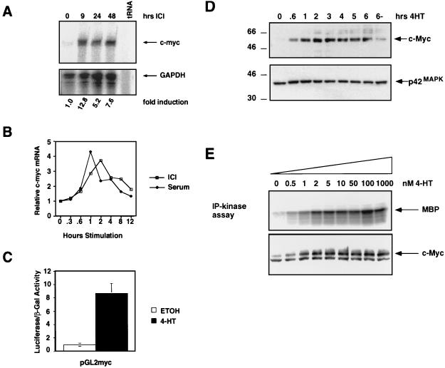
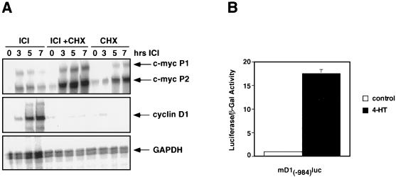
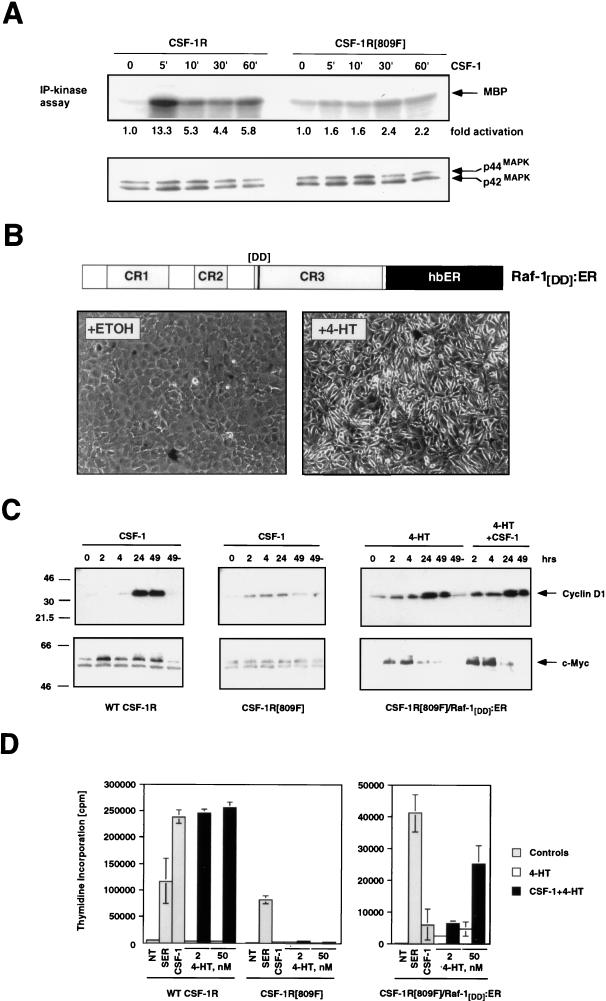
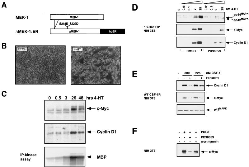
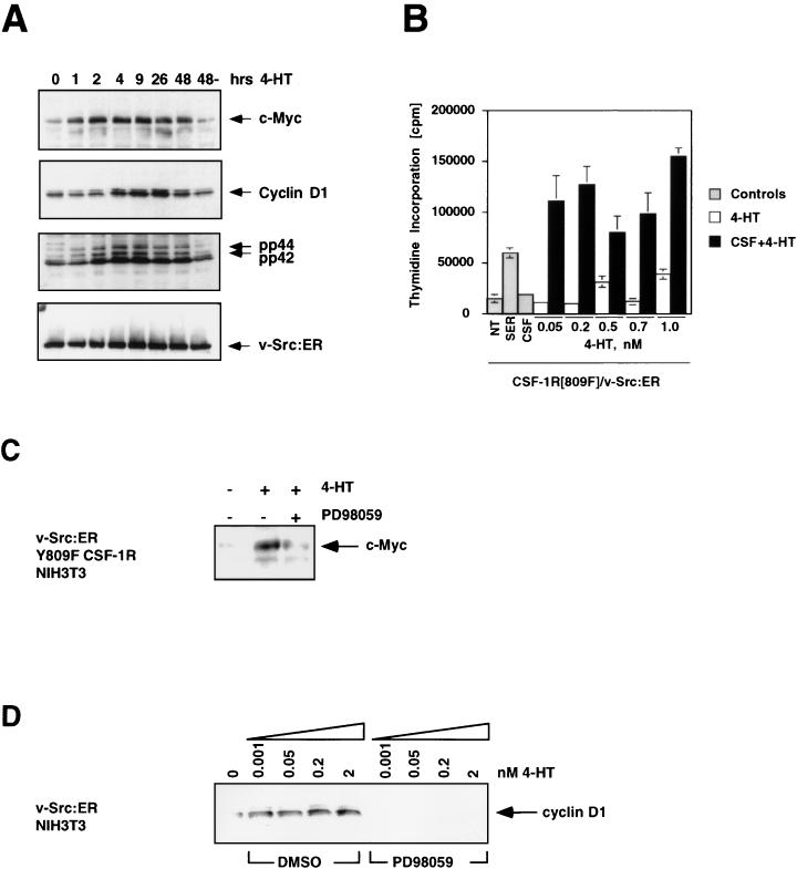
Ras-activated signal transduction pathways are implicated in the control of cell proliferation, differentiation, apoptosis, and tumorigenesis, but the molecular mechanisms mediating these diverse functions have yet to be fully elucidated. Conditionally active forms of Raf, v-Src, and MEK1 were used to identify changes in gene expression that participate in oncogenic transformation, as well as in normal growth control. Activation of Raf, v-Src, and MEK1 led to induced expression of c-Myc and cyclin D1. Induction of c-Myc mRNA by Raf was an immediate-early response, whereas the induction of cyclin D1 mRNA was delayed and inhibited by cycloheximide. Raf activation also resulted in the induction of an established c-Myc target gene, ornithine decarboxylase (ODC). ODC induction by Raf was mediated, in part, by tandem E-boxes contained in the first intron of the gene. Activation of the human colony-stimulating factor 1 (CSF-1) receptor in NIH 3T3 cells leads to activation of the mitogen-activated protein (MAP) kinase pathway and induced expression of c-Fos, c-Myc, and cyclin D1, leading to a potent mitogenic response. By contrast, a mutated form of this receptor fails to activate the MAP kinases or induce c-Myc and cyclin D1 expression and fails to elicit a mitogenic response. The biological significance of c-Myc and cyclin D1 induction by Raf and v-Src was confirmed by the demonstration that both of these protein kinases complemented the signaling and mitogenic defects of cells expressing this mutated form of the human CSF-1 receptor. Furthermore, the induction of c-Myc and cyclin D1 by oncogenes and growth factors was inhibited by PD098059, a specific MAP kinase kinase (MEK) inhibitor. These data suggest that the Raf/MEK/MAP kinase pathway plays an important role in the regulation of c-Myc and cyclin D1 expression in NIH 3T3 cells. The ability of oncogenes such as Raf and v-Src to regulate the expression of these proteins reveals new lines of communication between cytosolic signal transducers and the cell cycle machinery.


Figures

FIG. 1
FIG. 1
Induction of c-Myc by Raf in NIH 3T3 cells. (A) c-Myc mRNA regulation. NIH 3T3 cells expressing ΔB-Raf:ER* were cultured in 0.5% serum for 16 h, and RNA samples were prepared at different times after the addition of 1 μM ICI to activate ΔB-Raf:ER*. The expression of c-Myc (upper panel) and GAPDH (lower panel) mRNAs was detected by simultaneous RNase protection assay. The fold induction of c-Myc mRNA was quantitated by obtaining the ratio of c-Myc to GAPDH mRNAs by PhosphorImager analysis. (B) Comparison of c-Myc mRNA induction by serum versus ΔB-Raf:ER* activation. NIH 3T3 cells expressing ΔB-Raf:ER* were cultured in 0.5% serum for 16 h, and RNA samples were prepared at different times after stimulation with 10% FCS (closed diamonds) or the activation of ΔB-Raf:ER* with 1 μM ICI (open squares). c-Myc and GAPDH mRNAs were detected by RNase protection, and data were quantified by PhosphorImager analysis. Results are presented as the fold induction over the baseline level of expression. (C) Activation of the c-Myc promoter. NIH 3T3 cells expressing ΔB-Raf:ER* were transiently transfected with reporter constructs consisting of the promoter region of human c-Myc linked to luciferase (pGL2-myc) and pSRαβ-Gal as a control for transfection efficiency. Transfected cells were treated with either ethanol (solvent control, open bar) or 500 nM 4-HT (shaded bar) in 0.5% serum for 36 h, at which time the luciferase and β-galactosidase activities were measured. Results are presented as the ratios of the luciferase to β-galactosidase activities. (D) Induction of c-Myc protein expression. NIH 3T3 cells expressing ΔB-Raf:ER* were cultured in 0.5% FCS for 16 h, and cell extracts were prepared at different times after the addition of 500 nM 4-HT. The expression of p62c-Myc was detected by Western blotting with a specific antiserum (upper panel). The same Western blot was reprobed for the expression of p42 MAP kinase as a control for equal loading in each lane (lower panel). (E) Effect of increasing ΔB-Raf:ER* activity on c-Myc expression. NIH 3T3 cells expressing ΔB-Raf:ER* were cultured in modified DSFM for 24 h and stimulated with different concentrations of 4-HT, and cell extracts were prepared 4.5 h later. MAP kinase activity was measured by performing p42MAPK immune complex kinase assays with MBP as a substrate (upper panel). c-Myc expression was analyzed by Western blotting.
FIG. 2
FIG. 2
Regulation of cyclin D1 and c-Myc expression by Raf. (A) Cycloheximide sensitivity of Raf-induced cyclin D1 and c-Myc mRNA expression. NIH 3T3 cells expressing ΔB-Raf:ER* were cultured in 0.5% FCS for 24 h and were then either left untreated or treated with 25 μg of cycloheximide per ml for 1 h. These cells were then either left untreated (CHX) or treated with 1 μM ICI to activate ΔB-Raf:ER* for different lengths of time as indicated. The expression of cyclin D1, c-Myc, and GAPDH mRNAs were quantitated by RNase protection assays. (B) Cyclin D1 promoter activation. NIH 3T3 cells expressing ΔB-Raf:ER* were transiently transfected with reporter constructs consisting of the promoter region of mouse cyclin D1 linked to luciferase [mD1(-984)Luc] and pSRαβ-Gal as a control for transfection efficiency. Transfected cells were treated with either ethanol (solvent control, open bar) or 1 μM 4-HT (closed bar) for 41 h, at which time luciferase and β-galactosidase activities were measured. Results are presented as the ratios of the luciferase to the β-galactosidase activities.
FIG. 3
FIG. 3
Induced expression of ODC after Raf activation. (A) Induction of ODC mRNA by Raf. NIH 3T3 cells expressing ΔRaf-1:ER were cultured in DMEM containing 0.5% serum for 16 h, and RNA samples were prepared at different times after the addition of 1 μM ICI. Expression of the mRNAs encoding ODC (upper panel), heparin-binding epidermal growth factor (HB-EGF, middle panel), and GAPDH (lower panel) was quantitated by using RNase protection assays. (B) ODC promoter activation. NIH 3T3 cells expressing ΔB-Raf:ER* were transiently transfected with reporter constructs consisting of the promoter region of human ODC linked to luciferase (ODCΔLuc) or a form of this promoter containing a point mutation in each of the two E-boxes located in the first intron of the gene (ODCΔLucS-5A). pSRαβ-Gal was cotransfected with the ODC reporter plasmids as a control for transfection efficiency. Transfected cells were treated with either ethanol (solvent control, open bars) or 500 nM 4-HT (shaded bars) for 40 h, at which time the luciferase and β-galactosidase activities were measured. Results are presented as the ratios of the luciferase to the β-galactosidase activities.
FIG. 4
FIG. 4
Complementation of the mitogenic and signaling defect of the CSF-1R[809F] by Raf. (A) Comparison of CSF-1-dependent MAP kinase activity in CSF-1R and CSF-1R[809F]-expressing cells. NIH 3T3 cells expressing either the wild-type human CSF-1 receptor (WT CSF-1R) or a mutated form of the receptor encoding a single tyrosine-to-phenylalanine mutation CSF-1R[809F] were cultured in DSFM for 24 h, at which time 300 nM CSF-1 was added for different periods of time as indicated. The activities of the p42 and p44 MAP kinases were measured by an immune complex kinase assay with MBP as a substrate, and the fold MAP kinase activation was quantified by PhosphorImager analysis (upper panel). Equal amounts of p42 and p44 MAP kinases in each immunoprecipitation were confirmed by Western blotting with an antiserum that recognizes p42 and p44 MAP kinases (lower panel). (B) Construction of a conditionally active form of full-length Raf-1. DNA sequences encoding a form of full-length human Raf-1 containing two activating point mutations (Y304D and Y341D) were fused in frame to sequences encoding the hormone-binding domain of the human estrogen receptor (hbER) to generate Raf-1[DD]:ER (diagram). NIH 3T3 cells infected with a replication-defective retrovirus encoding Raf-1[DD]:ER were treated with ethanol (solvent control) or 1 μM 4-HT for 48 h as indicated, at which time photomicrographs were taken. (C) Induced expression of c-Myc and cyclin D1. NIH 3T3 cells expressing either the wild-type CSF-1 receptor (left panel) or CSF-1R[809F] (middle panel) were cultured in DSFM for 40 h and treated with 300 nM CSF-1 for different periods of time as indicated. NIH 3T3 cells expressing both the CSF-1R[809F] and Raf-1[DD]:ER (right panel) were cultured in DSFM for 40 h, at which time they were treated with 50 nM 4-HT in the absence or presence of 300 nM CSF-1 for different periods of time as indicated. Expression of c-Myc and cyclin D1 was detected by Western blotting. For cyclin D1 detection, ECL exposures were all for the same length of time; for c-Myc, wild-type CSF-1R, and Raf-1[DD]:ER/CSF-1R[809F], Western blots were exposed for the same length of time but the CSF-1R[809F] Western blot was deliberately overexposed in order to detect the lower level of basal c-Myc expressed in these cells. (D) Proliferation of CSF-1R-expressing cell lines. NIH 3T3 cells expressing the wild-type CSF-1 receptor CSF-1R[809F] (left panel) or both CSF-1R[809F] and Raf-1[DD]:ER (right panel) were cultured in DSFM for 28 h and then either left untreated (NT) or treated with 10% FCS (SER), 300 nM CSF-1 (gray bars), 4-HT (2 or 50 nM, open bars), or CSF-1 plus 4-HT (solid bars). Cell proliferation was determined by measuring the incorporation of [3H]thymidine over a period of 36 h.
FIG. 5
FIG. 5
MEK activity is sufficient and necessary for the induction of c-Myc and cyclin D1. (A) Construction of ΔMEK1:ER. DNA sequences encoding a constitutively active form of MEK1 (ΔN3, S218E, and S222D) were fused to the hormone-binding domain of the HE14 allele of the human estrogen receptor and cloned into the pBabepuro3 replication-defective retroviral expression vector (64, 72). (B) Morphological transformation of NIH 3T3 cells after activation of ΔMEK1:ER. NIH 3T3 cells expressing ΔMEK1:ER were cultured in DSFM for 24 h and then treated with either ethanol (solvent control) or 1 μM 4-HT for 48 h as indicated, at which time photomicrographs were taken. (C) MAP kinase activity and expression of c-Myc and cyclin D1 after ΔMEK1:ER activation. NIH 3T3 cells expressing ΔMEK1:ER were cultured in DSFM for 24 h and then treated with 1 μM 4-HT for various lengths of time. Expression of c-Myc (top panel) and cyclin D1 (middle panel) was detected by Western blotting, and MAP kinase activity was assessed by p42 and p44 immune complex kinase assay with MBP as a substrate (lower panel). (D) Effect of inhibiting MEK activity on Raf induction of c-Myc and cyclin D1 expression. NIH 3T3 cells expressing ΔB-Raf:ER* were cultured in DSFM for 24 h and then treated with either DMSO (solvent conrol) or 100 μM PD098059 for 40 min. Different concentrations of 4-HT were then added, and cells were harvested after either 6 h (top and middle panels) or 25 h (bottom panel). The activities of the p42 and p44 MAP kinases (top panel) were measured by immune-complex kinase assay, and expression of c-Myc and cyclin D1 (middle and bottom panels) was assessed by Western blotting. (E) Effect of inhibiting MEK activity on CSF-1 induction of c-Myc and cyclin D1. NIH 3T3 cells expressing the wild-type CSF-1 receptor (WT CSF-1R) were cultured in DSFM for 20 h and then treated with either DMSO or 100 μM PD098059 for 40 min. Cells were then treated with 300 or 225 nM CSF-1 and harvested after either 1 h (middle and bottom panels) or 22.5 h (top panel). c-Myc and cyclin D1 expression (top and middle panels) was assessed by Western blotting. The c-Myc blot was reprobed with an anti-p42 MAP kinase antiserum as a control for equal loading. (F) Effect of inhibiting MEK activity on PDGF induction of c-Myc. Parental NIH 3T3 cells were cultured in DSFM for 16 h and then treated with either 100 μM PD098059, 10 nM wortmannin, or DMSO as a solvent control for 40 min. Cells were then treated with 10 ng of PDGF per ml and harvested 2 h later. c-Myc expression was assessed by Western blotting.
FIG. 6
FIG. 6
Construction of v-Src:ER. (A) Construction of v-Src:ER. DNA sequences encoding the SRA form of v-Src were fused in frame to the hormone-binding domain of the human estrogen receptor and cloned into a series of replication-defective retrovirus vectors. (B) Morphological transformation of NIH 3T3 cells after activation of v-Src:ER. NIH 3T3 cells expressing v-Src:ER were cultured in DSFM for 16 h and treated with either ethanol (solvent control) or 1 μM 4-HT (to activate v-Src:ER) for 48 h, as indicated, at which time photomicrographs were taken. (C) Tyrosine phosphorylation of cellular proteins after activation of v-Src:ER. Control NIH 3T3 cells (control cells, two left lanes) or NIH 3T3 cells expressing v-Src:ER (three right lanes) were cultured in 10% FCS and treated with 1 μM 4-HT for various lengths of time as indicated. Tyrosine phosphorylation of cellular proteins was assessed by Western blotting with the 4G10 antiphosphotyrosine monoclonal antibody.
FIG. 7
FIG. 7
Complementation of the CSF-1R[809F] mitogenic and signaling defect by v-Src:ER. (A) Activation of the MAP kinases and induction of c-Myc and cyclin D1 by v-Src:ER. NIH 3T3 cells expressing v-Src:ER were cultured in DSFM for 2 days and treated with 500 nM 4-HT for various lengths of time. Expression of c-Myc, cyclin D1, activated p42 and p44 MAP kinases, and v-Src:ER was assessed by Western blotting. (B) Proliferation of v-Src:ER-expressing cells. NIH 3T3 cells expressing the CSF-1R[809F] and v-Src:ER were cultured in DSFM for 24 h and then either left untreated (NT) or treated with 10% FCS, 300 nM CSF-1 (gray bars), 4-HT (0.05 to 1 nM, open bars), or CSF-1 plus 4-HT (solid bars). Cell proliferation was determined by measuring the incorporation of [methyl-3H]thymidine over a period of 48 h. (C) Effect of inhibition of MEK activity on the induction of c-Myc by v-Src:ER. NIH 3T3 cells expressing CSF-1R[809F] and v-Src:ER were cultured in DSFM for 36 h, treated with 100 μM PD098059 for 1 h, and then stimulated with 50 nM 4-HT for 4 h. Expression of c-Myc was assessed by Western blotting. (D) Effect of inhibition of MEK activity on the induction of cyclin D1 by v-Src:ER. NIH 3T3 cells expressing v-Src:ER were cultured for 36 h in DSFM, treated with DMSO (solvent control) or 100 μM PD098059 for 40 min, and then stimulated with various concentrations of 4-HT (0 to 2 nM) for 24 h. Expression of cyclin D1 was assessed by Western blotting.

References

    1. Abrahamsen M S, Li R S, Dietrich-Goetz W, Morris D R. Multiple DNA elements responsible for transcriptional regulation of the ornithine decarboxylase gene by protein kinase A. J Biol Chem. 1992;267:18866–18873. - PubMed
    1. Albanese C, Johnson J, Watanabe G, Eklund N, Vu D, Arnold A, Pestell R G. Transforming p21ras mutants and c-Ets-2 activate the cyclin D1 promoter through distinguishable regions. J Biol Chem. 1995;270:23589–23597. - PubMed
    1. Alberts A S, Geneste O, Treisman R. Activation of SRF-regulated chromosomal templates by Rho-family GTPases requires a signal that also induces H4 hyperacetylation. Cell. 1998;92:475–487. - PubMed
    1. Alessi D R, Cuenda A, Cohen P, Dudley D T, Saltiel A R. PD 098059 is a specific inhibitor of the activation of mitogen-activated protein kinase kinase in vitro and in vivo. J Biol Chem. 1995;270:27489–27494. - PubMed
    1. Amati B, Land H. Myc-Max-Mad: a transcription factor network controlling cell cycle progression, differentiation and death. Curr Opin Genet Dev. 1994;4:102–108. - PubMed

Publication types

MeSH terms