. 1999 Feb;19(2):1202-9.
doi: 10.1128/MCB.19.2.1202.

p53 sites acetylated in vitro by PCAF and p300 are acetylated in vivo in response to DNA damage

Affiliations

p53 sites acetylated in vitro by PCAF and p300 are acetylated in vivo in response to DNA damage

L Liu et al. Mol Cell Biol. 1999 Feb.

Abstract

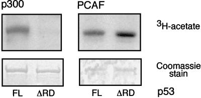
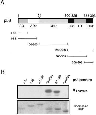
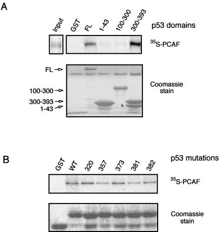
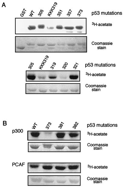
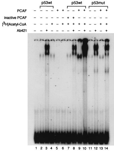
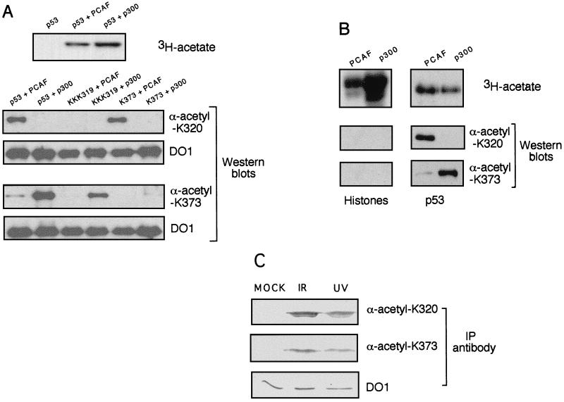
The p53 tumor suppressor protein is a sequence-specific transcription factor that modulates the response of cells to DNA damage. Recent studies suggest that full transcriptional activity of p53 requires the coactivators CREB binding protein (CBP)/p300 and PCAF. These coactivators interact with each other, and both possess intrinsic histone acetyltransferase activity. Furthermore, p300 acetylates p53 to activate its sequence-specific DNA binding activity in vitro. In this study, we demonstrate that PCAF also acetylates p53 in vitro at a lysine residue distinct from that acetylated by p300 and thereby increases p53's ability to bind to its cognate DNA site. We have generated antibodies to acetylated p53 peptides at either of the two lysine residues that are targeted by PCAF or p300 and have demonstrated that these antibodies are highly specific for both acetylation and the particular site. Using these antibodies, we detect acetylation of these sites in vivo, and interestingly, acetylation at both sites increases in response to DNA-damaging agents. These data indicate that site-specific acetylation of p53 increases under physiological conditions that activate p53 and identify CBP/p300 and PCAF as the probable enzymes that modify p53 in vivo.


Figures

FIG. 1
FIG. 1
Acetylation of p53 by PCAF and p300 in vitro. GST fused to either full-length p53 (p53FL) or a p53 truncation mutant (p53ΔRD) lacking 25 amino acids from its C terminus was acetylated with either p300 HAT (left) or PCAF HAT (right). Reaction products were separated by SDS-PAGE. The gel was stained with Coomassie blue to demonstrate that equivalent substrate was used in each reaction (lower panels), and [3H]acetate reaction products were subsequently visualized by autoradiography of the identical gel (upper panels).
FIG. 2
FIG. 2
Identification of p53 domains acetylated by PCAF. (A) Schematic diagram of p53 domain structure. A series of GST-p53 constructs are depicted containing functional domains of p53. AD1 and AD2, activation domains; DBD, DNA-binding domain; TD, tetramerization domain; RD1 and RD2, regulatory domains. (B) Acetylation of p53 functional domains by PCAF. GST bead slurry of each domain indicated that p53 truncation protein was subjected to acetylation assays with PCAF HAT. Reactions were analyzed as described in the legend for Fig. 1.
FIG. 3
FIG. 3
Interaction of p53 and PCAF in vitro. (A) Interaction of PCAF with p53 domains. GST-p53 truncation proteins, as shown in Fig. 1A, were incubated with in vitro-translated full-length [35S]methionine-labeled PCAF protein. Following incubation, the beads were washed and then subjected to SDS-PAGE, Coomassie staining, and autoradiography. PCAF input (2 μl) represents 10% of actual reaction volume. Unfused GST is present at the bottom of the gel in lane 1. The asterisk indicates a breakdown product of GST-p53(300–393). (B) Interaction of PCAF with GST-p53(300–393) bearing lysine substitutions, as indicated. Binding reactions were performed as described for panel A.
FIG. 4
FIG. 4
Determination of lysine residues acetylated by PCAF and p300. (A) PCAF acetylation of p53 lysine substitution mutants in RD1 and TD of p53. Lysine residues in the RD1 region (residues 300 to 324) and the tetramerization domain were mutated to arginine (aa 305, 319, 320, 321, 351, and 357). Each mutant was introduced within the 300 to 393 aa region of p53 fused to GST and named according to the position of the mutated amino acid(s). KKK319 stands for triple mutant at residues 319 to 321. Experimental details were the same as those described in the legend for Fig. 2. (B) Acetylation of p53 lysine substitution mutations in RD2 by PCAF and p300. GST fusion proteins containing lysine-to-arginine mutations at residues 373, 381, and 382 were analyzed for acetylation p300 (upper panel) or PCAF (lower panel). Experimental details were the same as those described in the legend for Fig. 2.
FIG. 5
FIG. 5
The effect of acetylation by PCAF on p53 sequence-specific binding to DNA. Purified recombinant human wild-type p53 (p53wt) or the triple lysine-to-arginine mutant (319/320/321; p53mut) was incubated with or without [3H]acetyl-CoA and either active PCAF or heat-treated PCAF (inactive). The reaction products were then incubated with a 32P-labeled p53 cognate site in binding buffer, and binding was determined by DNA mobility shift on a native polyacrylamide gel. The polyclonal antibody Ab421 was added as indicated. The mobility of the DNA probe alone is shown in lane 1. Multiple repetitions of the assay were performed, with quantitatively similar effects of K320 acetylation on DNA binding by p53.
FIG. 6
FIG. 6
Transcriptional activity of p53 K320 substitutions in Saos-2 cells. Cells were transfected with 0.1 μg of p53 wild type or mutant expression plasmid and 29 μg of pEp21-TK-SEAP. The level of activity of each p53 construct was compared to that of wild-type p53.
FIG. 7
FIG. 7
Acetylation of p53 in response to DNA-damaging agents. (A) Specificity of antibodies raised to p53 peptides encoding acetylated lysines 320 or 373. Recombinant p53 was incubated with either PCAF or p300 in the presence of [3H]acetyl-CoA. A sample of each reaction was subjected to SDS-PAGE and analyzed by autoradiography to determine the level of acetylation (upper panel). Samples of each reaction were then tested by Western analysis with the anti-K320-acetyl antibody (lower left) and anti-K373-acetyl antibody (lower right). The Western blot membranes were then reprobed with the anti-p53 monoclonal antibody DO1 to compare the amounts of p53 proteins used in each reaction. (B) Specificity of antibodies raised to acetylated p53 peptides against acetylated core histones. Equal amounts of free core histones or p53 were acetylated by PCAF or p300. Samples were treated the same way as described for panel A. (C) Effect of DNA-damaging agents on the acetylation state of p53. Whole cell extracts were prepared from untreated cells or cells treated with either ionizing radiation (IR) or UV radiation. Extracts were immunoprecipitated (IP) with anti-ac-K320, anti-ac-K373, or anti-p53 monoclonal DO1 antibody. The amount of p53 in each immunoprecipitate was then analyzed by Western blotting with DO1 antibody.

References

    1. Anderson, C. Personal communication.
    1. Arany Z, Sellers W R, Livingston D M, Eckner R. E1A-associated p300 and CREB-associated CBP belong to a conserved family of coactivators. Cell. 1994;77:799–800. - PubMed
    1. Arias J, Alberts A S, Brindle P, Claret F X, Smeal T, Karin M, Feramisco J, Montminy M. Activation of cAMP and mitogen responsive genes relies on a common nuclear factor. Nature. 1994;370:226–229. - PubMed
    1. Avantaggiati M L, Ogryzko V, Gardner K, Giordano A, Levine A S, Kelly K. Recruitment of p300/CBP in p53-dependent signal pathways. Cell. 1997;89:1175–1184. - PubMed
    1. Bannister A, Kouzarides T. The CBP co-activator is a histone acetyltransferase. Nature. 1996;384:641–643. - PubMed

Publication types

MeSH terms

LinkOut - more resources