. 1999 Feb;19(2):1301-12.
doi: 10.1128/MCB.19.2.1301.

Extracellular signal-regulated kinase 7 (ERK7), a novel ERK with a C-terminal domain that regulates its activity, its cellular localization, and cell growth

Affiliations

Extracellular signal-regulated kinase 7 (ERK7), a novel ERK with a C-terminal domain that regulates its activity, its cellular localization, and cell growth

M K Abe et al. Mol Cell Biol. 1999 Feb.

Abstract

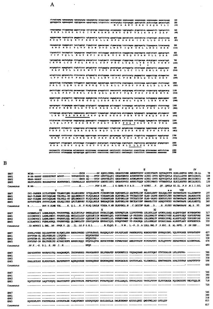
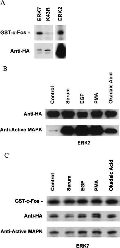
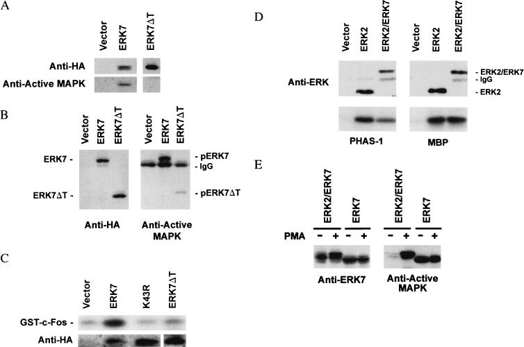
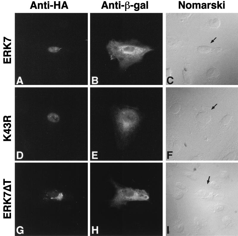
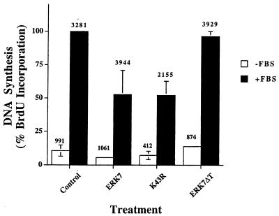
Mitogen-activated protein (MAP) kinases play distinct roles in a variety of cellular signaling pathways and are regulated through multiple mechanisms. In this study, a novel 61-kDa member of the MAP kinase family, termed extracellular signal-regulated kinase 7 (ERK7), has been cloned and characterized. Although it has the signature TEY activation motif of ERK1 and ERK2, ERK7 is not activated by extracellular stimuli that typically activate ERK1 and ERK2 or by common activators of c-Jun N-terminal kinase (JNK) and p38 kinase. Instead, ERK7 has appreciable constitutive activity in serum-starved cells that is dependent on the presence of its C-terminal domain. Interestingly, the C-terminal tail, not the kinase domain, of ERK7 regulates its nuclear localization and inhibition of growth. Taken together, these results elucidate a novel type of MAP kinase whereby interactions via its C-terminal tail, rather than extracellular signal-mediated activation cascades, regulate its activity, localization, and function.


Figures

FIG. 1
FIG. 1
Primary structure of ERK7 and comparison with other members of the MAP kinase family. (A) The nucleotide and deduced protein sequence of ERK7 cDNA. The threonine and tyrosine residues within the activation motif are marked with asterisks. Putative SH3 binding motifs, PXXP, in the C-terminal region are underlined. The putative nuclear localization sequence, PARKRGP, is doubly underlined. (B) Computer-generated alignments (GeneWorks, Oxford Molecular Group, Inc., Campbell, Calif.) of ERK7, ERK1, ERK2, and ERK5/BMK1. The C-terminal regions of ERK7 and ERK5 were not aligned. Roman numerals indicate the 11 conserved protein kinase regions. The conserved threonine/tyrosine regulatory residues are marked with asterisks.
FIG. 2
FIG. 2
Tissue distribution of ERK7. (A) A rat multiple tissue poly(A)+ RNA Northern blot (Clontech) was probed with radiolabeled ERK7. The positions of the RNA size markers in kilobases are illustrated on the right. (B) Lysates (100 μg) from multiple rat tissues, H19-7 and PC12 cells, were immunoblotted with the anti-ERK7 antiserum. Whole-cell lysates (50 μg) from COS cells transiently transfected with either pcDNA3 or HA-ERK7 are shown in the two left-hand lanes. The exposure time needed to detect endogenous ERK7 was considerably longer (4 min) than that needed to detect the ectopically expressed HA-ERK7 (<2 s).
FIG. 3
FIG. 3
ERK7 is TEY phosphorylated and has constitutive activity in COS cells. (A) Ectopically expressed ERK7 in mammalian cells is constitutively TEY phosphorylated. COS cells were transfected with epitope-tagged ERK7, the empty mammalian expression vector pcDNA3, or various ERK7 mutants. A kinase-inactive ERK7 mutant, K43R, was made by replacing the highly conserved lysine in the ATP binding region with arginine. The TEY activation domain was mutated by either changing the threonine residue to alanine, T175A, changing the tyrosine residue to phenylalanine, Y177F, or using both mutations, T175A Y177F. Lysates from serum-starved transfected cells were prepared and analyzed by immunoblotting with either the anti-HA 12CA5 monoclonal antibody (left) or the anti-active MAP kinase antibody (right). The data shown are representative of three experiments. (B) COS cells were transiently transfected with either pcDNA3, HA-ERK7, or K43R, serum starved for 24 h, and lysed with 1% TLB. Following immunoprecipitation with the anti-HA 12CA5 monoclonal antibody, enzyme activity was measured by an immune complex kinase assay with 2 μg of either GST–c-Fos, GST–c-Myc, GST-Elk1, PHAS1, GST–c-Jun, GST-ATF2, or MBP as noted on the right. The immunoprecipitated proteins were analyzed by Western blotting with the anti-HA 3F10 monoclonal antibody. A representative blot of immunoprecipitated protein is displayed at the bottom. ERK7 is seen as a single band as a result of the shorter duration of electrophoresis and the higher percentage acrylamide gel used for the in vitro kinase assays. (C) COS cells were transiently transfected with pcDNA3, ERK7, K43R, or the TEY mutants, serum starved for 24 h, and lysed with 1% TLB. Following immunoprecipitation with anti-HA 12CA5 monoclonal antibody, enzyme activity was measured by an immune complex kinase assay with 2 μg of GST–c-Fos (top). Western analysis of the immunoprecipitated proteins was performed with the anti-HA 3F10 monoclonal antibody (second row), the anti-ERK7 antiserum (third row), and the anti-active MAP kinase antibody (bottom). The data shown are representative of three experiments.
FIG. 4
FIG. 4
ERK7 is TEY phosphorylated and has constitutive activity in CV-1 cells. (A) CV-1 cells were transiently transfected either with pcDNA3, ERK7, K43R, or ERK2. Following serum starvation for 24 h, the cells were lysed with 1% TLB. Whole-cell lysates (left) were subjected to Western analysis with either anti-HA monoclonal antibody (top) or anti-active MAP kinase antibody (bottom). CV-1 cells transiently transfected with ERK2 (right) were treated with 10% FBS for 5 min or left untreated. Epitope-tagged ERK2 was immunoprecipitated with the anti-HA 12CA5 monoclonal antibody and analyzed by immunoblotting with either anti-HA monoclonal antibody (top) or anti-active MAP kinase antibody (bottom). (B) CV-1 cells were transiently transfected with pcDNA3, ERK7, or K43R, serum starved for 24 h, and lysed with 1% TLB. Following immunoprecipitation with the anti-HA 12CA5 monoclonal antibody, enzyme activity was measured by an immune complex kinase assay with 2 μg of GST–c-Fos. ERK7 is seen as a single band as a result of the shorter duration of electrophoresis and the higher-percentage acrylamide gel used for the in vitro kinase assays. The data shown are representative of three experiments.
FIG. 5
FIG. 5
ERK7 and ERK2 are differentially regulated. (A) The relative kinase activity of ERK7 in serum-starved COS cells is significantly greater than that of ERK2. COS cells were transiently transfected with equal amounts of either HA-ERK7 or HA-ERK2. Following serum starvation for 24 h, the cells were lysed with 1% TLB. The ERKs were immunoprecipitated with anti-HA 12CA5 monoclonal antibody and then either assayed directly for kinase activity under equivalent conditions with 2 μg of GST–c-Fos as a substrate (top) or immunoblotted with anti-HA 3F10 monoclonal antibody (bottom). ERK7 is seen as a single band as a result of the shorter duration of electrophoresis and the higher-percentage acrylamide gel used for the in vitro kinase assays. (B) Phosphorylation of the activation domain of ERK2 is stimulated by extracellular factors. COS cells were transiently transfected with ERK2. The cells were pooled and split equally. Following serum starvation for 24 h, the cells were treated with 20% FBS, 50 ng of EGF per ml, 200 nM PMA, or 200 nM okadaic acid or left untreated. The cells were lysed with 1% TLB, and the epitope-tagged proteins were immunoprecipitated with the anti-HA 12CA5 monoclonal antibody. Following separation by SDS-PAGE and transfer to nitrocellulose, ERK2 was subjected to immunoblotting with either anti-HA monoclonal antibody (top) or anti-active MAP kinase antibody (bottom). (C) Phosphorylation of the activation domain of ERK7 is unaffected by extracellular factors. COS cells were transiently transfected with ERK7. The cells were pooled and split equally. Following serum starvation for 24 h, the cells were treated with 20% FBS, 50 ng of EGF per ml, 200 nM PMA, or 200 nM okadaic acid or left untreated. The cells were lysed with 1% TLB, and the epitope-tagged proteins were immunoprecipitated with the anti-HA 12CA5 monoclonal antibody. Following immunoprecipitation, the enzyme activity of ERK7 was measured by an immune complex kinase assay with 2 μg of GST–c-Fos (top). Western analysis of the immunoprecipitated proteins was performed with the anti-HA 3F10 monoclonal antibody (middle). TEY phosphorylation of ERK7 was analyzed by immunoblotting with anti-active MAP kinase antibody (bottom).
FIG. 6
FIG. 6
The C-terminal domain is required for ERK7 activity. COS cells were transiently transfected with either pcDNA3, ERK7, or an epitope-tagged ERK7 mutant lacking the C-terminal domain, ERK7ΔT. The cells were serum starved for 24 h and lysed with 1% TLB. (A) Whole-cell lysates were evaluated by Western analysis with either the anti-HA 12CA5 monoclonal antibody (top) or anti-active MAP kinase antibody (bottom). (B) Epitope-tagged proteins immunoprecipitated with the anti-HA 12CA5 monoclonal antibody were detected with either the peroxidase-conjugated anti-HA monoclonal antibody (left) or anti-active MAP kinase antibody (right). (C) The epitope-tagged wild-type ERK7, kinase-inactive K43R mutant, and truncated ERK7ΔT mutant were immunoprecipitated with the anti-HA 12CA5 monoclonal antibody. Immune complex in vitro kinase activity was measured with 2 μg of GST–c-Fos as a substrate. (D) COS cells were transiently transfected with ERK2 or ERK2/ERK7, a chimera consisting of ERK2 and the C-terminal region of ERK7. Following stimulation with serum, the epitope-tagged proteins were purified by immunoprecipitation with the anti-HA 12CA5 monoclonal antibody. Immune complex in vitro kinase activity was measured using 2 μg of either PHAS1 (left) or MBP (right) as a substrate. (E) COS cells were transiently transfected with ERK2/ERK7 or ERK7. Following serum starvation for 24 h, the cells were stimulated without or with PMA. Whole-cell lysates were subjected to Western analysis with either anti-ERK7 antiserum (left) or anti-active MAP kinase antibody (right). The data shown are representative of three experiments.
FIG. 7
FIG. 7
MKP-1 dephosphorylates ERK7. COS cells were cotransfected with either the wild-type HA-ERK7 or HA-ERK2 and either MKP-1 or a control plasmid. Following serum starvation for 24 h, the cells were stimulated with 10% FBS for 5 min and cell lysates were prepared. Epitope-tagged ERK7 and ERK2 were immunoprecipitated with the 12CA5 monoclonal antibody and assessed by Western analysis with either anti-HA 3F10 monoclonal antibody (top) or anti-active MAP kinase antibody (bottom). Whole-cell lysate (left) from ERK7-transfected COS cells was also assessed by Western analysis with either anti-HA 3F10 monoclonal antibody (top) or anti-active MAP kinase antibody (bottom). The data shown are representative of three experiments.
FIG. 8
FIG. 8
Cellular localization of ectopically expressed ERK7, K43R, or ERK7ΔT in serum-starved CV-1 cells. CV-1 cells were cotransfected with either epitope-tagged ERK7 (A to C), K43R (D to F), or ERK7ΔT (G to I) and pCMV–β-gal. (A, D, and G) Immunofluorescent staining for HA-tagged proteins; (B, E, and H) the same cells after immunofluorescent staining for β-galactosidase; differential interference contrast (Nomarski) micrographs showing cells with double immunofluorescent staining (arrows) (C, F, and I). The data shown are representative of three experiments.
FIG. 9
FIG. 9
Effect of ERK7 on the proliferation of CV-1 cells. CV-1 cells were transfected with either pcDNA3, ERK7, K43R, or ERK7ΔT and pCMV–β-gal. In situ assay of β-galactosidase and immunostaining of BrdU incorporation were performed as described in Materials and Methods. The β-galactosidase-expressing cells were scored as proliferating (BrdU-positive) or nonproliferating (BrdU-negative) cells, without (□) and with (■) serum stimulation for 24 h. The total number of cells counted in each group is noted at the top of each column. The data represent the change in fractional BrdU labeling of serum-stimulated cells for each treatment group normalized to that of serum-stimulated control cells, which was defined as 100%. The data are derived from three independent experiments, and the means and standard deviations are indicated. Where not shown, the error bars were too small to be seen on the graph. Analysis by the one-tailed Student t test indicates that the inhibition by ERK7 (P < 0.01) and K43R (P < 0.002) is statistically significant.

References

    1. Abe J, Kusuhara M, Ulevitch R J, Berk B C, Lee J-D. Big mitogen-activated protein kinase 1 (BMK1) is a redox-sensitive kinase. J Biol Chem. 1996;271:16586–16590. - PubMed
    1. Abe J, Takahashi M, Ishida M, Lee J-D, Berk B C. c-Src is required for oxidative stress-mediated activation of big mitogen-activated protein kinase 1. J Biol Chem. 1997;272:20389–20394. - PubMed
    1. Abe M K, Chao T S, Solway J, Rosner M R, Hershenson M B. Hydrogen peroxide stimulates mitogen-activated protein kinase in bovine tracheal myocytes: implications for human airway disease. Am J Respir Cell Mol Biol. 1994;11:577–585. - PubMed
    1. Altschul S F, Gish W, Miller W, Myers E W, Lipman D J. Basic local alignment search tool. J Mol Biol. 1990;215:403–410. - PubMed
    1. Anderson N G, Maller J L, Tonks N K, Sturgill T W. Requirement for integration of signals from two distinct phosphorylation pathways for activation of MAP kinase. Nature. 1990;343:651–653. - PubMed

Publication types

MeSH terms

Substances

Associated data