. 1999 Feb;19(2):1401-9.
doi: 10.1128/MCB.19.2.1401.

A single amino acid substitution in the v-Eyk intracellular domain results in activation of Stat3 and enhances cellular transformation

Affiliations

A single amino acid substitution in the v-Eyk intracellular domain results in activation of Stat3 and enhances cellular transformation

D Besser et al. Mol Cell Biol. 1999 Feb.

Abstract

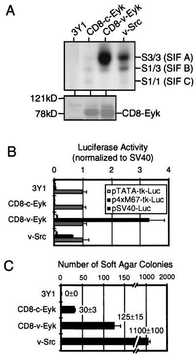
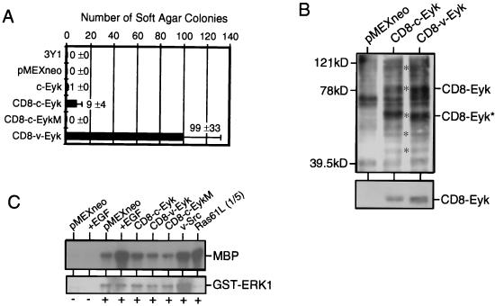
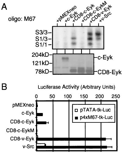
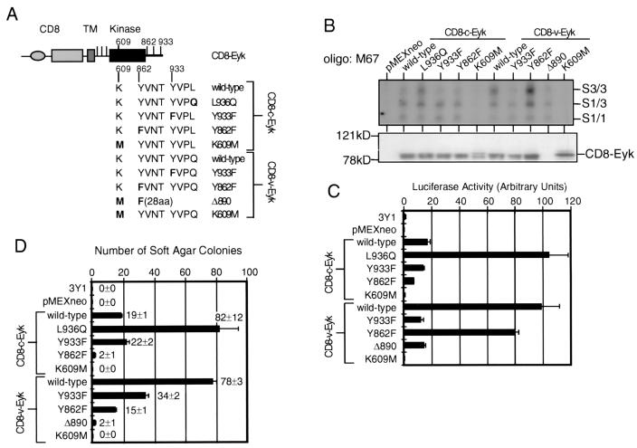
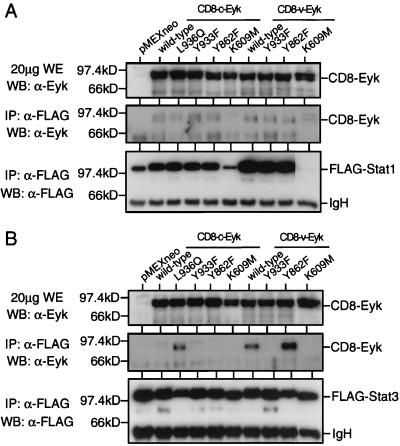
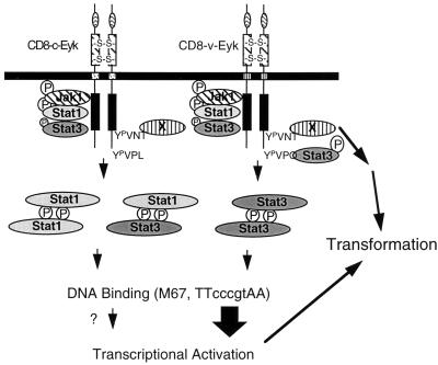
The receptor tyrosine kinase Eyk, a member of the Axl/Tyro3 subfamily, activates the STAT pathway and transforms cells when constitutively activated. Here, we compared the potentials of the intracellular domains of Eyk molecules derived from c-Eyk and v-Eyk to transform rat 3Y1 fibroblasts. The v-Eyk molecule induced higher numbers of transformants in soft agar and stronger activation of Stat3; levels of Stat1 activation by the two Eyk molecules were similar. A mutation in the sequence Y933VPL, present in c-Eyk, to the v-Eyk sequence Y933VPQ led to increased activation of Stat3 and increased transformation efficiency. However, altering another sequence, Y862VNT, present in both Eyk molecules to F862VNT markedly decreased transformation without impairing Stat3 activation. These results indicate that activation of Stat3 enhances transformation efficiency and cooperates with another pathway to induce transformation.


Figures

FIG. 1
FIG. 1
v-Eyk-transformed cells show higher binding activity primarily to Stat3 and have higher STAT-specific transcription compared to nontransformed cells. (A) Nuclear extracts (2 μg) from 3Y1 fibroblasts (3Y1) or 3Y1 cells stably transformed with CD8–c-Eyk, CD8–v-Eyk, or v-Src (0.32 × 10⁶ cells each) were used in an EMSA with 2 ng of 32P-labeled STAT-specific oligonucleotide M67 (upper panel). The positions of oligonucleotides shifted by a homodimer of Stat3 [S3/3 factor (SIF A)], by a heterodimer of Stat3 and Stat1 [S1/3 (SIF B)], or by a homodimer of Stat1 [S1/1 (SIF C)] are indicated. Eyk expression levels were detected in cytoplasmic extracts from these cells by Western blotting (lower panel). The positions of CD8-Eyk molecules and of the molecular weight standards are indicated. (B) Cell lines (0.06 × 10⁶ cells each) described above were transfected with 100 ng of luciferase expression vector pTATA-tk-Luc, p4xM67-tk-Luc (STAT-responsive promoter), or pSV40-Luc. Luciferase activity in 10 μg of extract was determined as described in Materials and Methods. The values were normalized with respect to that of pSV40-Luc in each cell line. Each column represents the average of duplicate samples, with the positive SD indicated as an error bar (n = 2). (C) Cells (104) from the cell lines used for panel A were plated in soft agar, and colonies were counted after 2 weeks. Each column represents the average of two samples, with the positive SD indicated as an error bar (n = 2). The original numbers are indicated beside the columns.
FIG. 2
FIG. 2
Transformation efficiency and activation of different signaling events in 3Y1 cells transiently expressing different Eyk constructs. (A) 3Y1 cells (0.32 × 10⁶) were transfected with 2 μg of the indicated constructs and 2 days later transferred to soft agar; colony numbers were determined 3 weeks later. Each column represents the average of two samples, with the positive SD indicated as an error bar (n = 2). The original numbers are indicated beside the columns. (B) 3Y1 fibroblasts were transfected with 15 μg of pMEX/neo, pMEX/CD8–c-Eyk (CD8–c-Eyk), or pMEX/CD8–v-Eyk (CD8–v-Eyk). Two days later, whole extracts were prepared and tyrosine-phosphorylated proteins (asterisks in the upper panel) and Eyk protein levels (lower panel) were detected in 20 μg of whole extract by Western blotting using a phosphotyrosine-specific antibody or an Eyk-specific antibody, respectively. (C) 3Y1 fibroblasts (0.15 × 10⁶ cells) were transfected with 100 ng of pEBG (−) or pEBG/erk1 (+) together with 500 ng of plasmid expressing the indicated protein. Two days later, the cells were either treated with 50 ng of EGF per ml (+EGF) for 10 min or left untreated, and whole-cell extracts were prepared. GST-ERK1 was precipitated by using glutathione-beads, and kinase activity toward MBP was determined (upper panel). GST-ERK1 was detected with a GST-specific antibody (Santa Cruz) (lower panel). In the rightmost lane, one-fifth of the kinase reaction for Ras61L was loaded to avoid overexposure.
FIG. 3
FIG. 3
Constitutively active Eyk constructs induce Stat3 DNA binding and STAT-specific transcriptional activation to different levels. (A) 3Y1 cells were transfected without or with 2 μg of plasmids expressing the indicated proteins. Two days after transfection, the cells were harvested and nuclear extracts were prepared. DNA binding activity (upper panel) and Eyk expression levels (lower panel) were determined as described for Fig. 1A. Western blotting was performed with an Eyk-specific antibody. Oligo, oligonucleotide. (B) 3Y1 cells were transfected with 100 ng of indicated luciferase reporter construct alone or together with 50 ng of the indicated construct. Luciferase activity was determined as described for Fig. 1B. Each column represents the average of two samples, with the positive SD indicated as an error bar (n = 2).
FIG. 4
FIG. 4
A YVPQ sequence in v-Eyk is responsible for increased DNA binding of Stat3 and transcriptional activation; tyrosine 862 is not responsible for Stat3 activation; Stat3 activation by CD8–v-Eyk and signaling events downstream of tyrosine 862 cooperate in transformation efficiency. (A) Graphic representation of the constructs used in for panels B to D. The CD8α extracellular domain (CD8), the transmembrane domain (TM), and the intracellular domain including the kinase domain (kinase) and tyrosine residues (vertical bars) are shown. Point mutations in four different sites are indicated in the context of wild-type CD8–c-Eyk and CD8–v-Eyk: leucine in CD8–c-Eyk 936 to glutamine and tyrosine 933 to phenylalanine, tyrosine 862 to phenylalanine, and lysine 609 to methionine (kinase dead) in both CD8-Eyk constructs. Δ890 denotes a COOH-terminal deletion construct of CD8–v-Eyk displaying the Y862F mutation followed by 28 unrelated amino acids (aa) and a stop codon. (B) 3Y1 cells were transfected with 2 μg of the indicated construct. Two days after transfection, the cells were harvested and nuclear and cytoplasmic extracts were prepared. EMSA using oligonucleotide M67 as the probe and Western blot analysis for Eyk expression were carried out as described for Fig. 1A. (C) 3Y1 cells were transfected with 100 ng of p4xM67-tk-Luc together without or with 50 ng of the indicated construct. Luciferase activity was determined 2 days later as described for Fig. 1B. (D) 3Y1 cells were transfected with 2 μg of the indicated construct. The transformation efficiency was determined in a soft agar assay as described for Fig. 3D.
FIG. 5
FIG. 5
The YVPQ sequence in v-Eyk is responsible for increased Stat3 but not Stat1 binding of Eyk. (A) 293T cells were transfected with 1 μg of pRcCMV/stat1-FLAG together with 1 μg of the indicated construct. Two days after transfection, the cells were harvested and whole-cell extracts were prepared. FLAG-tagged Stat1 was immunoprecipitated (IP) from 400 μg of extract, using the FLAG-specific antibody M2 (α-FLAG). Whole-cell extracts (WE; upper panel) and immunoprecipitates (middle and lower panels) were analyzed by Western blotting (WB) for expressed or coprecipitated Eyk levels (upper and middle panel) or FLAG-tagged Stat1 levels as described for Fig. 1A. The positions of FLAG-tagged Stat1 (Stat1) and of the immunoglobulin heavy chain (IgH) are indicated. (B) Same as panel A but with FLAG-tagged Stat3 (pRcCMV/stat3-FLAG).
FIG. 6
FIG. 6
A model for signaling and transformation events downstream of CD8-Eyk. The constitutively dimerized and activated Eyk constructs activate Jak1 (74) and Stat1 and slightly activate Stat3 via an unidentified region NH2 terminal to tyrosine 862 (Fig. 4, Δ890 construct). The Stat3-binding site Y933VPQ is present only in CD8–v-Eyk and leads to increased Stat3 DNA-binding activity, 10-fold-higher activation of STAT-responsive transcription, and a 10-fold-higher number of soft agar colonies, indicating that Stat3 enhances transformation. The influence of the Stat1 homodimer and the Stat1-Stat3 heterodimer on transcriptional activation is not clear. Another protein (X) that binds to Y862VNT is activated downstream of CD8–c-Eyk and CD8–v-Eyk and leads to activation of other signaling pathways independent of Stat1 or Stat3. The activation of this pathway is required for transformation by both CD8–c-Eyk and CD8–v-Eyk.

Similar articles

Cited by

References

    1. Bellosta P, Costa M, Lin D A, Basilico C. The receptor tyrosine kinase ARK mediates cell aggregation by homophilic binding. Mol Cell Biol. 1995;15:614–625. - PMC - PubMed
    1. Bellosta P, Zhang Q, Goff S P, Basilico C. Signaling through the ARK tyrosine kinase receptor protects from apoptosis in the absence of growth stimulation. Oncogene. 1997;15:2387–2397. - PubMed
    1. Besser D, Presta M, Nagamine Y. Elucidation of a signaling pathway induced by FGF-2 leading to uPA gene expression in NIH 3T3 fibroblasts. Cell Growth Differ. 1995;6:1009–1017. - PubMed
    1. Biscardi J S, Denhez F, Buehler G F, Chesnutt D A, Baragona S C, O’Bryan J P, Der C J, Fiordalisi J J, Fults D W, Maness P F. Rek, a gene expressed in retina and brain, encodes a receptor tyrosine kinase of the Axl/Tyro3 family. J Biol Chem. 1996;271:29049–29059. - PubMed
    1. Boccaccio C, Ando M, Tamagnone L, Bardelli A, Michieli P, Battistini C, Comoglio P M. Induction of epithelial tubules by growth factor HGF depends on the STAT pathway. Nature. 1998;391:285–288. - PubMed

Publication types

MeSH terms